Prominent ears are a fairly common and emotionally charged facial variation. Both children and adults may suffer a damaged psyche secondary to outside ridicule and self-criticism from ears that “stick out.” The external ear anatomy is intricate, with thin skin and resilient cartilage. These underlying characteristics make the ears prone to the overt display of surgical correction. This article details the various acute and chronic complications of otoplasty and includes tips and pearls to help prevent and treat these occurrences.
Key points
- •
Do not overtighten antihelix horizontal mattress sutures to avoid the hidden helix.
- •
Take full-thickness cartilage bites including the anterior perichondrium to avoid cartilage pull-through of mattress sutures and relapse.
- •
Avoid external canal narrowing by placing posteriorly oriented concha-mastoid sutures or excising cartilage.
- •
Cartilage splitting or scoring techniques run the risk of visible cartilage irregularities or sharp edges.
- •
Do not rely on skin excision to hold ear position.
Introduction
Auricular proportions and defining characteristics of the pinna have been well documented and defined in the literature. Although the function of the pinna in regards to hearing is minor, the social and psychological impact of having protruding or prominent ears is profound. This anatomic variation occurs in approximately 5% of the population, and usually by school age, children become very self-conscious of their “Dumbo or mouse ears.” Correcting this deformity can help children gain self-esteem and prevent social ridicule.
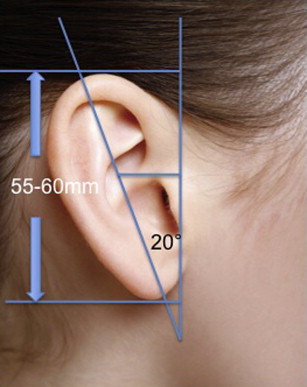
The earliest auricular surgery dates back to seventh century in the writings of Sushruta, an Indian healer whose teachings were rooted in Ayurveda. In 1848, JF Dieffenbach published a novel otoplasty technique using sutures to pin the ears back through an incision made in the post-auricular sulcus. Otoplasty surgery is usually directed toward 2 specific areas, the conchal bowl, which is often hypertrophied, and the anti-helix, which may be flattened and/or underdeveloped, or a combination of both. The aesthetically pleasing auricle projects approximately 20° to 30° from the skull ( Fig. 1 ). The length of the ear is approximately 55 to 60 mm when fully developed, and the width is approximately 55% of the length. Furthermore, along its vertical axis, the auricle sits a gentle 20° posterior.
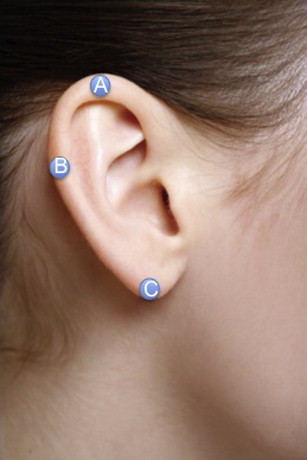
One of the better objective measurements is the helical to scalp distance, which is measured at 3 points, the superior point of the helical rim, the midpoint of the helix, and the lobule ( Fig. 2 ). These distances range from 10 to 12 mm, 16 to 20 mm, and 20 to 22 mm, respectively. In bilateral otoplasty, these measurements serve as a guide for the surgeon, and documentation of these distances is important in creating symmetry. In the unilateral otoplasty, measurements from the aesthetically better ear can be used to gauge setback of the protruding one.
Currently, 2 schools of thought predominate regarding otoplastic surgery summarized broadly as cartilage cutting and cartilage sparring techniques. The former involves removal of cartilage and the scoring of cartilage, whereas the latter involves contouring by placement of either permanent or absorbable sutures. Cartilage cutting techniques tend to be favored in Europe and are liked due to the durability of the correction over time, although there exists a higher risk of anterior cartilage irregularities. Typically, the otoplasty is performed with wedge excisions, scoring, abrasion, and cartilaginous incisions on either the anterior or the posterior cartilage surface in an attempt to counteract the spring of the unfurled antihelix. In North America, cartilage sparring techniques predominate, namely the Furnas and the Mustarde, or variations on these techniques. The advantages include less scaring, allowance of easy suture adjustment, preservation of the cartilage framework, and prevention of contour irregularities. Where it may fall short is with stability of the operation over time.
Fortunately, complications of otoplasty are relatively uncommon and can usually be avoided with meticulous preoperative, intraoperative, and post-operative care from both the patient and surgeon perspective. The surgeon must be familiar with the range of complications and must appropriately counsel the patient before surgery about these risks, in addition to being ready to confidently handle them if they arise. The cumulative rate of early complication has been cited from 0% to 8.4%, with information on late complications varying greatly between 0% and 47.3%. Granted, the literature is varied and most studies are retrospective reviews of surgical cases for individual institutions or surgeons. Most important to the otoplastic surgeon is a depth of understanding of the various techniques and complications so that the best result is obtained ( Table 1 ).
| Early | Late |
|---|---|
| Hematoma | Hypertrophic scar/keloids |
| Infection/Perichondritis | Suture complications |
| Cartilage/Skin necrosis | Recurrence |
|
Introduction
Auricular proportions and defining characteristics of the pinna have been well documented and defined in the literature. Although the function of the pinna in regards to hearing is minor, the social and psychological impact of having protruding or prominent ears is profound. This anatomic variation occurs in approximately 5% of the population, and usually by school age, children become very self-conscious of their “Dumbo or mouse ears.” Correcting this deformity can help children gain self-esteem and prevent social ridicule.
The earliest auricular surgery dates back to seventh century in the writings of Sushruta, an Indian healer whose teachings were rooted in Ayurveda. In 1848, JF Dieffenbach published a novel otoplasty technique using sutures to pin the ears back through an incision made in the post-auricular sulcus. Otoplasty surgery is usually directed toward 2 specific areas, the conchal bowl, which is often hypertrophied, and the anti-helix, which may be flattened and/or underdeveloped, or a combination of both. The aesthetically pleasing auricle projects approximately 20° to 30° from the skull ( Fig. 1 ). The length of the ear is approximately 55 to 60 mm when fully developed, and the width is approximately 55% of the length. Furthermore, along its vertical axis, the auricle sits a gentle 20° posterior.
One of the better objective measurements is the helical to scalp distance, which is measured at 3 points, the superior point of the helical rim, the midpoint of the helix, and the lobule ( Fig. 2 ). These distances range from 10 to 12 mm, 16 to 20 mm, and 20 to 22 mm, respectively. In bilateral otoplasty, these measurements serve as a guide for the surgeon, and documentation of these distances is important in creating symmetry. In the unilateral otoplasty, measurements from the aesthetically better ear can be used to gauge setback of the protruding one.
Currently, 2 schools of thought predominate regarding otoplastic surgery summarized broadly as cartilage cutting and cartilage sparring techniques. The former involves removal of cartilage and the scoring of cartilage, whereas the latter involves contouring by placement of either permanent or absorbable sutures. Cartilage cutting techniques tend to be favored in Europe and are liked due to the durability of the correction over time, although there exists a higher risk of anterior cartilage irregularities. Typically, the otoplasty is performed with wedge excisions, scoring, abrasion, and cartilaginous incisions on either the anterior or the posterior cartilage surface in an attempt to counteract the spring of the unfurled antihelix. In North America, cartilage sparring techniques predominate, namely the Furnas and the Mustarde, or variations on these techniques. The advantages include less scaring, allowance of easy suture adjustment, preservation of the cartilage framework, and prevention of contour irregularities. Where it may fall short is with stability of the operation over time.
Fortunately, complications of otoplasty are relatively uncommon and can usually be avoided with meticulous preoperative, intraoperative, and post-operative care from both the patient and surgeon perspective. The surgeon must be familiar with the range of complications and must appropriately counsel the patient before surgery about these risks, in addition to being ready to confidently handle them if they arise. The cumulative rate of early complication has been cited from 0% to 8.4%, with information on late complications varying greatly between 0% and 47.3%. Granted, the literature is varied and most studies are retrospective reviews of surgical cases for individual institutions or surgeons. Most important to the otoplastic surgeon is a depth of understanding of the various techniques and complications so that the best result is obtained ( Table 1 ).
| Early | Late |
|---|---|
| Hematoma | Hypertrophic scar/keloids |
| Infection/Perichondritis | Suture complications |
| Cartilage/Skin necrosis | Recurrence |
|
Related posts:

Stay updated, free articles. Join our Telegram channel

Full access? Get Clinical Tree


