Complications and prevention of complications in brow lift are presented. A discussion of anatomic features of the brow introduces the article in keeping with the focus that a thorough understanding of the anatomy, patient variations, and potential complications is requisite for surgeons performing forehead rejuvenation. The varying approaches to brow lift are discussed. Complications reviewed are bleeding, nerve injury, scarring, alopecia, brow asymmetry, and brow elevation overcorrection or undercorrection.
Key points
- •
A thorough understanding of brow anatomy, patient variations, and potential complications is requisite for surgeons performing forehead rejuvenation.
- •
As with every aspect of facial plastic surgery, the prevention of postoperative patient dissatisfaction begins with a careful patient history inclusive of psychiatric history, motivation and expectations for surgery, and investment in the outcome.
- •
In-depth knowledge of a patient’s medical history is important to identify patients who are poor surgical candidates and are at high risk of medical complications.
- •
Overall, complications can be minimized by a thorough preoperative history and physical examination combined with meticulous technique, conservative suspension, and a tension-free wound closure.
Introduction
Patients often present with the complaint of tired-appearing eyes with excessive eyelid skin. The astute aesthetic surgeon may note on examination the contribution of brow ptosis to the overall heavy appearance of the patient’s upper eyelids with or without true dermatochalasis. Rejuvenation of the forehead may result in a more youthful appearance and may reduce or negate the necessity of blepharoplasty in some patients. A thorough understanding of the anatomy, patient variations, and potential complications is requisite for surgeons performing forehead rejuvenation.
Forehead anatomy
Critical to facial rejuvenation is an understanding of the anatomy.
Brow Soft Tissue
In the forehead, the soft tissue layers include skin, subcutaneous tissue, muscle, fibrous galea, loose areolar tissue, and pericranium. The superficial musculoaponeurotic system in the inferior two-thirds of the face continues over the zygoma as the superficial temporoparietal fascia, which merges with the galea in the forehead. The temporal line, which is formed by the convergence of the deep and superficial layers of the deep temporal fascia investing the temporalis muscle with the frontal periosteum, meets the lateral margin of the galea to form the conjoined tendon. Another important landmark in brow lifting procedures is the densely adherent arcus marginalis along the supraorbital rim, which is where the frontal periosteum fuses with the galea. Release of these dense fibrous attachments in forehead rejuvenation is vital to produce the desired elevation and stabilization of the brow.
Brow Muscles
The muscles important to brow surgery are the frontalis muscle, paired corrugator supercilii muscles, and procerus muscle. The frontalis, which has a natural dehiscence in the midline, is a thin, fan-like muscle that acts largely to elevate the brow. This muscle is responsible for the development of horizontal rhytids with age.
The paired corrugator supercilii muscles have two heads, including transversely oriented fibers along the medial supraorbital rim acting to pull the brows medially, and oblique fibers that act to depress the medial brow. The corrugator muscles cause the vertically oriented rhytids that develop in the glabellar region.
The procerus muscle runs vertically from the nasal bones to the frontalis muscle and depresses the medial brow. It is responsible for the transversely oriented rhytids in the region of the radix. The medial brow is also depressed by the medial fibers of the orbicular is oculi muscles. Superior fibers of the latter depress the brow along its length.
Brow Innervation
Sensory innervation to the lateral and central forehead come from branches of the trigeminal nerve and run to the vertex of the scalp with subgaleal extensions. The ophthalmic division of the trigeminal nerve gives off the supratrochlear and supraorbital branches, whereas the maxillary division provides temporal sensation by the auriculotemporal and zygomaticotemporal branches. Motor innervation to the forehead is by the temporal branch of the facial nerve. The main trunk of the facial nerve exits the stylomastoid foramen, divides within the parotid, and travels deep to the superficial musculoaponeurotic system, giving off its terminal branches. The temporal branch is located just superficial to the zygomatic arch periosteum in the middle third, approximately 1.5 cm lateral to the lateral orbital rim. From here, it continues in the temporoparietal fascia to enter the undersurface of the brow musculature approximately 1 cm above the supraorbital rim. Appreciating and protecting the motor innervation is of the utmost importance during any approach to forehead lifting.
Brow Vasculature
There is a robust blood supply to the forehead from the internal and external carotid systems. Contributions of the internal carotid artery by the ophthalmic branch are the supratrocheal and supraorbital bundle, which travel with similarly names sensory nerves. These neurovascular bundles are found deep to the frontalis muscle until approximately 1 cm above the supraorbital rim when they pierce this muscle to run more superficially. The remainder of the blood supply can be found in the subcutaneous plane. The location of the vascular supply should be considered when deciding on a given approach to forehead lifting. Key venous anatomy is the sentinel vein, which runs superficial to the deep temporal fascia near the region of the frontozygomatic suture. Identification of this vein, particularly in endoscopic approaches, assists the surgeon in locating and avoiding the temporal branch of the facial nerve as it runs in close approximation and superficial to this vessel. In addition, the sentinel vein can pose a significant bleeding problem if not properly identified and preserved or cauterized.
Prevention of complications in brow surgery
Patient History
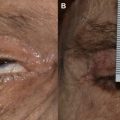
As with every aspect of facial plastic surgery, the prevention of postoperative patient dissatisfaction begins with a careful patient history inclusive of psychiatric history, motivation and expectations for surgery, and investment in the outcome. The presence of body dysmorphic disorder should be identified preoperatively and cosmetic surgical procedures in this patient group should be avoided. History of prior surgery should be elucidated. Concurrent desire for upper and/or lower eyelid rejuvenation should be discussed, and an ocular history taken to determine the presence of vision loss, dry eye syndrome, use of lubricating ophthalmic drops, or prior ocular surgery. Often, the symptoms may be subtle and include burning of eyes or a foreign body sensation. This history is key for avoidance of postoperative ocular complications.
In-depth knowledge of a patient’s medical history is important to identify patients who are poor surgical candidates and are at high risk of medical complications. Elective operations in high-risk patients should be carefully considered. Any personal or family history of coagulopathies should be noted, as should a personal history of a bleeding complication after prior surgery. An evaluation for bleeding disorders should be completed in these patients. Patients should be informed to avoid blood thinners and herbal supplements with blood-thinning properties.
Physical Examination
A thorough physical examination is requisite and knowledge of ideal brow position is vital. The distance from the midpupil to the upper edge of the eyebrow is 2.5 cm. The ideal brow position in females is considered to be above the supraorbital rim with a gentle arc and a peak located over the lateral limbus. The feminine brow is typically thicker medially and tapers laterally. For males, brow position is generally more horizontal, located at the supraorbital rim, and thicker. The presence of orbital and brow asymmetries should be noted, photographed, and discussed with the patient preoperatively.
A careful examination of the upper and lower eyelids should also be performed to identify dermatochalasis and blepharoptosis, which if not addressed concurrently may produce dissatisfaction in the postoperative forehead rejuvenation patient. Ptosis may be defined as palpebral fissure width measured at the central lid of less than 10 mm. Another method of assessing for ptosis is with the margin reflex distance-1, which measures the distance from the pupillary reflex to the central upper eyelid margin. Distances less than 4 mm may signify ptosis.
In addition to the patient history, the examination should assess for signs of dry eye syndrome, including a roughened or irritated cornea, mucoid deposition along the lid margin, and signs of chronic irritation. If there is any concern for dry eye syndrome, one should consider preoperative ophthalmologic evaluation and a Schirmer or tear breakup test should be performed. These symptoms may be exacerbated by periorbital surgery and forehead lifting.
Finally, the complete examination should include an assessment of skin type, photoaging status, presence of hair loss or thinning, location of the hairline, and presence of static and dynamic glabellar and forehead rhytids. The ideal hairline is approximately 5 cm from the upper edge of the brow. The length of the forehead and height of the hairline may influence the approach selected for rejuvenation.
Related posts:

Stay updated, free articles. Join our Telegram channel

Full access? Get Clinical Tree


