Lower eyelid blepharoplasty and midface lifting share a complex anatomy, which should be mastered before attempting these types of surgeries. In recent years, there have been significant contributions to rejuvenating this area. A thorough understanding of the rejuvenative approaches and their outcomes is imperative. The problem must be evaluated preoperatively to offer the appropriate technique and minimize complications.
Key points
- •
Performing the appropriate surgery could minimize the risk of complications.
- •
A mastery of the lower eyelid midfacial anatomy is essential.
- •
Each approach to lower lid blepharoplasty has its inherent complication.
- •
Understanding how complications are created is of the utmost importance.
Overview
Lower eyelid blepharoplasty and midface lifting share many details that must be mastered. A thorough understanding of lower eyelid and midfacial anatomy and their effects on aging is needed when addressing the rejuvenating needs of this complex location. Each individual’s anatomy must be considered when formulating a surgical plan, to minimize complications. Consideration should be given toward the underlying bone and the prominence of the globe/rim ratio, as well as the tone and elasticity of the eyelid, midface, and skin itself.
Anatomy
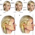
The superior border of the midface is defined as the line from the helix to the infraorbital rim, whereas the inferior border is defined as the line drawn from the oral commissure to the earlobe. The medial border is the nasolabial fold, whereas the lateral border is the masseter muscle.
The lower eyelid is divided into :
- •
Anterior lamellae: skin and orbicularis muscle. The orbicularis is divided into 3 sections:
- ○
Pretarsal section
- ○
Preseptal section
- ○
Orbital section
- ○
- •
Middle lamellae: septum separating orbital fat from the orbicularis muscle
- •
Posterior lamellae: conjunctiva and lower eyelid retractors inferiorly or tarsus superiorally
The lower eyelid fat pads are found between the orbital septum and lower eyelid retractors. The inferior oblique muscle separates the medial and central lower eyelid fat pads, and the arcuate expanse separates the central and lateral fat pads. Just below the orbital rim anatomically from anterior to posterior, we find the skin, orbital section of the orbicularis oculi, and the suborbicularis oculi fat pad (SOOF), periosteum, and bone. The zygomaticus muscles and the levator labii superioris muscles travel through the SOOF, inserting into the dermis, helping to define the nasolabial fold ( Fig. 1 ).
The surgeon must be familiar with the muscles of facial expression when approaching midface rejuvenation. These muscles include but are not limited to the:
- •
Levator labii superioris
- •
Levator labii superioris alaeque nasi
- •
Zygomaticus major
- •
Zygomaticus minor
The origin of the levator labii superioris muscle is just inferior to the inferior orbital rim inserting into the border of the upper lip, with its effect being elevation of the upper lip. The levator labii superioris muscle originates from the frontal process of the maxillary bone and inserts into both the upper lip and cartilage of the nasal alae. This muscle allows one to both raise the upper lip and flare the nostril. The zygomaticus major muscle arises lateral to the zygomaticus minor, whereas both muscles remain lateral to the levator labii superioris. The zygomaticus muscles insert at the upper lip corner, allowing for a superolateral movement of the mouth. These muscles lie in a plane superficial to the facial nerve.
The superficial musculoaponeurotic system (SMAS) is a musculofascial layer connecting the facial muscles with the dermis. This layer is continuous with the platysma inferiorly and the temporoparietal fascia superiorly.
Surgical options
In the past, blepharoplasty involved removing this herniated fat, leaving a concave appearance to the lower lid, as opposed to maintaining a uniform continuum between the lower eyelid and cheek junction, which represents a youthful appearance. Midface and lower eyelid aesthetic goals include decreasing the prominence of the nasolabial fold and restoring the lower eyelid-cheek junction into a single continuous unit.
Fillers
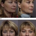
In addition to surgical techniques discussed later, fillers may be used to address the nasolabial and nasojugal fold and the overall volume loss that may occur with aging, which includes the cheek mound itself. Fillers currently used include autologous fat transfers, hyaluronic acid, silicone, and bovine collagen, polylactic acid, and hydroxyapatite. It is critical to ensure that the injections of the fillers are subdermal or submuscular. If the injection is too superficial, there is a risk for irregularities in the facial contour or even ulcerations. Fillers are not without inherent risks. Silicone injections can often migrate, cause severe granulomatous responses, and in some cases, lead to death. Skin testing is required for bovine collagen injections, because of potential allergic reactions. Hyaluronic acid has become the most widely used filler material but has been associated with arterial embolization. Autologous fat transfers are also commonly used, but the long-term viability of the fat transfers frequently varies. The concomitant use of facial implants, whether silicone, Medpor, or Gore-Tex, can easily increase the projection of the midface, adding to the aesthetic outcome and impression of the lift ( Fig. 2 ).
Transeyelid
The surgeon may access the midfacial structures through the eyelid either via a transconjunctival or subciliary incision; spacer grafts may be of use in the approach if necessary.
Transconjunctival Approach
- •
In the transconjunctival approach, an incision is made 3 to 4 mm below the caudal margin of the tarsal plate, because making the incision too close to the sulcus could lead to a contraction scar from the invasion of the orbital septum.
- •
The incision typically spans 1 mm lateral to the caruncle to the lateral canthus. In patients with significant lid laxity, lateral canthal resuspension may also be included.
- •
The orbital fat and its septa are noted and pushed posteriorly.
- •
Next, an incision is made above the arcus marginalis down to the periosteum of the orbital rim.
At this point, the surgeon may proceed either preperiosteal or subperiosteal.
Preperiosteal Approach
The preperiosteal dissection is adequate for patients with prominent tear trough deformities.
- •
If dissection is preperiosteal, one may then proceed with blunt dissection preperiosteally past the tear trough deformity.
- •
The SOOF is then identified, and a purchase is then secured to the arcus marginalis to provide the appropriate lift.
Subperiosteal Approach
If the surgeon opts for the subperiosteal approach, a more robust lift is achieved.
- •
An incision is made into the periosteum a few millimeters below the orbital rim.
- •
The periosteum is lifted off the maxilla, creating a subperiosteal plane ( Fig. 3 ).
Fig. 3
Transconjunctival midface lift with distal periosteotomy.
The dissection proceeds inferiorly to the nasal ala and laterally to the zygoma as allowed by the skin incision, with care taken at the insertion of the levator labii superioris as it encounters the infraorbital notch with all of its crucial contents.
- •
An incision is made in the periosteum in the form of a distal periosteotomy to allow for a complete release of the periosteum, at the inferior border (see Fig. 3 ).
- •
The distal end of the periosteum is then incised, allowing for free movement of the midface structures.
- •
The midfacial structures are then elevated superiorly, and the freely mobile periosteum is sutured to the periosteal cuff at the orbital rim to achieve the desired result.
- •
For a more robust lift, this release can be made continuous with a sublabial approach, in essence, degloving the entire midface, to allow for free mobility upward ( Fig. 4 ).
Related posts:
 An Algorithmic Approach to Multimodality Midfacial Rejuvenation Using a New Classification System for Midfacial Aging
 Dark Circles
Transconjunctival Lower Lid Blepharoplasty with and Without Fat Repositioning
The Transeyelid Midface Lift
Dark Circles
Transconjunctival Lower Lid Blepharoplasty with and Without Fat Repositioning
The Transeyelid Midface Lift
 Transtemporal Midface Lifting to Blend the Lower Eyelid-Cheek Junction
Autologous Fat Grafting for Midface Rejuvenation
Transtemporal Midface Lifting to Blend the Lower Eyelid-Cheek Junction
Autologous Fat Grafting for Midface Rejuvenation

Stay updated, free articles. Join our Telegram channel

Full access? Get Clinical Tree


