The transconjunctival technique for lower lid blepharoplasty, by virtue of a reduced rate of lower eyelid malposition, improved healing, and a concealed incision, has some appeal compared with the transcutaneous method. The transconjunctival approach to lower lid blepharoplasty provides access for both fat resection and transposition to soften tear trough deformities and correct infraorbital fat herniation. Transposing a contiguous orbital septum/fat unit is key for fat replacement and repositioning. Overall, there is a paradigm shift that has focused more on enhancing lower lid-cheek shape and contour and preserving orbital volume.
Key points
- •
The transconjuctival technique for lower eyelid blepharoplasty is a means of excising and repositioning lower eyelid fat with a decreased risk for eyelid retraction and ectropion.
- •
Lower lid laxity and tear trough deformities are important to evaluate preoperatively in a patient. Patients with a tear trough deformity require transposition rather than resection.
- •
Preseptal dissection exposes a contigous semptum/fat unit for transposition while a direct technique through the septum offers a resective approach.
- •
Ectropion can be corrected with conservative measures such as vertical massage and taping if identified early. More invasive interventions are necessary if rectraction is unappreciated.
Introduction
There are two main concerns in blepharoplasty procedures: the excess presence or malposition of fat and excess eyelid skin. The transconjunctival technique for lower lid blepharoplasty (TCB), by virtue of a reduced rate of lower eyelid malposition, improved healing, and a concealed incision, has some appeal compared with the transcutaneous method in addressing fat. Fat excision or reposition do not by themselves address dermatochalasis but can be combined with eyelid excision to acheive an optimal result.
Initial techniques of TCB focused on young patients with isolated fat prominence. The classic TCB concept of Castanares involved fat excision as the primary approach to address orbital fat herniation. Young patients with true excess or herniated orbital fat, a bulge in the lower eyelid, and minimal excess skin have had excellent results following fat removal. Determination of how much eyelid fat to resect in transconjunctival lower lid blepharoplasty was subjective, and over-resection of fat resection left a hollowed-out appearance that, paradoxically, created a more aged appearance. Older patients often have undesirable lower eyelid and midface contours following fat loss over the orbital rim, orbicularis ptosis, and malar descent, creating a tear trough deformity. In this setting, fat repositioning may avoid many of the disadvantages of transcutaneous skin-muscle flap techniques, while successfully correcting tear trough deformities using vascularized autogenous tissue. Fat can be repositioned or excised with transcutaneous surgery yet the risks of ectropion and cutaneous irregularities are greater.
Techniques to reposition infraorbital fat may involve either direct manipulation (redraping) of the pedicled fat from under the septum or fat repositioning through septal reset of the whole fat/septal unit. The fat can be repositioned into the supraperiosteal or subperiosteal plane with shorter time for supraperiosteal dissection versus the less traumatic and vascular dissection of the subperiosteal plane. A study by Yoo and colleagues concluded that, ultimately, the aesthetic results are comparable between the two approaches.
Septal reset involves the orbital septum and fat as one unit to be fixated to the periosteum, enabling the septum and periosteum to provide additional support for the underlying repositioned fat and avoid additional contour irregularities. Hamra originally described a technique incising the arcus marginalis and elevating the subseptal fat to the level of the orbital rim after partial resection of the orbital septum. In later modifications to the reset technique with sutures, the septum is advanced and tightened to the orbital rim. The integrity of the orbital septum is stated to be a key to fat replacement into the orbit. The main concern associated with septal reset is the disruption of the middle lamellae leading to contracture and shortening, but the extensile nature of the septum seems to make such a complication a rare coincident. Ptosis of the orbicularis oculi muscle is addressed by redraping the muscle via lateral plication with subsequent release of the orbicularis-retaining ligament, achieving significant softening of a visible orbital rim.
Anatomy
With age, the midfacial fat compartments deflate with progressive exposure of the underlying infraorbital rim, and an attenuated orbital septum allows fat to bulge forward above the orbital rim. As the supporting tissues of the eyebrow, cheek, and jaw descend, a double-convex pattern of the eyelid-cheek transition develops. The double convexity is composed of fat herniation above the orbital rim (the first convexity), a depression over the infraorbital rim, followed inferiorly by ptotic cheek tissue (the second convexity). A recognizable tear trough deformity emerges along the inferior orbital rim within the area of the septal attachment to the maxillary bone. The medial and central fat pads are often used as the pedicle for fat repositioning within the tear trough deformity, whereas the lateral fat pad is typically conservatively debulked.
Evaluation
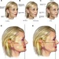
Clinical examination involves determining the degree of lower eyelid laxity, orbicularis oculi muscle tone, presence or absence of a nasojugal groove (tear trough deformity), excess skin or fat, contour of the lower lid-cheek interface, and asymmetries of eyelid or orbit (refer to the preoperative evaluation shown in Fig. 1 A).
Blepharoplasty procedures on a lax lower lid can increase chances of lid retraction and scleral show. Thus laxity must be evaluated preoperatively to preserve eyelid function, maintain appearance, and prevent malposition. Lower eyelid laxity should be assessed by the snap test, as well as evaluating for a negative vector eyelid, which is a particular and specific globe-orbital topographic relationship. A negative vector exists when the most anterior projection of the globe is anterior to the lower lid and malar eminence. Long and narrow eyes characterize youthful periorbital morphology, in contrast with round eyes, which are associated with age. Patients who exhibit excess fat and no tear trough deformity and have smooth skin can be treated with fat resection alone, whereas fat transposition is for those patients with a tear trough deformity.
Correction of the tear trough by blending of the lid-cheek junction is an important goal of transconjunctival blepharoplasty with the addition of fat transposition. Evaluation of the patient’s upward gaze can help reveal the anatomic location and amount of excess infraorbital fat (refer to the preoperative evaluation shown in Fig. 1 B). Preoperative judgment includes how much fat to reposition over the orbital rim and whether excess skin should be resected in conjunction with blepharoplasty via skin pinch to provide a clean, smooth lower lid contour.
Transconjunctival blepharoplasty has a low complication rate and is associated with a high patient satisfaction rate. Even so, patients should still be counseled regarding possible lid retraction, ectropion, hematoma, transient diplopia, and fat granuloma formation during the postoperative period.
Surgical procedure
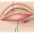
The nasojugal groove is demarcated with a marking pen with the patient in an upright, sitting position. Surgery can be performed with the patient under conscious sedation or general anesthesia. A drop of tetracaine ophthalmic solution is instilled in each eye and the conjunctiva and nasojugal groove are infiltrated with 1% lidocaine with 1:100,000 epinephrine. Eye shields can be placed per surgeon preference. An incision, using electrocautery, is then made just above the fornix through the conjunctiva. A traction suture placed through the posterior conjunctival flap is secured to the head drape for additional corneal protection and exposure of the fat pads ( Fig. 2 ). The eyelid edge of the divided conjunctiva is similarly retracted caudal with a Demar or custom-design Hidalgo retractor.
Related posts:
 An Algorithmic Approach to Multimodality Midfacial Rejuvenation Using a New Classification System for Midfacial Aging
 Dark Circles
 Transcutaneous Lower Blepharoplasty with Fat Transposition
The Transeyelid Midface Lift
Transcutaneous Lower Blepharoplasty with Fat Transposition
The Transeyelid Midface Lift
 Transtemporal Midface Lifting to Blend the Lower Eyelid-Cheek Junction
Autologous Fat Grafting for Midface Rejuvenation
Transtemporal Midface Lifting to Blend the Lower Eyelid-Cheek Junction
Autologous Fat Grafting for Midface Rejuvenation
Stay updated, free articles. Join our Telegram channel

Full access? Get Clinical Tree


