The transeyelid approach to midface lift is an elegant approach for mild descent of malar soft tissue. The subciliary approach is the most commonly used and technically less challenging for surgeons experienced in facelift techniques. This technique in midface rejuvenation also has the advantage of ease of combining with other periocular and mid and upper face rejuvenation, such as blepharoplasty and forehead lift. Complication is rare with lid malposition, scaring, and temporary nerve function impairment being the most common. This article describes midface surgical elevation techniques.
Key points
- •
Preoperative evaluation involves open communication and delineation of patient expectations and clinical assessment.
- •
Careful analysis of the patient’s individual anatomy in the periocular and midface region decreases complication risk.
- •
Creation of a composite flap in midface elevation can be achieved through a transeyelid or transconjunctival approach.
- •
Complete release of the orbicularis retaining ligament and zygomaticocutaneous ligament is required for elevation of the midface.
- •
Transeyelid midface lift can be combined with other periocular or facial rejuvenation procedures.
Introduction
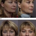
The midface extends from the lower eyelid to the oral commissure. There are three aging changes that occur in the midface: (1) skin aging, (2) soft tissue volume loss, and (3) gravitational descent. Surgical midface rejuvenation can be a powerful rejuvenation tool for volume loss and gravitational changes in the lower eyelid and midface. Shorr and Fallor first described transeyelid midface lifting in 1985 as a cosmetic and functional repair technique in patients with lower eyelid retraction. In addition to surgical elevation techniques, volume augmentation and skin rejuvenation of the midface play an equally important role in the overall management of the aging face. This article describes midface surgical elevation techniques.
Surgical anatomy
Transeyelid midface elevation requires thorough understanding of the anatomic planes and retaining ligaments of the lower eyelid and cheek. Immediately deep to the lower eyelid skin, the orbicularis oculi muscle consists of three parts (pretarsal, preseptal, and orbital) to form the palpebral fissure sphincter ( Fig. 1 ). Deep to the orbicularis oculi muscle is the suborbicularis oculi fat (SOOF), which drapes over the inferior and lateral three-quarters of the orbital rim ( Fig. 2 ). The SOOF extends inferiorly within the zygomaticus muscle system and is continuous with the superficial musculoaponeurotic system (SMAS) ( Fig. 3 ). The orbicularis retaining ligament extends from the dermis through the orbicularis muscle at the preseptal and orbital transition, and forms a fibrous insertion to the arcus marginalis, the fusion of maxillary bone periosteum and the orbital septum. This fibrous condensation divides the SOOF into superior and inferior compartments. The superior compartment contains the malar mound. This fibrous osseocutaneous retaining ligament extends caudally and laterally to become the zygomatic ligament. The strongest points of attachment for the midface soft tissue are at these osseocutaneous ligaments. This highlights the importance of releasing these ligaments to achieve effective elevation of the malar fat pad and midface soft tissue envelope.
During flap dissection for midface elevation, care must be taken to preserve the infraorbital nerve and zygomaticofacial branches of V2. The infraorbital nerve exits the infraorbital canal approximately 1 cm below the medial third of the inferior orbital rim. The zygomaticofacial foramen is approximately 6 mm inferior to the inferior orbital rim, but it is highly variable in location and number. The facial nerve has significant variation in distal branching pattern with conservation of branching of the main trunk only. The zygomatic and orbicularis branches of the facial nerve emerge from the parotid gland deep in the facial fascia approximately 34 mm anterior to the tragus. The temporal branches cross the zygomatic arch 32 to 37 mm lateral to the orbital rim and diverge ( Fig. 4 ). The orbicularis oculi, zygomaticus major, levator labii superioris, and depressor anguli oris are supplied from the deeper surface by the facial nerve. The superior aspect of the orbicularis oculi is innervated by the temporal braches 3 to 4 mm medial to the free edge of the muscle, whereas inferior aspect is innervated by the zygomatic branches. The superior of the zygomatic branches innervate the zygomatic major muscle in the lower third, whereas the lower branches run underneath the zygomatic major muscle to innervate the levator labii superioris in the undersurface. The lower zygomatic branches also innervate the buccinators and levator anguli oris from the superficial surface of the muscle.
Patient evaluation
A cardinal component of the cosmetic evaluation is management of patient expectations. It is important to clearly understand the goals of the patient and to build rapport through open communication. A thorough medical history should be obtained including risk factors for surgery and anesthesia. Active smokers are not ideal candidates for surgery because of the increased risk for anesthesia and poor wound healing. History of previous facial fractures, eyelid surgery, or midface surgery affects the anatomy of the midface and introduces the additional variables of scar tissue and orbicularis apraxia.
There are three main indications for surgical midface elevation: (1) gravitational descent, (2) anterior lamellar insufficiency with lid malposition, and (3) acquired soft tissue defect. Patients with gravitational descent as their primary midface aging change are good candidates and can be done in conjunction with volume augmentation or skin rejuvenation. Midface elevation can also address post blepharoplasty sequelae of lower eyelid retraction, anterior lamellar skin insufficiency, and volume collapse through recruitment of soft tissue from the midafce. These patients may also benefit from concurrent lower eyelid and canthal technique, such as hard palate grafting to the posterior lamella of the eyelid and canthoplasty, to provide additional support in countering lower lid retraction. Lastly, midface elevation can recruit soft tissue and skin to enable repair of acquired defect from Mohs micrographic surgery or trauma.
It is useful to highlight the three components of aging during initial consultation to the patient for realistic expectations. Besides gravitational descent and soft tissue volume loss, aging changes also include solar skin damage. Underlying bony asymmetry between the sides should be observed and discussed with the patient. In addition, the patient’s underlying maxillary bone prominence relative to the globe also contributes to the final effect of soft tissue lifting. When the relative position of the globe to the malar prominence creates a negative vector, with the most anterior portion of the globe protruding past the malar eminence, soft tissue lifting alone is not sufficient to produce malar projection. An adjunct with orbital rim or maxillary-zygomatic arch implant may be needed to achieve the desired elevation in these patients.
In the periocular area, assessment of lower lid position, lower lid laxity, canthal position and shape, upper lid dermatochalsis, upper lid ptosis, lagophthalmos, and orbicularis weakness should be recorded. Even in primary cosmetic cases, achieving optimal results may require concurrent canthoplasty, blepharoplasty, or lid tightening procedures. An external ocular examination to evaluate for pathology and dry eye syndrome should be performed. Pre-existing brow ptosis should be identified, as the elevation of the midface in these cases may cause the appearance of a smaller palpebral fissure.
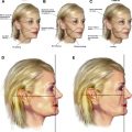
Careful, consistent preoperative photography documents these preoperative findings for subsequent postoperative comparisons. Standard photographic documentation is strongly recommended with and without flash in five cardinal positions: frontal, right/left lateral, and right/left oblique (45-degree angle). Photographs serve several purposes: critical analysis and preoperative planning, demonstration of asymmetry for counseling, insurance preauthorization, medicolegal documentation, and postoperative comparison.
Patient perspective
As with any surgery, thorough preoperative counseling is critical. Appropriate expectations for rejuvenation of the middle third of the face are lift in the cheek area, filling of the tear trough, and softening of the nasolabial prominence. Patients should understand that midface elevation does not change the appearance of the brow or upper eyelid, or address gravitational descent that leads to jowling along the jawline or neck laxity. In addition, midface elevation does not replace the role of volume augmentation.
The preoperative visit should also include detailed informed consent, ensuring that the patient understands fully the risks, benefits, indications, and alternatives of the procedure. Patients should be counseled that, although symmetry is certainly the goal, minor asymmetry after surgery should be the expectation. Congenital skeletal and soft tissue variations render perfect symmetry especially difficult.
Surgical procedure
Over the past decade, multiple surgical techniques have been described to achieve elevation of the midface through a transeyelid approach. Subciliary incision is most commonly described followed by transconjunctival approach; however, techniques on creation of flap, direction of elevation, and location of suture anchorage vary. The procedure is best performed under general anesthesia or monitored anesthesia care for patient comfort, but it can also be performed under oral sedation with local anesthesia in select cases. If no sedation is to be used, tumescent can be used in the midface area to reduce the risk of local anesthetic toxicity.
Transcutaneous Approach
The transcutaneous approach to midface elevation is performed when lower blepharoplasty requiring skin redraping and excision is performed concurrently.
- •
A subciliary incision is created with a #15 blade, CO 2 laser, “pure cut” electrocautery, Westcott scissors, or an inverted #11 blade in the standard fashion for lower blepharoplasty extending 7 to 10 mm beyond the lateral canthal angle or orbital rim.
- •
A traction suture is placed in the eyelid margin or preseptal orbicularis at the midpoint to provide superior traction in addition to protecting the cornea. Preservation of the pretarsal orbicularis is recommended to protect the functional integrity of the eyelids.
- •
The suborbicularis preseptal dissection can be carried inferiorly to the level of the orbital rim and arcus marginalis with blunt Stevens scissors either as a single skin-muscle flap or separately in layers. Care should be taken to avoid aggressive manipulation of the orbicularis muscle to avoid postoperative complications, such as lower eyelid retraction, ectropion, lagophthalmos, and inadequate blink leading to dry eye and exposure.
- •
Alternatively, the orbicularis muscle can be left intact, and the transconjunctival route can be used to manipulate the fat pads only.
- •
In either case, once the septum is exposed, the lower eyelid fat pockets are visible deep to the septum, which can be easily opened to access the three lower eyelid fat pads.
Surgical Insight: Depending on the patient’s anatomy, the fat can be redraped over the orbital rim or resected as needed. As described previously, the upper extent of the SMAS is the orbicularis muscle. It is important to obtain complete release of the orbicularis retaining ligament and zygomaticocutaneous ligament so that, with elevation of the orbicularis, the midface is elevated via the SMAS.
- •
Orbicularis retaining ligament and zygomaticocutaneous ligament are released via blunt dissection with Stevens scissors through a suborbicularis or preperiosteal dissection to 2 cm below the orbital rim into the area of the malar fat pad.
- •
The orbicularis/SMAS flap can be elevated in a predominantly vertical vector or in a superior-lateral vector. When elevating the flap in a pure vertical vector, purchase can be made with four 4-0 polypropylene sutures through the SOOF/orbicularis in a horizontal mattress fashion and sutured to the arcus marginalis.
- •
This vertical elevation, if used with a subperiosteal dissection plane for the composite, is best combined with release of the distal portion of the periosteum to allow proper elevation of the flap. This can be accomplished with a needle tip cautery.
- •
Alternatively a lateral retinacular suspension is accomplished by elevation of the lateral edge of the composite flap using a 4-0 polyglactin suture in an interrupted fashion through the orbicularis and suturing to the periosteum at the lateral orbital rim at the level of the lateral canthus or the level of the frontal zygomatic suture.
- •
When combined with upper blepharoplasty, the composite flap can be tunneled under the bridge of skin following blunt dissection with Stevens scissors to create a suborbicularis pocket along the area of the lateral orbital rim.
Surgical Insight: Our preference is for securing the composite flap to the deep temporalis fascia via this combined upper blepharoplasty incision option.
- •
After creating the composite flap, blunt dissection can be accomplished with Stevens scissors in a cephalic direction along the frontal process of zygoma to the level of the frontal-zygomatic suture. The dissection should be deep along the bone to avoid damage to the zygomatic branches of the facial nerve.
- •
Blunt dissection with cotton-tip applicators in a dorsal fashion at the level of the frontal-zygomatic suture unveils the temporalis fascia.
- •
The composite flap can be sutured in a superolateral direction using a 4-0 polyglactin or poliglecaprone suture in a horizontal mattress fashion to the deep temporalis fascia.
- •
After securing the malar soft tissue to a more youthful position, redundant skin and muscle may be noted.
- •
Excessive skin can be trimmed conservatively, and muscle redraped using small mattress sutures.
- •
The subciliary incision can be closed with interrupted or running 6-0 fast-absorbing gut suture.
Transconjunctival Approach
The transconjunctival approach can also allow for an effective midface lift. Options include subperiosteal midface elevation with multipoint fixation to the arcus marginalis, or SMAS elevation by release of the orbicularis retaining ligament.
- •
If the eyelid has sufficient laxity to allow for adequate visualization into the midface, then a simple conjunctival incision can allow access to the midface. However, if this is not the case, then an open lateral canthotomy/cantholysis (the “swinging eyelid approach”), or a closed cantholysis via an upper eyelid crease incision can be performed.
- •
The conjunctival incision is made with insulated needle tip monopolar cautery in the infratarsal space along the entire length of the lower lid.
- •
A malleable retractor or stainless steel eye shield is used to protect the globe.
- •
The lower eyelid retractor-septal-conjunctival complex can be brought up on stretch by temporary retaining sutures secured to the drape to further protect the globe, orbit, and expose the septum.
- •
A preseptal dissection can be extended to the orbital rim using blunt dissection with cotton tip applicators.
- •
At the orbital rim, after an incision is made with the sharp tip cautery, a subperiosteal dissection can be performed with a Freer elevator to release the orbicularis retaining ligaments and the periosteum medially to the piriform aperture/nasal bone and laterally toward the masseter muscle. Care is taken to avoid damage to the infraorbital and zygomatic nerve.
- •
The periosteum is incised inferiorly using a blade or cautery to allow the midface flap to elevate.
- •
Multipoint fixation of the midface periosteum can be performed to the arcus marginalis, where a rim of periosteum should be left intact.
Similarly to the subcutaneous approach, a preperiosteal dissection can also be accomplished through the transconjunctival approach. At the orbital rim, the SOOF is separated from the surrounding support structures via blunt scissor spreading dissection, to create a composite flap that includes the mimetic muscles. This composite flap can be elevated in a vertical fashion to the orbital rim periosteum-arcus marginalis complex.
- •
Closure of the conjunctiva is not necessary in cases where there is no tension on the conjunctiva. Furthermore, suture closure can result in postoperative patient discomfort and risk of corneal abrasions.
- •
If the canthus was released via an open or closed approach, it is then reconstructed at the end of the case. In most cases, the goal is to resuspend the canthus in the same location. If appropriate, the canthus can be resuspended in a more superior, a more inferior, or a more intraorbital location.
Combining with Endoscopic Forehead Lift
Components of the techniques described previously for transconjunctival approach can be combined with concurrent endoscopic forehead lift or as a stand-alone transtemporal midface lift. The standard temporal incision for endoscopic forehead lift creates a dissection plane along the deep temporalis fascia down to the lateral orbital rim. Once a temporal pocket is formed, the endoscope can be introduced. We limit our discussion to using the temporal incision for forehead lift to assist in midface elevation.
- •
This dissection along the lateral orbital rim or frontal process of the zygoma can be carried in the preperiosteum plane down to the anterior maxillary face, connecting with the previously dissected orbicularis/SOOF/SMAS flap. It is important to stay within the medial third of the zygomatic arch to avoid damage to the facial nerve.
- •
The zygomaticotemporal sentinel vein can be identified close to the frontozygomatic suture and cauterized when necessary.
- •
The flap can be sutured using 3-0 or 4-0 poliglecaprone suture at 5 to 5.5 cm from the midline at the level of the alar to the deep temporalis fashion in a superotemporal vector plane.
- •
Alternatively an additional suture can be placed through the lateral aspect of the inferior orbicularis oculi to the deep temporalis fascia.
Related posts:
 An Algorithmic Approach to Multimodality Midfacial Rejuvenation Using a New Classification System for Midfacial Aging
 Dark Circles
Transconjunctival Lower Lid Blepharoplasty with and Without Fat Repositioning
The Management of Festoons
Dark Circles
Transconjunctival Lower Lid Blepharoplasty with and Without Fat Repositioning
The Management of Festoons
 Transtemporal Midface Lifting to Blend the Lower Eyelid-Cheek Junction
Autologous Fat Grafting for Midface Rejuvenation
Transtemporal Midface Lifting to Blend the Lower Eyelid-Cheek Junction
Autologous Fat Grafting for Midface Rejuvenation
Stay updated, free articles. Join our Telegram channel

Full access? Get Clinical Tree


