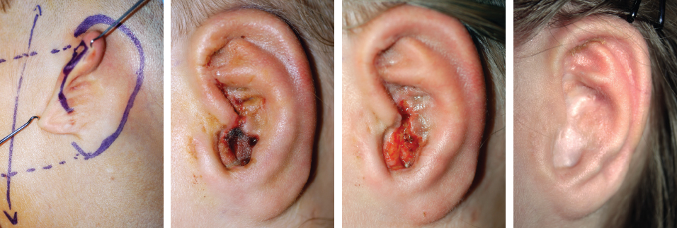
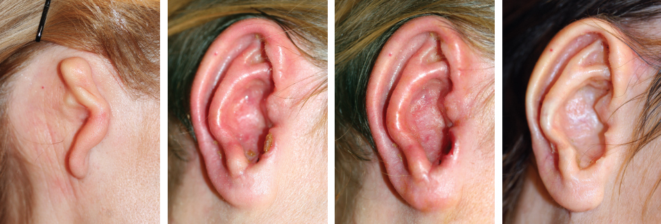
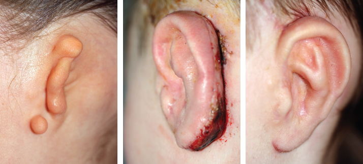
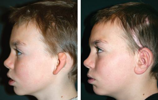
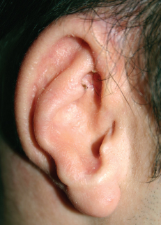
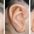
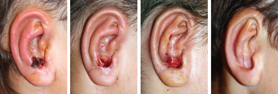
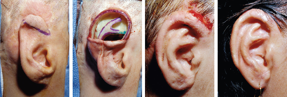
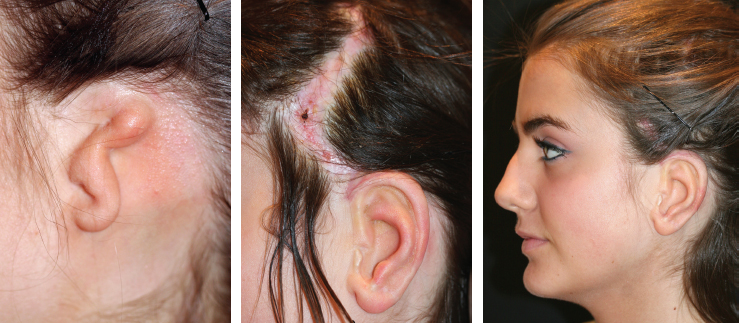
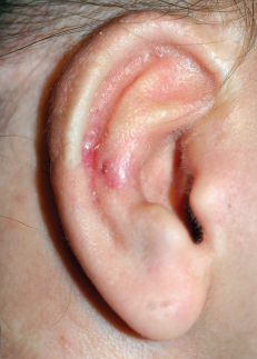
CHAPTER 9 “Neither myself nor anyone else has anything to learn from success, but only from failures and complications… Success always leads to self-satisfaction and thus no further improvement… Failures push us to analyze, though not necessarily leading us to the correct conclusion. We should live in a world of complications, provided we know, observe, and have the courage to investigate the causes that are not always flattering: incorrectly applied indications, disorganization, ignorance and haste.” –Paul Tessier Many different complications can occur after ear reconstruction. Managing them, analyzing why they occurred, and preventing them in the future are essential (see video 9-1). Management of particular complications is the same regardless of the circumstances, microtia or trauma. They occur more often after the first stage, because placement of a rigid, three-dimensional framework under the thin auricular skin can lead to problems with vascularity of the skin. The most frequent complication is skin necrosis, and the most severe is infection. We define small complications as ones for which no additional surgery is indicated, but for which a particular follow-up protocol is required. These are most often seen after the first stage. Auricular skin is well vascularized. However, skin pocket dissection in the auricular region must be performed with great care to preserve the subdermal plexus. Any injury to the vascular supply or excessive tension on the very thin auricular skin can lead to skin necrosis and exposure of the underlying framework. When the area of necrosis is small and occurs in a favorable area, it can be managed by spontaneous healing. The favorable area is mostly the area not covering cartilage, as in the depth of the concha. This patient’s ear was reconstructed with a type 3b skin approach and a TYPE I framework. A small area of skin necrosis was encountered in the inferior portion of the concha. No cartilage was exposed in the concave area of the conchal bowl; thus the area was allowed to heal by secondary intention, with a favorable outcome. The cause of the complication was incorrect dissection of the skin pocket in an adherent area of the lobular remnant, causing damage to the subdermal plexus. Fig. 9-2 This patient had a small area of skin necrosis overlying the antitragus. Excision of the exposed cartilage and regular dressings allowed healing by secondary intention. The consequence of this complication was that the antitragus almost disappeared, and a wide intertragal notch was created. This case shows a small exposure of the tragus after a type 3b skin approach and a TYPE I framework reconstruction. Reducing the projection of the tragus to facilitate skin closure primarily easily solved this problem. The final result shows an irregularity of the tragal contours, which could be addressed secondarily. Fig. 9-4 This posttraumatic case had already been reconstructed with a skin flap without any cartilaginous support. This should never be done. A single-stage secondary reconstruction (skin approach type 3a, framework TYPE III) was performed, and the anterior part of the helix became exposed postoperatively. Even though the necrosis was overlying the helix, no cartilage resorption resulted, and the final result was not affected. The cause of the necrosis was excessive tension resulting from excision of hair-bearing skin and previous scarring. The incisura is the curved notch between the tragus and antitragus. We have observed a particular small complication that can occur after a type 2 skin approach in which a deep retroauricular sulcus is created that may be difficult for the patient to clean (see Chapter 4, type 2 skin approach). Over time, the skin in this sulcus macerates and can develop an inflammatory process. When the adhesion is tight, the inflammation of the sulcus may come into contact with the skin, and cartilage of the intertragal notch and a small fistula can develop. Fig. 9-5 For this patient, the management is regular cleaning of the fistula to eliminate infection and closure at the second stage. Very occasionally, the skin graft used to create the posterior surface of the auricle and the retroauricular sulcus takes poorly. This usually occurs only at the tip of a temporofascial flap or if a mastoid fascial flap was raised to cover a cartilage block. It can usually be managed with dressings, although this may lead to reduced depth of the sulcus because of contraction of the area healed by secondary intention. This case demonstrates a lobular-type microtia reconstructed by a type 3b skin approach and elevated with a type A (temporofascial flap) second stage. The skin graft underwent partial loss over the tip of the temporofascial flap. With spontaneous healing, the sulcus epithelialized with a good final result. The tip of the fascia may have poor vascularization. Therefore we always elevate the inferior part of the framework with attached soft tissues covering the posterior surface of the lobule. This prevents exposure of the cartilage and allows spontaneous healing if the tip of the flap fails. Harvesting of a fascial flap from the temporal region may occasionally leave widened scars that can be noticeable. The patient may be able to hide this scar easily if the hair is long. However, in patients with thin or short hair, particularly males, the scar can be more visible than the reconstructed ear. Scar revision is possible, but the scar may still be visible in the long term. Fig. 9-7 This case demonstrates a young female patient with lobular-type microtia. She had a good final result, but a large scar was present in the temporal region. Although she could hide the scar with her hair, we chose to revise the scar to try to improve the overall cosmesis. This young male patient has a good result from his ear reconstruction using temporal fascia for the second stage (type A). The temporal scar is visible through his thin hair, and in hindsight, it may have been more prudent to perform a type D second stage using a tunnel technique. Thus a large cartilage graft and temporofascial flap would not have been needed in the second stage, and achieving symmetry may have been possible by setback otoplasty of the contralateral ear. Nagata was first to advocate using double-ended stainless steel wire sutures. They are the basis of our practice for stabilizing the many pieces of cartilage framework. We find the sutures to be convenient and very well tolerated by patients. Occasionally, a wire extrudes, which is not a problem if correctly managed. The solution is to remove the wire straightaway to prevent inflammation and cartilage resorption. Fig. 9-9 Five years after ear reconstruction, this patient presented with a late wire extrusion that he had noticed 2 weeks earlier.
Complications
SMALL COMPLICATIONS
After the First Stage
Skin Necrosis
Incisural Inflammation
After the Second Stage
Failure of the Skin Graft
Long-Term Problems
Temporal Scarring
Extrusion of Wire Sutures
Plastic Surgery Key
Fastest Plastic Surgery & Dermatology Insight Engine