A woman presents after Mohs excision of a basal cell carcinoma within the right alar. A composite defect of her right upper lip, cheek, and ala is present. Although distressed, her concerns are somewhat alleviated by the prior successful reconstruction of a full-thickness defect of her left ala, some years previously. This content presents the principles of the repair, the surgical plan, and details of the multiple procedures performed for successful reconstruction.
Analysis of the defect and decision making
On physical examination ( Fig. 1 ), a faint vertical left paramedian forehead scar, which followed the previous reconstruction, is visible. The forehead is otherwise high and expansive.
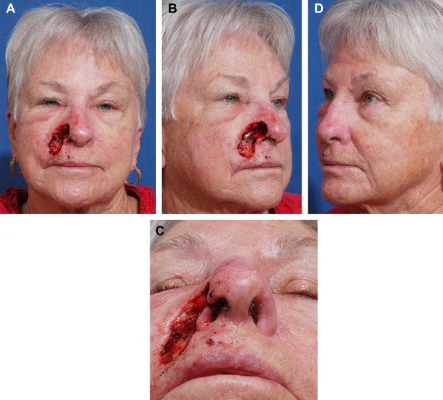
The left ala appears normal, although it is not. Some years previously, the patient presented with a full-thickness defect of the left entire ala and part of the inferior sidewall. Adjacent lip and cheek were uninjured. The defect had been repaired with a 3-stage folded forehead flap for cover and lining, with a delayed, primary, ear cartilage alar margin buttress graft to support, shape, and brace the left nostril margin. Following subunit principles of nasal reconstruction, the normal intact right ala had been used as a guide to design a template with the correct dimension and outline of the contralateral normal ala. The right ala was used to plan the exact replacement of the entire left ala and a few millimeters of the left tip subunit with a vertical forehead flap. A distal extension, about 1.5 cm wide and 7 mm long, was added to the distal covering flap. The extension was folded inward to replace the missing lining. During an intermediate operation 1 month later, the covering flap was elevated completely off the nose with 2 to 3 mm of subcutaneous fat. The distal folded lining was now healed to the adjacent residual nasal lining and was no longer dependent on the supratrochlear pedicle for blood supply. The underlying doubly layered excess of subcutaneous fat and frontalis areolar tissue was excised, exposing thin supple vascular lining. The contralateral normal alar template was then used to design a precise alar margin graft to shape the left nostril margin. The graft was fixed to the restored lining. The thin forehead flap was then returned to the recipient site. One month later, the pedicle of the flap was divided. During a subsequent revision, the left alar crease was further refined through a direct incision to sculpt a flat sidewall, a deep alar crease, and a convex alar contour. The slightly thick rim margin was thinned by excising excess soft tissue between the lining and cartilage graft through the old incision present along the nostril margin.
A short transverse scar is visible within the superior dorsum at the site of a previous skin cancer excision that was closed primarily.
The new defect involves several facial units but is more superficial.
Anatomically, the skin is missing over the entire ala, part of the inferior sidewall, and the adjacent medial cheek and lateral lip. Soft tissue within the cheek over the piriform aperture has been excised. The normal fibrofatty middle layer support of the ala is gone. Nasal lining is intact.
Aesthetically, the complex midface has been destroyed. The expected color and texture, landmark outline, and 3-dimensional shape are abnormal. Because the underlying orbicularis muscle is present, if skin is restored to cover the lip, the lip will function normally.
The face can be divided into geographic areas of characteristic skin quality, border outline, and 3-dimensional contour. The cheek is a peripheral unit, largely flat and expansive with a variable border outline that is not completely seen on frontal view. The nose and upper lip are central units and are exactly contoured and outlined. An abnormality in a part of the lip or cheek is quickly apparent because the contralateral remaining lip or cheek creates a visually disturbing comparison.
The nose sits on a facial platform of the cheek and lip. The nasolabial fold separates the round fullness of the medial cheek and the flat upper lip. The nasolabial fold does not extend into the alar crease but is separated from the crease by a hairless triangle of skin, which lies adjacent to the alar base inset. The nose sits on the facial platform in an exact position and projects with specific angles.
The nose is divided into subunits. The flat sidewall is separated from the round convex ala by the alar crease. The alar subunit is outlined by the nostril margin inferiorly, the alar crease superiorly, the slight alteration in contour with the soft triangle and tip subunits medially, and the alar groove laterally where the ala is inset into the lip at the alar base.
Each facial unit must be restored in terms of its own quality, outline, and contour and in relationship to the other facial units. The dimension, volume, position, projection, platform, skin quality, border outline, and 3-dimensional contour of the midface must be reestablished.
Principles of repair
- 1.
Patients wish to look normal.
- 2.
The “normal” is defined by visual skin quality, border outline, and 3-dimensional contour.
- 3.
Individual areas of the face can be described in terms of facial units and subunits.
- 4.
The restoration of these visual units defines the surgical result, which looks normal. The wound must be repaired anatomically and aesthetically.
- 5.
Missing tissues must be replaced in exact dimension and border outline to reestablish the normal and prevent distortion of adjacent structures.
- 6.
The contralateral normal or the ideal side should be used as a guide.
- 7.
Exact templates should be used to design covering and lining replacements, cartilage graft dimension and outline, and to determine the position of expected facial landmarks.
- 8.
The nose must be built on a stable platform. If a composite defect of the nose, cheek, and lip is present, the lip and cheek should be rebuild initially. If the new platform is unstable and may shift because of gravity, tension, or resolution of edema, the nasal repair should be delayed to avoid late shifting of the nasal reconstruction into an abnormal position on the face. If the defect is more superficial and the platform base is unlikely to become distorted during wound healing, the nose can be repaired simultaneously with platform restoration.
- 9.
Surgical staging should be used to advantage. The anatomic and aesthetic needs of the repair, priorities, quality of donor tissues, and ideal timing to transfer and modify materials with safety and precision should be designed. Although Gillies and Millard emphasize the use of “like tissue,” a flat thick forehead flap, an ear cartilage graft, or a cheek flap have little in common with the delicate outline or contour of the midface.
- 10.
It is often useful to repair a composite defect of the cheek, lip, and nose with individual grafts and flaps to position the final scars in the joins between units and restore 3-dimensional contour.
The surgical plan
The wound is clean, and early reconstruction is appropriate. The defect is debrided, and the wound margins are incised to create clean right-angled skin edges. Templates of the contralateral normal nose and upper lip are used to design the skin replacement and alar margin support and to determine the ideal position, in height and width, of the right alar base after restoration of the cheek and lip platform. The soft tissue deficiency in the medial right cheek is augmented with a Millard flip fat flap. The cheek and lip skin defect is repaired by advancing a cheek flap with a random extension to resurface the upper lip defect. Because this is a relatively superficial defect with intact lining, nasal repair begins simultaneously with the placement of a primary conchal cartilage alar margin cartilage graft and a right paramedian forehead flap to resurface the ala and part of the inferior sidewall. An intermediate operation is planned to allow adjustments and more precise soft tissue contouring before pedicle division. Later, the pedicle is divided and the nasal labial fold recreated. The patient is informed that a late revision some months later may be appropriate to improve the alar crease, revise the forehead scar, thin the nostril margin, and so forth. All surgical procedures are performed under general anesthesia to avoid soft tissue distortion and blanching resulting from the injection of local anesthesia and epinephrine. It is difficult to make precise intraoperative decisions in restoring contour or determining the viability of tissues if they are bloated or chemically constricted.
Operation 1
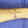
The hairline, frown lines, location of the supratrochlear vessels by Doppler, subunits of the nose, nasolabial folds, philtrum, vermilion, old scars within the forehead and nasal dorsum, midline of the lip, and outline of the old forehead flap that resurfaces the left ala and part of the sidewall are marked with ink ( Fig. 2 ).
The wound does not represent the true tissue loss and is expanded by edema, gravity, local anesthesia, or tension. If such a wound healed by secondary intention or was previously reconstructed, it may be contracted by scar or inadequate tissue replacement. Templates based on the contralateral normal permit exact replacement of missing tissues and dimension, outline, and position.
Quarter-inch Steri-Strips (3M Corporation, St Paul, MN, USA) are applied to the left nose and upper lip to create a paper pattern of the contralateral ala and hemilip. Collodion is applied externally with a Q-tip to further “glue” the paper tape strips together. Each pattern is then elevated. Because the ink applied to the skin subunit surface adheres to the undersurface of the Steri-Strips, the outline and dimension of the contralateral normal subunits are visible. Excess tape is trimmed, and the contralateral alar and hemilip outlines are transferred to the aluminum foil of a suture pack ( Fig. 3 ).
The right cheek skin, with 2 to 3 mm of fat, is undermined laterally for 5 to 8 cm. The medial border of the flap is incised directly in the residual nasolabial fold, inferiorly, and at the junction of the cheek and sidewall subunits, superiorly. Residual medial cheek fat, lateral to the piriform soft tissue deficiency, is marked with ink as a medially based hinge-over flap. Subcutaneous fat is hinged over, like a page of a book, and is fixed with absorbable sutures to fill the premaxillary soft tissue loss and reestablish medial cheek fullness in the nasal base platform height ( Fig. 4 ). Dog-ears are excised along the side of the nose and lateral to the commissure after advancement of the flap medially to resurface the cheek with a skin extension that replaces missing lateral upper lip skin. The deep surface of the flap is fixed with suture to the deep soft tissues along the nasal facial groove and piriform aperture. Any residual cheek soft tissue deficiency left in the area of the donor fat flap is obliterated with a few sutures by the soft tissues, which advance with the cheek flap. The skin is closed with subcuticular and fine skin sutures.
Related posts:
 Bilobed Flaps in Nasal Reconstruction
 Cartilage Grafting in Nasal Reconstruction
 Complex Nasal Reconstruction: A Case Study: Reconstruction of Full-Thickness Nasal Defect
Reconstruction of the Dorsal and Sidewall Defects
Complex Nasal Reconstruction: A Case Study: Reconstruction of Full-Thickness Nasal Defect
Reconstruction of the Dorsal and Sidewall Defects
 Reconstruction of Nasal Tip and Columella
 Microvascular Reconstruction of Nasal Defects
Stay updated, free articles. Join our Telegram channel

Full access? Get Clinical Tree


