Chapter 11 Combined abdominoplasty and breast enlargement by autologous tissue transfer or transabdominal implant placement
A Augmentation Mammaplasty by Reverse Abdominoplasty
Key Points
• Augmentation mammaplasty by reverse abdominoplasty (AMBRA) may be a viable option for patients with upper abdominal skin laxity and adiposity who desire autologous tissue breast augmentation and/or mastopexy combined with reverse abdominoplasty.
• Patients with epigastric or subcostal incisional scars that exclude them as candidates for conventional abdominoplasty, can safely undergo AMBRA.
Introduction
Relative to conventional abdominoplasty, reverse abdominoplasty has been less frequently studied.1–4 This is in part due to the fact that the reverse abdominoplasty technique necessitates an upper abdominal scar, and that most patients present with lower, rather than upper, abdominal laxity. In patients with relatively normal infraumbilical abdomen tissues who request that the lax epigastric tissues be addressed, or patients who are not candidates for conventional abdominoplasty because of previous subcostal incisional scars, reverse abdominoplasty may be the safer, better, or sole option.5
Preoperative Planning/Patient Selection
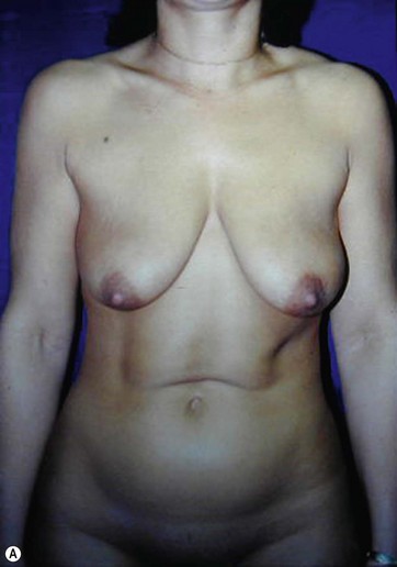
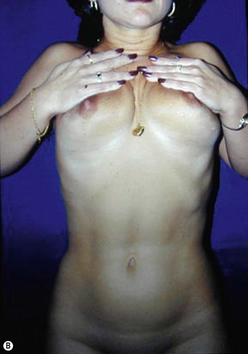
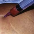
The key steps in determining a patient’s suitability for the AMBRA procedure are a careful evaluation of the soft tissues of the upper epigastrum and performing a “breast suspension” test (Fig. 11.1). The patient is asked to lift each breast with a cupped hand. This should restore an esthetic result to the upper abdomen and umbilicus. Some patients benefit from suction-assisted lipectomy to the lower abdomen for contouring. Other patients would be better served by a mini-abdominoplasty to address moderate skin laxity of the lower abdomen. These adjuncts can help optimize the final result of AMBRA, without undue added risk.
Surgical Technique
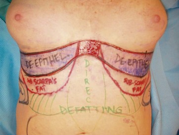
These preoperative markings are conducted while the patient is standing upright, to delineate the inframammary folds, and breast markings for mastopexy are performed concurrently. Then the patient is placed supine to allow the surgeon to retract the excess upper abdominal tissues cephalad, which is an adaptation of the Lockwood technique.6 The redundant soft tissues of the upper abdomen are then folded over to mark the “safe” zone of skin excision that permits tension-free closure with the inframammary fold. This area can be de-epithelialized without sacrificing a tension-free closure of the upper abdominal–IMF wound (Fig. 11.2). This de-epithelialized tissue provides the bulk for each breast’s augmentation.
Superiorly Based Augmentation Mammaplasty by Reverse Abdominoplasty
The patient is returned to a fully supine position, and with cephalic tension on the flap, the de-epithelialized border of the skin apron is anchored to the perichondrium, periosteum, and deep fascia along the lower border of the breast. A nipple–areola complex located slightly lower on the breast mound (1 to 2 cm) than ideal, to allow for lower pole descent, is preferred. The edge of the abdominal skin flap is then reinforced along its entire length with sutures to the periosteum. The central midline supraumbilical abdominal area is then liposuctioned to emphasize the median raphe. The closure of upper and lower wounds is the same except that Scarpa’s fascia is approximated in the lower abdominal closure, followed by dermal closure (Box 11.1).
Box 11.1
Optimizing outcomes pearls
1. AMBRA is a forgiving technique, if the surgeon preserves the dominant central abdominal perforators and avoids venous and lymphatic compromise.
2. In patients undergoing AMBRA with concomitant lower abdominal adiposity, a mini-abdominoplasty or panniculectomy can treat persistent lower abdominal skin and tissue redundancy without compromising central abdominal vascularity.
Peri- and Postoperative Care
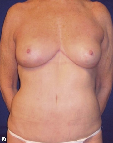
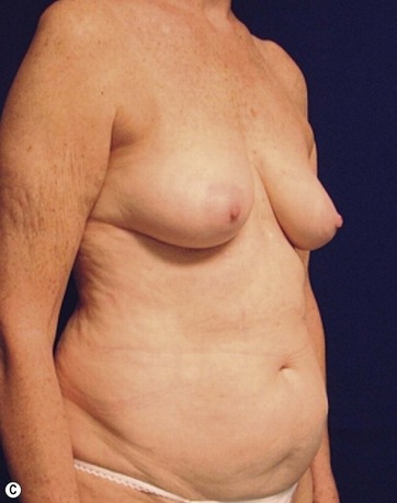
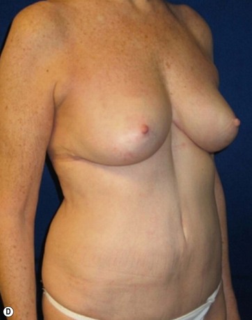
Case Examples
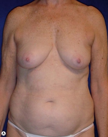
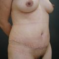
A 70-year-old patient desired fuller breasts and superior and central abdominal tightening (Fig. 11.3A, B, C, D).
< div class='tao-gold-member'>
Related posts:
Stay updated, free articles. Join our Telegram channel

Full access? Get Clinical Tree


