Introduction
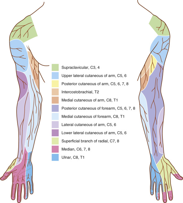
Reconstructive microsurgery of the upper extremity requires detailed knowledge of the regional anatomy of the arm, forearm and hand. Specific familiarity with the anatomy of the neurovascular structures allows safer, more expeditious flap elevation and recipient vessel exposure. This chapter reviews anatomy pertinent to upper extremity reconstruction, and describes the anatomy of common recipient vessel approaches.
Anatomy of the Arm
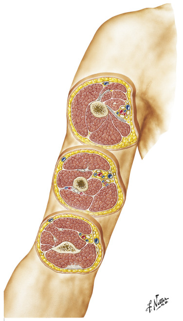
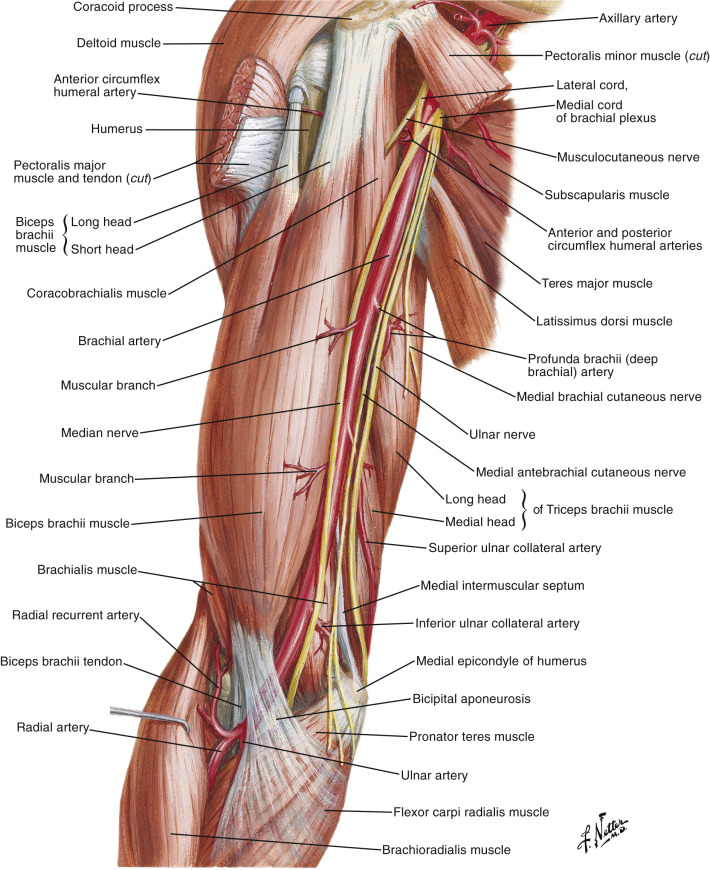
The arm is divided into anterior and posterior compartments by the medial and lateral intermuscular septa ( Fig. 12.1 ). The anterior compartment contains the biceps, brachialis and coracobrachialis muscles, the brachial artery and veins, and the median and musculocutaneous nerves ( Fig. 12.2 ). The posterior compartment contains the three heads of the triceps and profunda brachii ( Fig. 12.3 ). The ulnar and radial nerves traverse both compartments.
A line drawn from the lateral epicondyle of the humerus, to the insertion of the deltoid corresponds with the lateral intermuscular septum. This landmark is useful when marking the axis of the lateral arm flap, or for exposing the radial collateral artery.
The medial intermuscular septum runs along the medial epicondylar ridge, beginning at the medial epicondyle of the humerus distally and blending with the coracobrachialis fascia proximally. The anterior and posterior compartments in the arm are enveloped by a layer of deep fascia, and the only notable structures found superficial to this layer are the cutaneous nerves and superficial veins.
Vascular Anatomy of the Arm
The Brachial Artery
The brachial artery is the continuation of the axillary artery, beginning at the distal edge of the teres major and ending approximately 2–3 cm distal to the elbow where it bifurcates into the radial and ulnar arteries at the level of the radial neck ( Fig. 12.2 ). It is accompanied along its course by two interconnected venae comitantes, which are large enough to be used as recipient veins in microvascular reconstruction. The artery lies under the deep fascia of the arm and is easily palpable along its course commencing anteromedial to the triceps in the upper arm, gradually becoming more anterior to lie along the brachialis muscle, and eventually medial to the biceps. Its final position is anteromedial to the elbow joint, where it runs under the bicipital aponeurosis (lacertus fibrosis), entering the forearm superficial and medial to the biceps tendon ( Fig. 12.4 ).
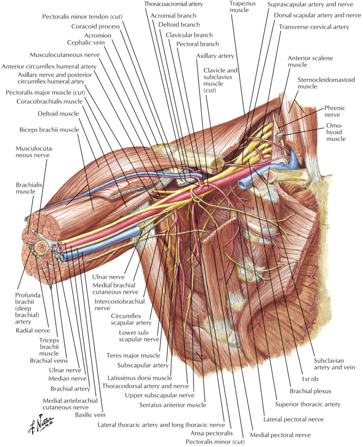
In the proximal arm the brachial artery is surrounded by the terminal branches of the brachial plexus ( Fig. 12.5 ). An appreciation of these neurovascular relationships is necessary to avoid intraoperative nerve injuries in recipient vessel exposure, and is also useful when performing complex peripheral nerve reconstruction and nerve transfers.
Along its entire course, the brachial artery remains intimately related to the median nerve. Proximally, the median nerve lies anterior to the artery but crosses it to lie medial to the brachial artery in the distal arm ( Fig. 12.3 ).
The ulnar nerve is initially found medial to the brachial artery in the proximal arm, along with the medial antebrachial cutaneous nerves. As the brachial artery gradually courses in an anterior direction towards the antecubital fossa, the ulnar nerve heads posteriorly away from the artery, piercing the medial intermuscular septum half way down the arm to enter the posterior compartment of the arm medial to the triceps ( Fig. 12.2 ). It eventually lies posterior to the medial epicondyle of the humerus where it can be reliably located, as it enters the forearm ( Fig. 12.6 ).
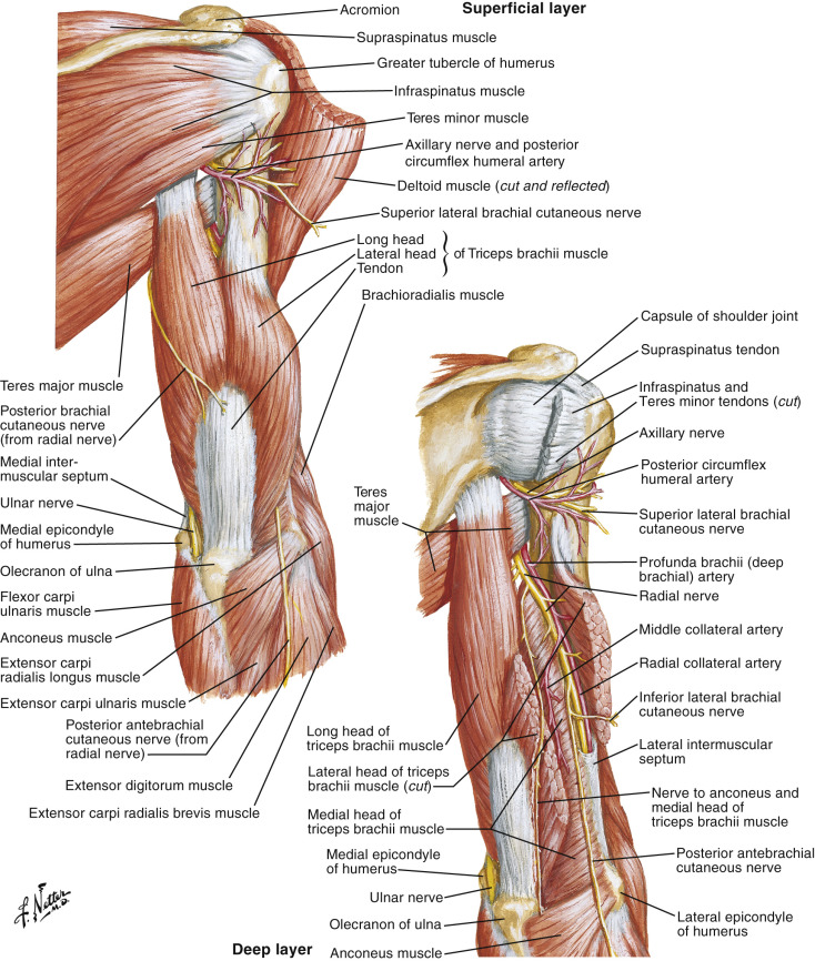
The radial nerve lies posterior to the brachial artery in its proximal portion ( Fig. 12.4 ) but rapidly heads in a much deeper and lateral direction. Along with the profunda brachii (deep brachial artery), it passes through the triangular interval between teres major, the long and lateral heads of triceps, to lie posterior to the humerus and away from the brachial artery for the remainder of its course in the arm ( Fig. 12.3 ). After the radial nerve winds posteriorly around the humerus in the spiral groove, it pierces the lateral intermuscular septum leaving the posterior compartment, to enter the anterior compartment in the distal arm. From here, it enters the forearm anterolaterally between the brachioradialis and brachialis ( Fig. 12.6 ), before dividing into its two terminal branches ( Fig. 12.4 ).
Branches of the Brachial Artery
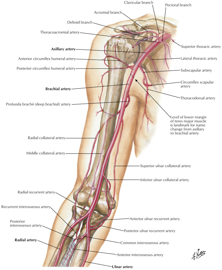
The brachial artery gives rise to four important named branches in the arm (the profunda brachii, the nutrient artery to the humerus, and the superior and inferior collateral arteries) ( Fig. 12.5 ).
The profunda brachii (the deep brachial artery) is the first and largest branch of the brachial artery arising just past the distal border of the teres major on the posteromedial aspect of the brachial artery. It eventually supplies the territory of the lateral arm flap via its radial collateral branch.
In its proximal portion the profunda brachii gives rise to a small branch that ascends between the long and lateral heads of the triceps (the ascending deltoid branch) that anastomoses with the posterior circumflex humeral artery. The profunda brachii then passes into the posterior compartment of the arm with the radial nerve between the lateral and long heads of the triceps, and teres major (triangular interval). It runs with the radial nerve in the spiral groove posterior to the humerus and eventually branches into two terminal divisions – the radial collateral artery and the middle collateral artery ( Fig. 12.3 ). The middle collateral artery enters the medial head of the triceps and ends in the olecranon plexus of the posterior elbow.
The radial collateral artery follows the course of the radial nerve before dividing into anterior and posterior divisions. The anterior division (ARCA) continues to accompany the radial nerve into the flexor compartment between brachialis and brachioradialis to eventually anastomose with the radial recurrent artery (RRA).
However, the posterior division of the radial collateral artery (PRCA) stays in the posterior compartment and eventually reaches the lateral intermuscular septum, where it can be found between the triceps posteriorly and the brachialis anteriorly. It gives rise to several septocutaneous perforators (1–15 cm proximal to the lateral epicondyle) that supply the overlying skin, which corresponds with the vascular territory of the lateral arm flap, before ending in a plexus of vessels around the lateral epicondyle and olecranon. This plexus is also supplied by the ARCA, the interosseous recurrent, inferior ulnar collateral, and the radial recurrent artery ( Fig. 12.7 ). These vessels provide retrograde flow to the PRCA giving the basis for the distally-based lateral arm flap.
The next branch of the brachial artery is the superior ulnar collateral artery. This arises from the brachial artery just distal to the mid-arm in 78% of specimens, or from the profunda brachii in 22%. It pierces the medial intermuscular septum to join the ulnar nerve in the posterior compartment medial to the triceps ( Fig. 12.1 ), and follows the nerve around the medial epicondyle into the forearm ( Fig. 12.2 ). It eventually anastomoses with either the posterior ulnar recurrent artery or a branch of the inferior ulnar collateral artery ( Fig. 12.7 ).
The last branch of the brachial artery in the arm is the inferior ulnar collateral artery. This arises from the medial aspect of the brachial artery 5 cm proximal to the elbow and divides into anterior and posterior branches. The posterior branch runs behind the humerus under the triceps, joining the middle collateral artery to form an anastomotic arch just proximal to the olecranon. The anterior branch passes anterior to the medial epicondyle to anastomose with the anterior ulnar recurrent artery ( Fig. 12.7 ).
Anastomoses Around the Elbow
Complex anastomoses around the elbow allow for excellent collateral circulation and can be simplified into those that run anterior versus posterior to either the medial or lateral epicondyle ( Table 12.1 , Fig. 12.7 ).
| Location | Vessels |
|---|---|
| Anterior to medial epicondyle | Anterior branch of inferior ulnar collateral Anterior ulnar recurrent Anterior branch of superior ulnar collateral |
| Anterior to lateral epicondyle | Radial recurrent artery (RRA) Anterior radial collateral branch of profunda (ARCA) |
| Posterior to medial epicondyle | Middle collateral branch of profunda Inferior ulnar collateral |
| Posterior to lateral epicondyle | Posterior radial collateral |
Exposure of the Brachial Artery in the Medial Arm
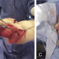
The brachial artery can be exposed easily and expeditiously anywhere along its course in the medial arm. With the patient supine, the arm extended on a hand table, and the forearm supinated, a longitudinal incision is marked over the medial biceps groove ( Fig. 12.6 ). Useful surgical landmarks during this dissection include the bicipital groove, the medial epicondyle, the basilic vein and the medial intermuscular septum. The incision can be made anywhere from the elbow crease to the axilla, depending on the surgical requirements ( Fig. 12.8A ). The skin and subcutaneous fat are incised and attention should be paid to avoiding injury to the basilic vein, which runs in the subcutaneous plane in the distal two-thirds of the arm, and is accompanied by the median antebrachial cutaneous nerve ( Fig. 12.8B ).
Distally, the brachial artery pulse is easily palpable and the overlying deep fascia is incised to expose the neurovascular bundle. The median nerve lies medial to the brachial artery at this level, and there are also usually two sizeable venae comitantes surrounding the vessel ( Fig. 12.8C ).
When a proximal approach is necessary, the basilic vein can be a useful landmark as it pierces the deep fascia of the arm to join the neurovascular bundle in the middle to proximal thirds of the arm. The vein can be followed proximally and the sheath incised at this point.
The brachial artery can be used in an end-to-side manner to provide excellent inflow to microvascular reconstructions, or large side branches can be sacrificed for an end-to-end anastomosis.
Gross Regional Anatomy of the Antecubital Fossa
The antecubital fossa is an anatomically complex area, and knowledge of this anatomy allows exposure of multiple structures that may be required for complex upper extremity reconstruction, including exposure of recipient vessels as well as nerves for complex peripheral nerve reconstruction and functional muscle transfer. Important anatomic landmarks include the elbow flexion crease proximally, the brachioradialis laterally, and the pronator teres medially ( Fig. 12.9 ). The floor of the fossa comprises the brachialis muscle for the most part.
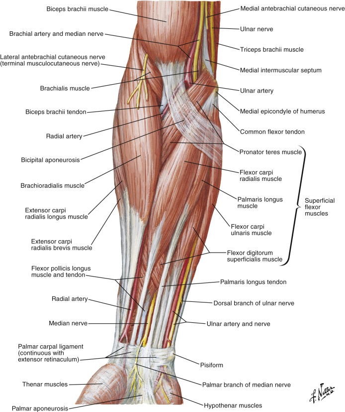
The biceps tendon and aponeurosis are centrally located and somewhat divide the fossa into medial and lateral compartments. In the superficial plane, the antecubital fossa has a rich subcutaneous veno us network that can be used for recipient venous anastomoses or vein grafts harvest ( Fig. 12.10 ). The lateral antebrachial cutaneous nerve also lies in the subcutaneous plane, on the lateral side of the fossa having exited the deep fascia of the distal arm just lateral to the biceps ( Fig. 12.11 ), and this nerve can be harvested as a nerve graft, although results in a distal sensory deficit ( Fig. 12.12 )
On the medial side of the fossa, lying deep to the fascia and lacertus fibrosus, are the median nerve and the brachial artery. Lateral to the artery is the biceps tendon that heads deep into the fossa to insert on the radial tuberosity, and this insertion may need to be exposed in functional muscle transfers for elbow flexion. Lateral to the biceps tendon, the radial nerve enters the antecubital region deep to the brachioradialis muscle belly, and divides into its two terminal branches ( Fig. 12.4 ).
Exposure of the Distal Brachial Artery and Its Bifurcation
At the elbow crease, the brachial artery becomes an anterior structure laying on the brachialis muscle in the middle of the antecubital fossa. At this level, it is covered only by the lacertus fibrosus, the median cubital veins and skin. It is located between the muscle bellies of the pronator teres and brachioradialis until it bifurcates approximately 2–3 cm distal to the elbow crease ( Fig. 12.7 ).
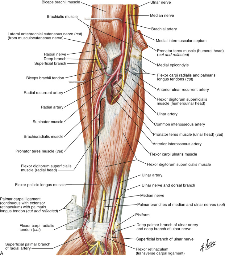
With the arm extended and the forearm supinated, a skin incision is made along the medial bicipital groove starting 4–5 cm proximal to the flexion crease of the elbow. At the crease, the incision continues obliquely or transversely, then distally along the medial border of the brachioradialis muscle ( Fig. 12.13A ). Subcutaneous veins can be ligated and divided, or preserved for use as recipient outflow vessels. Specific attention should be paid to sizeable cutaneous nerve branches that may be encountered during this approach, as injury leads to distal paresthesias and painful neuroma formation. The medial antebrachial cutaneous nerve lies with the basilic vein on the medial aspect of the exposure in the distal arm ( Fig. 12.13B ), and the lateral antebrachial cutaneous nerve is located on the lateral side of the exposure ( Fig. 12.13D ).
The lacertus fibrosus can be seen arising from the biceps tendon, fanning out medially over the pronator teres and flexor muscle mass ( Fig. 12.13B ). It is essentially continuous with the distal aspect of the deep fascia of the arm. To access the distal brachial artery, this deep fascia is incised and reflected, exposing the median nerve, which, by now has become medial to the brachial artery ( Fig. 12.13C ). Sizeable venae comitantes can be preserved and used for outflow anastomoses.
In order to access and expose the bifurcation of the brachial artery in the antecubital fossa, the fascial opening is continued distally by incising the lacertus longitudinally over the pronator and reflecting it proximally ( Fig. 12.13C ).
Directly underlying the lacertus is the continuation of the brachial artery, lying between the biceps tendon and the median nerve. The median nerve gives off several nerve branches medially, supplying the muscles of the flexor-pronator mass, then heads deep to pronator teres. The nerve and its branches should all be preserved, and the brachial artery can be bluntly dissected distally as it moves away from the nerve, running towards the biceps tendon to become centrally located in the fossa, between the pronator teres medially and the brachioradialis laterally. The lateral antebrachial cutaneous nerve also lies on the lateral side of the fossa and should be preserved.
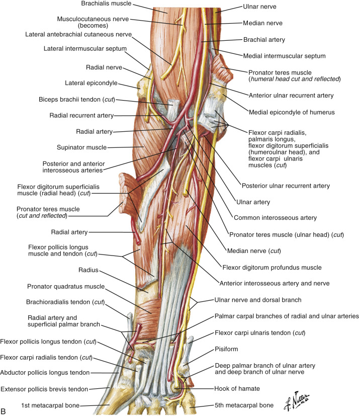
The deep fascial incision is extended distally and the pronator teres and brachioradialis muscles are separated and retracted, exposing the bifurcation approximately 2–3 cm distal to the elbow crease ( Fig. 12.13E ). The larger and more medial of the branches is the ulnar artery and this dives deep, under the flexor-pronator mass before giving off the common interosseous branch on its lateral and deep side ( Fig. 12.13F ).
The radial artery branch remains at a more superficial level, lying on or lateral to the pronator muscle belly, directly under and medial the brachioradialis. It gives off multiple branches on the radial side, the first and largest of which is the radial recurrent artery ( Fig. 12.13E ), arising just distal to the brachial artery bifurcation and is large enough to be used as a recipient vessel for microvascular tissue transfer.
Gross Regional Anatomy of the Forearm
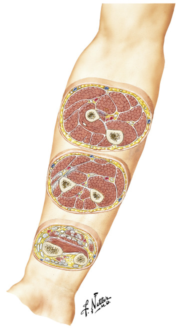
The arrangement of compartments in the forearm is more complex than the arm. There are three fairly well-defined compartments – the volar compartment, the dorsal compartment, and the mobile wad ( Fig. 12.8 ).
The volar compartment is further divided into superficial, intermediate, and deep layers. In the superficial layer ( Fig. 12.9 ), the pronator teres, flexor carpi radialis, palmaris longus and flexor carpi ulnaris arise from the common flexor origin (the medical epicondyle of the humerus). The middle layer is the flexor digitorum superficialis ( Fig. 12.10 ), and the deep layer ( Fig. 12.4 ) contains the flexor digitorum profundus, flexor pollicis longus, and the pronator quadratus. The major neurovascular bundles are present on the volar aspect of the forearm, lying deep in the proximal forearm, but are immediately subfascial in the distal third of the forearm.
The dorsal compartment is arranged in two layers. There are four superficial muscles that fan out from the lateral epicondyle and these are the: anconeus, extensor carpi ulnaris, extensor digiti minimi, and extensor digitorum communis. The deeper layer contains three muscles that power the thumb (abductor pollicis longus, extensor pollicis brevis, extensor pollicis longus), and the supinator and extensor indicis muscles. The posterior interosseous nerve innervates the extensor muscles and is the key anatomic structure from an operative standpoint ( Fig. 12.6 ). The posterior interosseous artery runs under the superficial layer, close to the interosseous membrane and gives off multiple septocutaneous perforators between the extensor digiti minimi and the extensor carpi ulnaris.
The mobile wad comprises three muscles on the radial side of the forearm – the brachioradialis, extensor carpi radialis longus, and brevis.
Vascular Anatomy of the Forearm
The Ulnar Artery
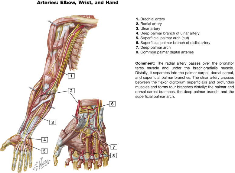
The ulnar artery is a direct continuation of the brachial artery and is the larger of its two terminal branches. It originates at the level of the radial neck approximately 2–3 cm distal to the elbow crease, and is accompanied by two venae comitantes. It reaches the ulnar margin of the forearm approximately half way between the elbow and the wrist ( Fig. 12.4 ).
Proximally, it is deep to the pronator teres, flexor carpi radialis, and flexor digitorum superficialis muscles, lying initially on the brachialis then flexor digitorum profundus muscle bellies. Distally, it continues to lie superficial to the flexor digitorum profundus muscle belly between the flexor carpi ulnaris (ulnarly) and the flexor digitorum superficialis (radially), and is quite superficial in the distal third of the forearm, covered only by deep and superficial fascia and skin ( Fig. 12.9 ), allowing for expeditious exposure with minimal dissection. The ulnar artery gives rise to an average of three (1–6) perforators in its mid-portion. Most of the septocutaneous perforators can be found along a line drawn from the medial epicondyle to the pisiform, ~7–15 cm proximal to the pisiform, or within a 3 cm radius of the midpoint of the forearm. Additional perforators may be located more proximally, however these are usually musculocutaneous through the flexor carpi ulnaris muscle belly and are of limited use in microvascular reconstruction. However, the flexor carpi ulnaris can be used as a pedicled turnover muscle flap based on one or more of these perforators for coverage of small elbow defects.
The ulnar nerve joins the ulnar artery in the proximal forearm and accompanies it on the ulnar side throughout its course ( Fig. 12.4 ). Both structures descend the forearm superficial to the flexor digitorum muscle belly, eventually lie ulnar to the flexor tendons, and pass across the wrist entering Guyon’s canal superficial to the flexor retinaculum.
Branches of the Ulnar Artery
The ulnar artery gives rise to several named branches (anterior ulnar recurrent, posterior ulnar recurrent, common interosseous) and multiple muscular branches ( Fig. 12.7 ).
The anterior ulnar recurrent artery is the most proximal branch of the ulnar artery arising medially just distal to the origin of the ulnar artery, heads proximally and anteriorly between the brachialis and pronator muscle bellies. It anastomoses anterior to the medial epicondyle with the inferior ulnar collateral artery.
The posterior ulnar recurrent artery is usually larger than the anterior ulnar collateral, arises more distally and heads posteriorly and proximally between the deep and superficial finger flexors. It ascends posterior to the medial epicondyle to anastomose with the superior and inferior ulnar collaterals.
The common interosseous artery is the third and largest branch of the ulnar artery, although it can occasionally arise from the brachial artery. It has a short course of about 1 cm, arising from the posterolateral aspect of the ulnar artery at the level of the bicipital tuberosity of the radius ( Fig. 12.7 ). It divides early into anterior and posterior branches that pass to their respective sides of the interosseous membrane.
The anterior interosseous artery courses through the deep flexor compartment accompanied by the anterior interosseous nerve, lying on the interosseous membrane ( Fig. 12.4 ). Distally, it divides into terminal palmar and dorsal branches. The palmar branch contributes to the palmar carpal arch and eventually anastomoses with recurrent vessels off the deep palmar arch ( Fig. 12.15 ), while the dorsal branch pierces the interosseous membrane just proximal to the DRUJ to communicate with the posterior interosseous artery and eventually the dorsal branch of the radial artery, supplying the dorsal carpal arches. These arches ( Fig. 12.16 ) form the basis of several pedicled distal radius vascularized bone grafts, and also provide retrograde flow to the distally-based posterior interosseous artery flap.