Introduction
The structure of the chest and back serves multiple functions that are important to be aware of and maintain during reconstruction. The chest and back protect the vital heart, lungs, great vessels, and spine, and provide a pressure environment for inspiration and expiration. This structure also serves as the foundation for upper limb movement and support of the head and trunk.
Chest and back defects have various causes and each scenario requires careful thought about the etiology and possible effects of the reconstructive process. Defects may be caused by tumor resection infection, specifically, sternal wound infections, chronic empyema, and bronchopleural fistulas, osteoradionecrosis, trauma, congenital, and exposed hardware.
Preoperative Considerations
As with any surgical patient, the surgeon should complete a full history and physical exam and identify any medical comorbidities that need to be optimized prior to reconstruction. Certain comorbidities, such as end-stage renal disease and chronic obstructive pulmonary disease, have been associated with increased mortality in patients undergoing chest reconstruction. With back reconstruction, it is important to assess whether the patient is ambulatory. While indicated in both the ambulatory and the non-ambulatory patient, perforator flap reconstruction is preferred over muscle flap reconstruction in the ambulatory patient.
It is also important to understand all previous operations, as this can affect which flap options are available. If the patient has had a previous thoracotomy or axillary irradiation, the latissimus dorsi may be affected and would not be an ideal reconstructive option. Previous laparotomies may have compromised the rectus abdominis or omentum. Prior internal mammary artery harvest does not preclude a superior-based rectus abdominis flap, as there is collateral flow through the musculophrenic and lower intercostal vessels, however there may be a need to supercharge the outflow and/or inflow. In previous spine surgery, the posterior intercostal perforators may have been divided and their presence should be confirmed with Doppler prior to performing a paraspinous or latissimus dorsi flap reconstruction.
When approaching the defect, one should consider what needs to be addressed prior to definitive reconstruction. Prior irradiation may have rendered tissue rigid and scarred and led to further healing complications if not adequately debrided. Similarly, infected wounds should be precisely debrided prior to definite reconstruction. Any exposed prostheses or hardware need to be assessed and it needs to be determined whether the hardware must be removed or if it remains a critical structure to work around and properly cover. If hardware is well fixed and the infection is early, the area should be scrupulously debrided and the hardware can be salvaged. Spinal hardware infection that has been present for longer than 6 months typically requires removal, due to prolonged microbial contamination. In back wounds, any dural tears need to be identified and treated appropriately with the involvement of neurosurgery.
Imaging may be helpful, but is not essential. Chest and spine X-rays can give information on hardware and general lung condition. One should consider obtaining a CT scan if spinal hardware is present and there is concern for infection or fluid collections. In these cases, fluid collections deep to the muscle are usually an indication of deep hardware infection.
Indications
There are six main objectives of reconstruction:
- 1.
Debride devitalized and/or infected tissues
- 2.
Obliterate dead space
- 3.
Cover exposed prostheses, hardware, and vital structures
- 4.
Cover large surface area defects
- 5.
Buttress organ repairs
- 6.
Structural support.
The aims of providing structural support are to maintain shoulder function and eliminate any discomfort from resected structures. Skeletal reconstruction of the ribcage is indicated if more than four consecutive ribs are resected, or there is >5 cm of lateral chest wall resected. Otherwise, the patient may experience flail chest and severe respiratory compromise. This number can be increased to five consecutive ribs if the patient had previous radiation due to increased fibrosis of the chest wall. There are many options for skeletal reconstruction, including synthetic or biologic meshes, titanium ribs, bone grafts or vascularized osseous flaps. With sternal wounds, one should also consider sternal plating as this may reduce postoperative pain. Contraindications to sternal plating are osteoporosis, infection, extreme obesity, or significant bone loss.
Free flap reconstruction should be considered if:
- 1.
Prior reconstructions, ablations, or radiation have rendered regional options useless
- 2.
Large surface area defects are not adequately covered with regional options
- 3.
No locoregional flap options are available based on location:
- •
Lower cervical back (large defects)
- •
Lower lumbar/sacral midline (large defects)
- •
Posterolateral iliac crest.
- •
Reconstruction by Region
Anterolateral Chest
Pectoralis Major
Coverage:
Advancement flap – upper two-third sternum
Island – complete sternum
Clavicle
Axilla
Turnover flap – lower two-third sternum
Advancement/Island Flap
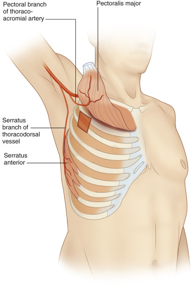
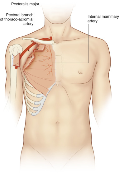
Details of pectoralis flap anatomy and dissection can be found in Chapter 34 . Briefly, the pectoralis major is accessed via the midline sternotomy wound and the muscle edge is identified by dissecting the skin approximately 1 cm ( Fig. 17.1A ). Dissection then proceeds on the undersurface of the muscle superior, to the clavicle, inferior, with elevation of the rectus abdominis fascia, and lateral, until there is enough advancement to midline ( Fig. 17.1B ). This usually extends to between the midclavicular and anterior axillary line. The thoracoacromial pedicle is encountered on the deep surface of the muscle at the junction of middle and lateral third of the clavicle. If more advancement is needed, the muscle can be released from the origin on the clavicle and the insertion on the humerus. When the pectoralis major is being used to reconstruct an area on the anterolateral chest, a similar dissection is performed with rotation into the defect. For axillary defects, an island flap can be used.
Turnover Flap
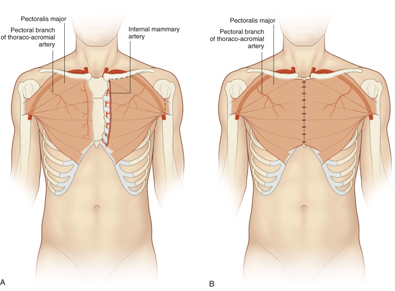
The pectoralis major muscle is exposed to the lateral extent needed for coverage. Next, the muscle and thoracoacromial pedicle are split laterally. The anterior axillary line may be preserved by splitting the muscle vertically at the lateral border of pectoralis minor and suturing the remaining lateral pectoralis major to the underlying pectoralis minor and rib periosteum or by making an L-shaped cut preserving the inferolateral pectoralis major. The undersurface of pectoralis major is then dissected free and the origin at the clavicle is divided. The internal mammary perforators pierce the muscle in the intercostal space 1.5–2 cm from the sternal border and dissection is stopped once these are encountered. The muscle can be split horizontally to interdigitate with the contralateral pectoralis major flap if needed for coverage ( Fig. 17.2 ). This is helpful for small (<5 cm) remaining inferior sternal defects.
Rectus Abdominis
Coverage:
Sternum up to sternal notch
Anterolateral chest
Details of rectus abdominis flap anatomy and dissection can be found in Chapter 37 . Briefly, the rectus abdominis muscle is dissected out via a midline abdominal incision. The deep inferior epigastric vessels and the inferior tendonous insertion to pubic symphysis are divided. The muscle is then passed either subcutaneously or transdiaphragmatically to fill the defect. As mentioned previously, this flap may still be used despite prior internal mammary harvest, but dissection should only extend up to the costal margin, which according to some reports, will only allow coverage of the inferior wound. If skin is needed, a vertical rectus abdominis myocutaneous flap can be performed which includes an ellipse of skin overlying the muscle ( Fig. 17.3 ).
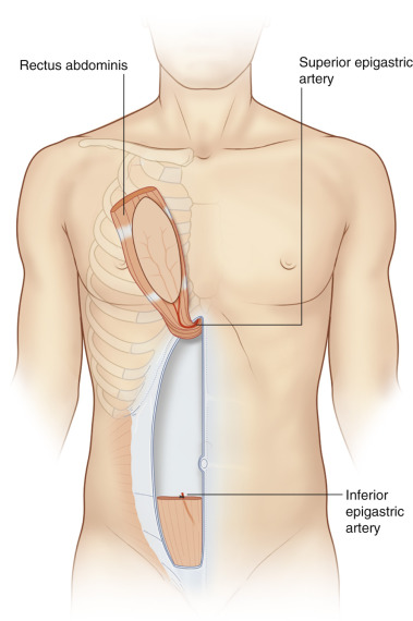
A fasciocutaneous flap, sparing the underlying rectus abdominis, is an alternative. Perforating vessels at the costal margin should be preserved. If a greater arc of rotation is needed, a cuff of rectus abdominis, including the superior epigastric vessels can be included at the costal margin.
Omentum
Coverage:
Sternum
Anterolateral chest up to root of neck
Axilla
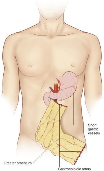
Details of omentum anatomy and dissection can be found in Chapter 36 . Once the omentum is dissected free, the flap is then passed into the chest defect via either a subcutaneous tunnel, which creates a hernia, or preferably via a right transdiaphragmatic route with the liver buttressing the defect. Use of the right gastroepiploic artery provides 5–10 cm greater arc of rotation and if the pedicle needs to be lengthened, internal longitudinal dissection can be performed due to the secondary arcade (Barkow’s arch) ( Fig. 17.4 ). Transdiaphragmatic harvest is also an option to avoid laparotomy for harvest.
Latissimus Dorsi
Coverage:
Sternum
Anterolateral chest
Axilla
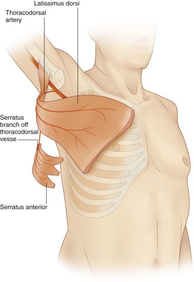
Details of latissimus dorsi anatomy and dissection can be found in Chapter 41 . Briefly, the flap is raised based on the thoracodorsal vessels and a subcutaneous tunnel is created in the axilla to the chest defect. If more reach is needed, the flap can be tunneled under pectoralis major or detached from the humeral insertion to gain approximately 5–10 cm. If a larger flap is needed, the latissimus dorsi can be taken with the serratus anterior ( Fig. 17.5 ). If a smaller flap is needed, the muscle can be split by taking muscle overlying only one of the thoracodorsal branches, thus preserving the other portion. The latissimus dorsi can be completely spared by raising a thoracodorsal artery perforator flap.
Serratus Anterior
Coverage:
Anterolateral chest
Axilla
Some detail on the serratus muscle flap can be found in Chapter 41 . An oblique incision over the lateral chest wall is made and dissection proceeds between the anterior border of latissimus dorsi and inferolateral border of pectoralis major. The thoracodorsal vessels are identified and followed proximal to identify the branch to serratus anterior. Special care should be taken not to damage the pedicle, which lies superficial to the muscle. The long thoracic nerve is identified medial to the pedicle and preserved. The lower 3–4 segments of serratus anterior are harvested by dissecting anterior to posterior in a supraperiosteal plane. The upper segments of muscle are kept attached to the scapula to prevent winging of the scapula.
External Oblique
Coverage:
Superior extent – 3rd intercostal space, reports of up to clavicle
Medial extent – 5 cm past midline
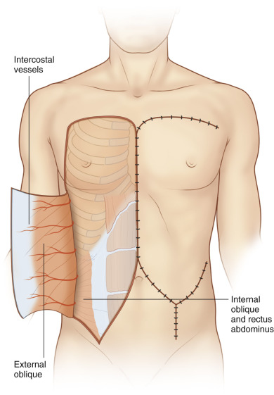
The incision is made extending inferior from the defect with the inferior limb making a gentle horizontal curve from just above the umbilicus to the anterior superior iliac spine. Dissection proceeds in the subcutaneous plane to linea semilunaris, where the anterior rectus sheath is incised. Dissection then continues between the external oblique and internal oblique to the midaxillary line, where the intercostal vessels enter. The external oblique is then divided from the anterior iliac crest and costal margin. When closing the abdomen, if a portion of the anterior sheath was taken with the flap, the internal oblique is advanced and sutured to linea alba to avoid potential weakness of the abdominal wall. Larger flaps with greater arcs of rotation can be created by making an oblique inferior backcut for more cephalad movement and closing the donor site in a V-Y fashion ( Fig. 17.6 ). The external oblique muscle can be spared and the flap raised on thoracoabdominal perforators as an island flap.
Intercostal Perforator Flap
Coverage:
Anterolateral chest
Axilla
The flap can be based off either the lateral or anterior intercostal perforator vessels depending on the coverage needed ( Fig. 17.7 ).
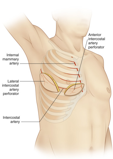
For lateral intercostal perforator flaps, the pedicle used is usually from the 4th to 8th intercostal space, with the 6th space being the largest. The skin paddle is usually oriented parallel to the rib with the posterior border based at least 5 cm behind the posterior axillary line. Flaps as large as 25 × 20 cm may be raised, but in order to close the donor site primarily the size taken is usually smaller. The posterior incision is made first and dissection proceeds subfascial along the latissimus dorsi. As the dissection continues, the perforator is identified. The 6th intercostal perforator is located 3 cm anterior to the border of latissimus dorsi. Once the vessel is identified, the intercostal muscle is freed from the lower border of the rib and the anterior extension of the posterior intercostal vessel is divided to allow up to 180° of rotation into the defect.
For anterior intercostal perforator flaps, the largest anterior intercostal pedicles are located in the 2nd–3rd intercostal spaces 1–3 cm lateral to the sternum. The flap design is usually longitudinal or oblique towards the ipsilateral shoulder. Dissection is performed in a latero-inferior to medio-superior direction and may need to proceed through the pectoralis major or rectus abdominis muscles.
Scapular/Parascapular
Coverage:
Axilla
Details of scapular/parascapular perforator flap anatomy and dissection can be found in Chapter 40 . For axilla defects, a flap based on the parascapular axis is preferred. This flap is especially useful in release of contractures from axillary burns.
Trapezius
Coverage:
Anterior superior chest
Axilla
Details of trapezius flap anatomy and dissection can be found in Chapter 39 . The dorsal scapular perforator flap variation is the best option for defects of the anterior chest and axilla (see Fig. 17.9B ). This flap is based on a skin paddle near the inferior portion of the muscle.
Intrathoracic
There are many regional flap options to cover exposed vascular grafts or buttress intrathoracic repairs. The most common used flaps are: pectoralis major, latissimus dorsi, and serratus anterior. Each flap offers different advantages depending on the defect characteristics. Latissimus dorsi offers more bulk to fill large dead space, whereas serratus anterior is thin and pliable, which is advantageous for buttressing bronchopleural fistula repairs. The muscles are dissected free from their origins and insertions around the pedicle and portions of the ribs near the pedicle are removed to allow intrathoracic passage ( Fig. 17.8 ). For buttressing intrathoracic repairs, the muscle is draped over the repair rather than being sewn to the potentially delicate, friable repair.