Introduction
Reconstruction of the torso is a broad topic, which in and of itself includes techniques of local tissue rearrangement, skin grafting, local and regional flaps, as well as complex microsurgical reconstruction. The defects to be reconstructed are equally diverse and can include breast reconstruction, wounds related to spinal surgery, cardiothoracic surgery, abdominal wall reconstruction, or inguinal vessel exposure. The majority of these techniques are addressed in specific chapters throughout this book; however, the purpose of this chapter is to approach the torso in a broad manner, specifically addressing the anatomic considerations of muscle, vessel, nerve and lymphatic anatomy, which play a significant role in the understanding of torso reconstruction as a whole.
Muscular Anatomy by Region
The Chest
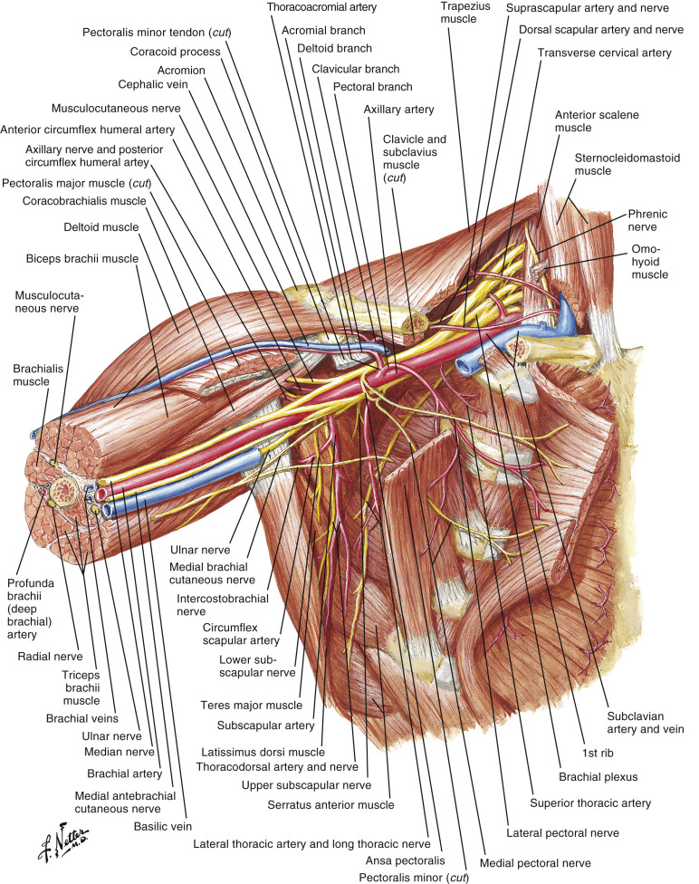
The workhorse muscle of the thorax is the pectoralis major. It has a dual origin from both the clavicle and the sternum and inserts on the intertubercular groove of the lateral humerus. Owing to its predictable, dual blood supply through the thoracoacromial and internal mammary perforators, as well as its broad surface area, the pectoralis major can be useful in many forms of reconstruction. The pectoralis minor sits under its major counterpart and is much more diminutive in nature ( Figs 11.1 , 11.2 ).
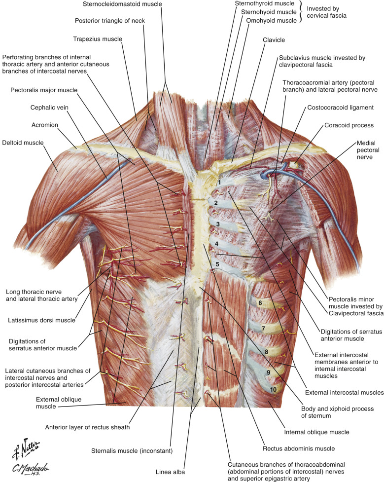
The serratus anterior originates from the lateral, anterior surfaces of ribs 1 through 8 and inserts on to the anterior surface of the medial border of the scapula. Injury to its innervation, the long thoracic nerve, results in “winging” or protrusion of the scapula off of the chest wall. Elevation of the anterolateral origins of the muscle is very useful in inferolateral muscular coverage of an expander for breast reconstruction.
The intercostal muscles are comprised of the external intercostal, internal intercostal, and innermost intercostal, stacked on top of one another from superficial to deep. Their membranous coverings are each synonymous with, and contiguous with, the corollary muscle of the abdominal wall. These muscles are intimately involved in respiration.
The Abdomen
The rectus abdominis is a vertically oriented, paired muscle of the anterior abdominal wall. The pair is separated in the midline by a dense fibrous line called the “linea alba.” The pair originates on the costal margin bilaterally and inserts on the symphysis pubis. The lateral abdominal wall is comprised of three muscles, the external oblique, internal oblique and transversus abdominis. The external oblique is the largest and most superficial of the three with fibers oriented in a superolateral to inferomedial direction. It originates on the external surfaces of the 5th through 12th ribs and inserts on the linea alba, pubic tubercle and iliac crest. The inferior edge is rolled over on itself and makes the inguinal ligament. The internal oblique is deeper and more vertically oriented with an origin on the thoracolumbar fascia, iliac crest, and inguinal ligament with an insertion on the inferior border of the 10th through 12th ribs. The transversus abdominis is the deepest of the three with a transverse orientation as the name would suggest. It originates laterally on the thoracolumbar fascia, inguinal ligament and iliac crest and inserts on the linea alba.
The rectus sheath is a strong fibrous layer surrounding the rectus abdominis muscles. It is a fusion of the aponeuroses of the three lateral muscles. For the majority of the sheath, the posterior sheath is comprised of the transversus abdominis and half of the internal oblique. The anterior sheath is comprised of half of the internal oblique and the entire external oblique. That is to say that the internal oblique bifurcates anterior and posterior to the rectus abdominis muscles and all three merge in the midline to form the linea alba. Midway between the umbilicus and pubic symphysis however, is the arcuate line. Below this line, the aponeurosis of all three muscles passes anterior to the rectus muscles. Thus, there is no posterior rectus sheath below the arcuate line.
The Back
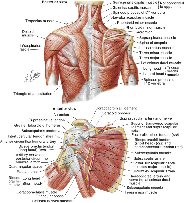
The most superficial, but dominant muscle of the upper back is the trapezius ( Fig. 11.3 ). It has broad attachments medially along the ligamentum nuchae, occipital protuberance and along the spinous processes of C7 through T12. These allow for an expansive and strong action on the shoulder via its lateral attachments to the lateral third of the clavicle, acromion and spine of the scapula. It is innervated by the accessory nerve (CN XI) and cervical roots of C3 and C4.
Inferiorly, the lower back is covered by the latissimus dorsi, a broad, fan-like muscle incredibly versatile for local and free flap reconstruction. It originates medially along the spinous processes of T6 through T12, as well as the thoracolumbar fascia, iliac crest and inferior 3 or 4 ribs. It inserts along the intertubercular groove of the humerus. Innervated by the thoracodorsal nerve, this muscle is the main actor for adduction and medial rotation of the humerus, as well as shoulder extension. With arm extension, this muscle is the primary driver to lift the torso while climbing.
Additional players of shoulder motion include the levator scapulae, rhomboid major and rhomboid minor. All three originate along the spine and insert along the medial border of the scapulae to aid in scapular stability, retraction and elevation ( Fig. 11.4 ).
The paraspinous muscles are a group of muscles including but not limited to the spinalis, longissimus and iliocostalis which aid in spinal extension and stability. These long muscles located adjacent to the spinous processes of the spinal column are useful in spinal hardware coverage.
Arterial Anatomy by Region
The Chest
The most commonly used arteries for chest reconstruction include the thoracodorsal, thoracoacromial, and internal mammary systems (see Figs 11.3 , 16.1 , 16.2 , 16.4 ). Less commonly used arteries include the circumflex scapular, the lateral thoracic ( Fig. 11.1 ), and a perforating artery of the internal mammary. The thoracodorsal system is used most commonly in pedicled reconstruction using the latissimus dorsi muscle or myocutaneous flap. The thoracoacromial or internal mammary arteries are useful for pedicled pectoralis major advancement or turnover muscle flaps. While all of these arteries are useful in regional reconstruction, the thoracodorsal and internal mammary arteries are the most useful as recipient vessels in microsurgical cases.
The Anatomy and Exposure of the Subscapular System
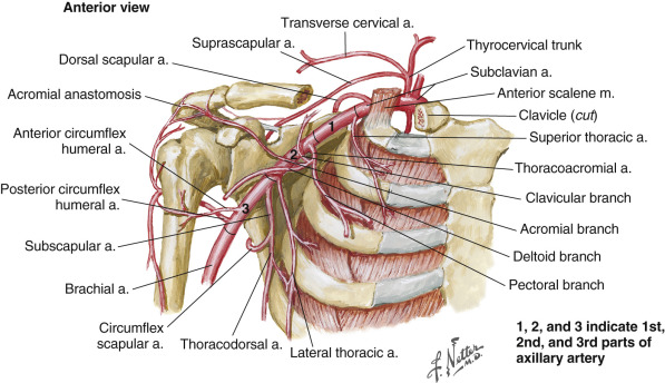
The subscapular system begins as a short artery arising from the axillary artery, which divides into the thoracodorsal and circumflex scapular arteries. The thoracodorsal artery continues for 2–3 cm before it gives off one or two branches to the serratus muscle. For free tissue transfer, these serratus branches or the terminal branches of the thoracodorsal artery can be used when the donor pedicle artery is of a smaller caliber. The circumflex scapular courses laterally as it originates from the subscapular artery ( Figs 11.1, 11.4, and 11.5 ) and passes through the triangular space to become a cutaneous artery that supplies the scapular and parascapular fasciocutaneous flaps.
If an axillary dissection has been part of the surgery that has immediately created the chest defect, the thoracodorsal vessels are usually exposed and require little additional dissection to prepare them as recipient vessels. In this setting, the thoracodorsal nerve usually crosses anterior to the artery and vein. This nerve should be dissected off the vessels and the vessels dissected from several small branches so as to free the thoracodorsals from their origin to the first or second serratus branch. In addition, the vein should be separated from the artery for several centimeters as well. If the axilla has not been dissected as part of the immediate defect, the thoracodorsal vessels are approached through a transverse incision between the anterior and posterior axillary folds. The level of the incision is just below the lower limit of the axillary hair.
The lateral border of the pectoralis muscle is defined and the axillary fat is entered directly posterior to this border of the muscle. The lateral thoracic vein is usually first encountered within the axillary fat and this vein can be traced superiorly to identify the axillary vein. Spreading the axillary fat just posterior to the origin of the lateral thoracic vein usually reveals the proximal portion of the thoracodorsal vessels and nerve. The axillary fat is usually easily dissected off these vessels. Self-retaining retractors are helpful in holding back the axillary fat so that these vessels can be dissected and prepared. There is usually a single perforating branch to the axillary fat at the proximal portion of the thoracodorsal vessels. This branch should be ligated before separating the thoracodorsal vessels from the axillary fat. With proper retraction, it is unnecessary to remove any of the axillary lymphatics in preparing these recipient vessels.
If the axilla has been previously dissected, the thoracodorsal vessels may still be usable as recipient vessels. The skin incision is made as above. The lower portion of the latissimus muscle border is defined by separating the lateral chest skin off the chest wall musculature, well below the axillary vessels. With the lower lateral border of the latissimus defined, the vessels are identified as they enter the muscle and then traced proximally. This allows for safe dissection up to the proximal thoracodorsal vessels as well as the axillary vein and thoracodorsal nerve.
The circumflex scapular vessels lie just posterior to the origin of the thoracodorsal vessels and then travel laterally just below the inferior border of the axillary vein toward the triangular space. Small branches coming off these vessels are divided and the distal ends of the artery and vein can be divided near the triangular space. This allows for rotation of these circumflex scapular vessels medially and anteriorly, so as to make them useful as recipient vessels within the axilla. If the thoracodorsal vessels are found to be not useful as a result of previous axillary dissection, the circumflex scapular vessels should be examined since they are not usually injured by previous axillary surgery.
The Anatomy and Exposure of the Internal Mammary Artery
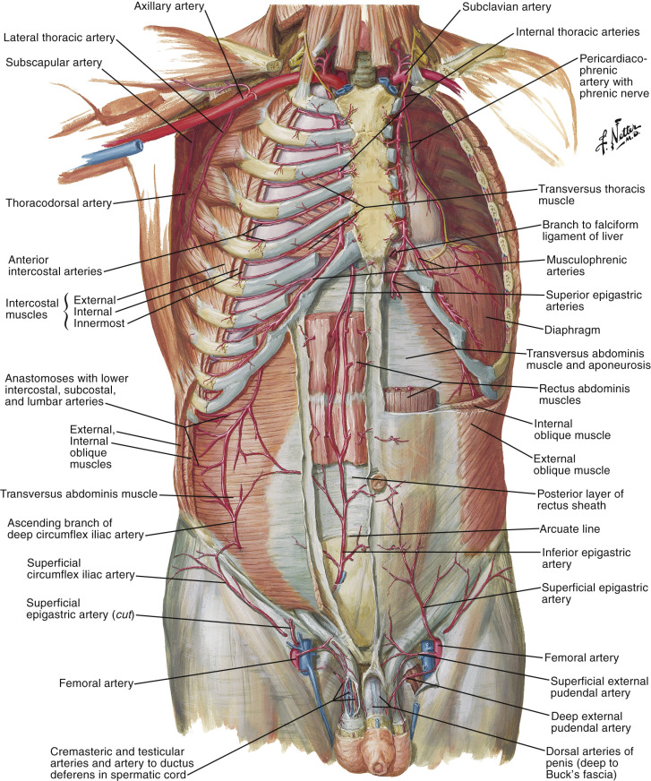
The internal mammary artery originates from the corresponding brachiocephalic or subclavian artery and courses inferiorly, deep to the costal cartilages and superficial to the pleura. As this vessel exits underneath the costal margin, it becomes the superior epigastric artery (see Fig. 11.3 ). The internal mammary artery is usually accessed by removing the third costal cartilage. Perforating vessels just above the second, third or fourth costal cartilages are common and can be used as recipient vessels in this region if large enough.
A direct incision may be made overlying the third costal cartilage ( Fig. 11.2 ). This can be identified by palpating the sternal angle which is at the level of the second costal cartilage. Moving one cartilage lower identifies the third costal cartilage. The skin and soft tissues are dissected just above the pectoralis muscle fascia. The pectoralis muscle is separated on top of this cartilage and the cartilage is removed through a subperichondrial dissection. The deep perichondrium is incised and reflected lateral to medial to expose the internal mammary vessels and lymphatics. The intercostal musculature is divided up to the bottom of the second costal cartilage. This muscle can also be divided to the top of the fourth costal cartilage if additional pedicle length is required. There is usually a perforating branch at the level of each costal cartilage that needs to be divided in order to free up these vessels. There are also other small branches that will need to be divided as well. The artery and vein are usually easily separated from each other. At this level, there is usually only one vein on each side, with the left vein tending to be smaller than the right. The vein is usually thin walled and the artery is also subject to easy trauma, resulting in segmental thrombosis. These vessels, in particular, require gentle dissection. The artery’s intima separates easily and the surgeon must be assured that the sutures capture arterial adventitia in order to obtain adequate strength for the anastomosis.
Perforators off the internal mammary system should be evaluated as the skin and soft tissues are elevated off the pectoralis muscle. A sufficiently large perforator can be considered as a recipient vessel, avoiding further soft tissue dissection and removal of the costal cartilage. These perforating vessels can be traced up into the subcutaneous tissue, dividing small branches and developing a reasonable length of recipient vessels.
The Anatomy and Exposure of the Lateral Thoracic Artery
The lateral thoracic artery ( Figs 11.1 , 11.5 ) is a direct branch off the axillary artery and follows with the lateral thoracic vein ( Fig. 11.8 ). This vessel is more superficial within the axilla as compared to the thoracodorsal artery. The same incision and approach are used to harvest the lateral thoracics, as done in dissecting the thoracodorsal vessels. The lateral thoracic vein is usually easily identified within the more superficial axillary fat. The lateral thoracic vein is typically larger than the thoracodorsal vein. The lateral thoracic artery is just posterior to the vein and is considerably smaller in caliber. On occasion, this artery is as large as the thoracodorsal artery.
The Anatomy and Exposure of the Thoracoacromial Artery
The thoracoacromial artery comes off the subclavian artery to supply the pectoralis major muscle ( Figs 11.1 and 11.5 ). It typically enters the undersurface of the pectoralis major between the middle and lateral thirds of the muscle. An incision is made just below the lateral half of the clavicle. The skin and soft tissue are elevated to expose the pectoralis major muscle. The clavicular portion of the pectoralis muscle is divided to expose the thoracoacromial vessels. Their location can first be identified with a handheld Doppler. After dividing the muscle, the vessels are usually within a sheath of fatty tissue with two accompanying veins. The vessels can be dissected away from the muscle and then toward their origin on the axillary vessels.
Advantages and Disadvantages of Recipient Arteries for Reconstruction of the Chest Region
The advantages of the thoracodorsal artery include dependable anatomy and a generally consistent caliber. The artery is usually easily identified along with the vein and, more importantly, the thoracodorsal nerve. The thoracodorsal artery is easily dissected and is not sensitive to manipulation during dissection. It is generally useful even in the setting of previous axillary dissection. The main disadvantage is the need to dissect through a moderate amount of axillary fat and lymphatic contents to expose this artery when an axillary dissection has not been part of the procedure. Although this artery can be transposed to a more superficial position after dissection and distal division, the axilla still tends to be a deeper, confining recipient site requiring longer instruments and additional skill to perform anastomoses.
The advantages of the internal mammary artery include dependable anatomy, a consistently large vessel caliber, and its superficial location, which allows greater ease for performing anastomoses. This artery provides for an excellent alternative to the thoracodorsal artery in the setting of previous axillary dissection. The disadvantages include this artery’s susceptibility to injury even with gentle dissection. The attachments of its intima are also fragile, with intimal separation and injury occasionally occurring during routine anastomoses. The accompanying vein, particularly on the left side, can be too small despite a very adequate recipient artery, necessitating a move to another recipient site. The internal mammary perforators have the advantage of eliminating the need for the removal of a costal cartilage. Their primary disadvantage is a small caliber.
The advantages of the circumflex scapular include consistent anatomy and a large-caliber vessel, which is usually larger than the accompanying thoracodorsal artery. On occasion, the thoracodorsal artery is injured as a result of the axillary surgery; however, it would be rare to injure the circumflex scapular in this particular setting. The circumflex scapular artery can be dissected as it courses toward the triangular space and transposed into a more superficial location in the axilla. The disadvantages include the same depth issues in performing the anastomosis as in the thoracodorsal vessels. Congenital absence of the circumflex scapular artery is uncommon but has been reported.
The advantages of the lateral thoracic artery are its superficial location and its very large accompanying vein. The lateral thoracic vein is usually larger than the thoracodorsal vein. The main disadvantage of the lateral thoracic artery is its routinely small caliber, making the thoracodorsal artery a better choice in this setting.
The Abdomen
The Anatomy and Exposure of the Inferior Epigastric Artery
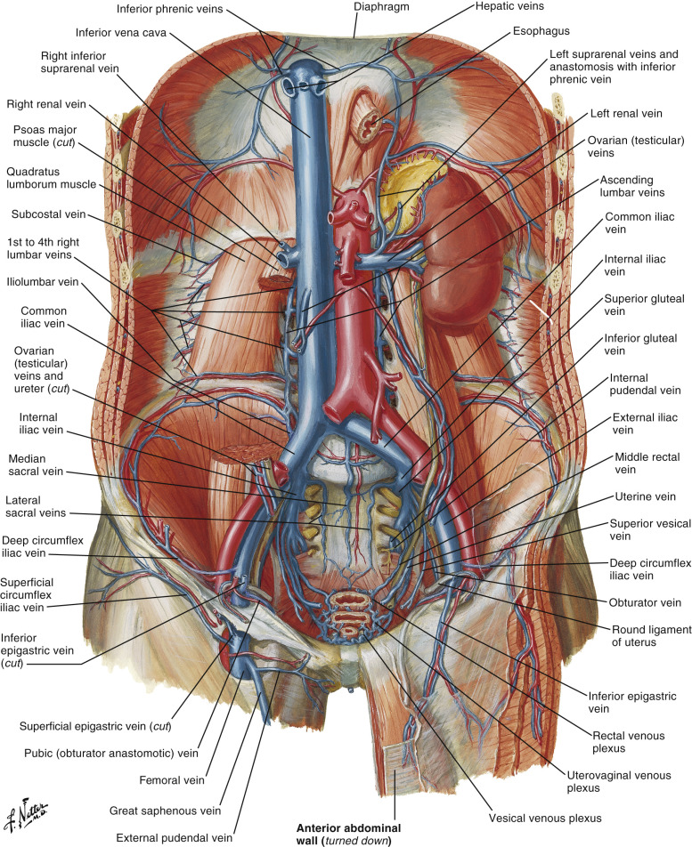
The deep inferior epigastric artery is a direct branch off the external iliac artery. This artery continues moving superiorly and medially for 4–6 cm before it enters the rectus abdominis muscle. The artery gives off several small branches as it runs from the external iliac artery to the muscle ( Fig. 11.6 and see Fig. 11.3 ).