Cleft lip with or without cleft palate is the most common congenital malformation of the head and neck. Orofacial clefting could significantly affect the quality of life of the child and requires multiple steps of care to obtain an optimal outcome. Each patient should be evaluated for congenital anomalies, developmental delay, neurologic disorders, and psychosocial concerns. A multidisciplinary team is necessary to ensure that every aspect of the child’s care is appropriately treated and coordination between providers is achieved. This article discusses the assessment and treatment recommendations for children born with cleft lip and/or cleft palate.
Key points
- •
Cleft lip with or without cleft palate is the most common congenital malformation of the head and neck.
- •
Each patient should be evaluated for congenital anomalies, developmental delay, neurologic disorders, and psychosocial concerns before surgery.
- •
A multidisciplinary team is necessary to ensure that every aspect of the child’s care is treated.
- •
The surgeon should be aware of the needs of the cleft patient and be able to educate and assist caretakers as necessary.
- •
A fundamental understanding by the surgeon of the surgical options for cleft repair is warranted.
Overview
Cleft lip with or without cleft palate is the most common congenital malformation of the head and neck. The impact on quality of life for the child and the family can be severe, particularly in unsuspecting families. Emotional and psychological needs must be recognized and addressed, in addition to surgical care, for all those involved with the patient. Assessment and treatment of those with cleft lip and/or palate requires a multidisciplinary approach. Access to and evaluation by speech-language pathology, surgery, psychology, psychiatry, social work, audiology, genetics, dentistry, otolaryngology, and pediatric primary care are all recommended by the American Cleft Palate–Craniofacial Association. The recommendation for a team approach allows the child to be able to obtain complete and coordinated care. This article discusses the assessment and treatment recommendations for children born with cleft lip and/or cleft palate. This article focuses on the surgical management and treatment of these special patients.
Incidence and Genetics
Clefts of the lip and/or palate affect approximately 1 in 700 live births. The incidence varies widely depending on geographic origin, racial and ethnic group, environmental exposures, and socioeconomic status. Asian and Native American populations have reported prevalence rates as high as 1 in 500. European populations are approximately 1 in 1000, whereas African populations have a reported prevalence close to 1 in 2500. Clefts of the lip and/or palate can be categorized as syndromic or nonsyndromic. Syndromic clefts are those that occur in association with a recognized pattern of human malformation or syndrome. The cause of a syndromic cleft may be associated with gene transmission, chromosomal aberrations, teratogens, or environmental factors. Identifying an associated syndrome is important, because it can have prognostic implications that may be helpful to the patient and the family.
Classification
Orofacial clefts include all variations of cleft lip and cleft palate. A variety of classification schemes have been suggested and recommended for typical and atypical orofacial clefts. The features used to initiate the classification of an orofacial cleft include the laterality, completeness, severity (wide vs narrow), and presence of any abnormal tissue. Diminutive orofacial clefts may also be described as microform, occult, or minor. The cleft lip laterality includes unilateral and bilateral. A complete cleft lip extends through the lip and the nasal sill, whereas an incomplete cleft lip extends through the orbicularis oris and skin, but intact lip tissue persists. The cleft alveolus can also be considered complete or only notched. A weblike piece of tissue may extend from the lip’s cleft side to the noncleft side at the nasal sill. This abnormal tissue is called a Simonart band and, if present, it is not considered the same as an incomplete cleft.
A cleft palate can be unilateral, when 1 palatal shelf attaches to the nasal septum, or bilateral. The classification scheme introduced by Victor Veau is the most popular system. Clefts of the palate were placed into 4 groups. A group I defect includes a cleft of the soft palate only. Group II clefts exist when the defect involves the soft palate and the hard palate to the incisive foramen (secondary palate). Groups III and IV are unilateral and bilateral defects extending through the entire palate and alveolus, respectively.
Patient assessment
Multidisciplinary Care
Patients with cleft lip and/or palate may present as early as during the prenatal period. Surgical consultation before birth is becoming more common because of the ability to make the diagnosis on prenatal ultrasonography. Discussion regarding general care issues and surgery can help improve some of the anxiety the expecting mother may be experiencing. After birth, the initial management includes ensuring proper feeding of the neonate and evaluation for other comorbidities. As previously discussed, a multidisciplinary approach should be used in the assessment of the child. The patient should undergo initial evaluations by a pediatrician, geneticist, surgeon, feeding specialist, and social worker. Such services allow immediate education and support for the caretakers and the patient. Future assessments need to be performed by audiology, otolaryngology, dental, maxillofacial, speech-language pathology, and psychosocial practitioners.
Surgical Assessment
Immediately after birth, the surgeon should evaluate and examine the neonate. The various anatomic sites for clefting are assessed, including the upper lip, alveolar arches, nostrils, and primary and secondary palates. These areas should be palpated and inspected under direct visualization. Microform cleft lip and submucous cleft palate can often present with only subtle findings on clinical examination. Particular attention should be given to the nasal characteristics in the setting of a cleft lip. The nasal alar symmetry, tip projection, and alar base position and width should all be assessed. The extent of the clefting can be classified, as described previously. A thorough physical examination is necessary to evaluate for any signs of dysmorphia that may lead to the identification of other congenital anomalies or a syndromic diagnosis.
Regular clinic visits with the patient and caretakers allow the surgeon to provide counsel and guidance before surgery. Feeding and weight gain are important aspects to monitor with each visit. Associated congenital anomalies, developmental delay, and neurologic disorders should also be followed. Any concern for cardiac defects or airway obstruction needs to be identified and evaluated promptly. A multidisciplinary approach is warranted to ensure proper management of all of the needs of the child. The surgeon should be aware of these needs and assist with any referrals to the necessary specialists.
Unilateral cleft lip and nasal deformity
Preoperative Planning and Preparation
Before surgical treatment of a unilateral cleft lip, adequate weight should be established, with the child weighing at least 4.5 kg (10 lb). Breast feeding is recommended, when possible, but often this is difficult for the infant. However, pumped breast milk may be taken via bottle feeds with the use of a specialized nipple that controls the flow rate, such as a Haberman or pigeon-type nipple based on an evaluation by speech therapy. Most surgeons prefer an average of 28 g (1 ounce) of weight gain per day beginning 2 after birth.
Adequate management for any cardiopulmonary anomalies should be ensured. If there are any concerns with the ability of the child to tolerate general anesthesia, evaluation by an anesthesiologist before surgery is warranted. The anesthesiologist should be knowledgeable and experienced in pediatric anesthesia.
Consideration must be given to the overall width of the cleft. The presence of a wide cleft may make the repair difficult and place undesirable tension on the closure. Excess tension after lip repair can lead to lip breakdown and scarring. Presurgical devices to improve the success of the repair by narrowing the width of the cleft include lip taping, use of an oral appliance (eg, Latham appliance), and nasoalveolar molding. Surgical options may include performing a 2-staged repair with primary lip adhesion or delaying the repair in order to allow increased growth of the tissues.
Timing of Repair
The surgical repair of the lip is usually performed during the first year of life and may be performed as early as is considered safe for the patient. In utero surgery for cleft lip has been contemplated and attempted because it may potentially provide a scarless repair of the deformity. However, this option must become safer for the fetus and the mother before it can be an acceptable practice. In the 1960s, the general so-called rule over 10 was proposed as criteria for the timing of lip surgery. This rule requires the patient’s weight to be more than 10 lbs (4.5 kg), have a hemoglobin more than 10 g, and be more than 10 weeks of age. Millard recommended postponing repair of a unilateral cleft to at least 3 months of age and preferably 4 or 5 months when possible. Many consider that waiting 3 months allows for a safer anesthesia, improved accuracy of the repair, and parental acceptance of the malformation.
Surgical Technique
As mentioned earlier, multiple techniques and options have been described to restore function and anatomic contour to a cleft lip. These options include the straight line closure, geometric lip repair, the rotation-advancement technique, and variations on each of these techniques. The ultimate goal is to have a patient with a balanced and symmetric face. The principles of advancement and rotation are common practice in repairing clefts. These principles were first made apparent in the Millard technique. Since Millard’s description, a variety of techniques for cleft lip and nasal repair have been described, all with excellent results. Each of these repairs attempts to lengthen the noncleft side by placing a scar at the base of the columella (Millard, geometric) or above the vermilion (straight line). This article provides a detailed description of the Millard rotation advancement, which has provided excellent and reproducible results. However, the various options should be known by the surgeon to allow intraoperative changes because of the unique anatomy that may present with each patient.
Patient positioning
The patient is positioned supine on the operating room bed. Preoperative checklists are performed, correct patient ensured, and the consent is verified. General anesthesia is performed by the anesthesia care team and the patient is intubated with either a standard endotracheal tube or a RAE tube. The tube is taped in the middle of the lower lip on the chin. Tegaderm dressings (3M Health Care, St Paul, MN) are placed to protect both eyes. The surgical bed is typically rotated at least 90° from the anesthesiologist and a shoulder roll is placed. Infraorbital nerve blocks are performed with 0.25% bupivacaine. Intravenous antibiotics are administered. The surgical site is prepared and cleaned with Betadine. The surgical site is then toweled off and draped in a standard fashion.
Procedural design and markings
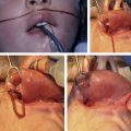
The incisions are planned and marked using methylene blue or gentian violet on a sharply pointed wooden end of a cotton tip applicator. The markings are similar to those described originally by Millard ; however, each patient’s malformation is unique and every situation calls for a certain amount of improvisation and artistry by the surgeon ( Fig. 1 ). The peak of Cupid’s bow is identified and marked on the noncleft side, followed by marking of the midline of the bow. A mark is placed equidistant from the midline of the Cupid’s bow and the peak on the noncleft side. This mark is the point at which the Cupid’s bow will be created on the cleft side. On the cleft side, the point corresponding with the new peak is also marked. This point is determined by identifying the end of the white roll and following the white roll laterally until the lip appears to have maximal vermilion height and muscle bulk, usually 1 to 2 mm. Both points corresponding with the new Cupid’s peak are tattooed at the white roll with either methylene blue or gentian violet using a 27-gauge needle. The tattooed marks are visible subdermally and aid in exact white roll approximation during closure.
The philtral lengths on the cleft and noncleft sides are assessed. A caliper can be used to determine the discrepancy and the amount of necessary rotation of the noncleft flap can be predicted. An incision is marked along the mucocutaneous junction extending from the tattooed Cupid’s peak to the medial nasal floor. This mark should not include any mucosa and should attempt to preserve as much skin as possible, depending on the patient’s anatomy and the surgeon’s artistic eye. Based on the amount of necessary rotation, a curvilinear incision is marked, extending from tattooed Cupid’s peak to 1 mm inferior to the subnasale. The arc of rotation should be inferior to the columellar crease and its length should be equal to the philtral height on the noncleft side. This incision usually needs to proceed to the midline of the columella and can proceed up to, but not across, the noncleft philtral ridge. This philtral ridge should be kept in continuity in order to preserve its appearance.
On the cleft side, a similar curvilinear incision is marked along the mucocutaneous junction, extending from the tattooed white roll and then curving to near the alar base superiorly. The advancement flap is marked at a perpendicular angle from the alar base, but there is no need to continue the incision around the alar base, because this may result in an unsightly scar. Markings are then made extending from both tattooed marks perpendicular to the white roll line toward the gingivolabial sulcus. Using 0.5% lidocaine with 1:100,000 epinephrine, the lip, gingivolabial sulcus, pyriform aperture, supraperiosteal maxilla, and nasal septum are injected.
Incisions and flap creation
The lip is grasped between the thumb and index finger and pressure is applied to minimize blood flow from the labial artery. Using a #15 blade scalpel, preferably on a round knife handle for easier mobility, the lateral incision is made and incised down to the labial sulcus, preserving as much of the orbicularis muscle as possible. The incision is extended, as described previously, to the lateral alar base. A back cut is made near the labial sulcus to aid in medial advancement of the lateral lip and to allow improved access to the alar base. Through this incision, the alar base can be released from the premaxilla and orbicularis oris in a supraperiosteal fashion, allowing alar base repositioning superiorly and medially. Using the scalpel, the edge of the orbicularis muscle is skeletonized from the overlying dermis and underlying mucosa.
The marked incisions are similarly incised medially with a #15 blade scalpel. However, on this side the vermilion mucosa is pedicled on the red lip and can be tailored at the end to assist with providing increased fullness to prevent notching. Careful attention is made to preserve the columellar based skin flap, also known as the c flap, and to separate it from the underlying orbicularis muscle fibers. A back cut is then made in the labial sulcus up to the nasal spine, including the release of the upper labial frenulum. This allows access to release and rotate the orbicularis oris fibers from off the nasal spine in order to provide rotation of the medial lip and lengthening of the lip on the noncleft side. In similar fashion to the contralateral side, the orbicularis muscle is released from the dermis and mucosa of the lip ( Fig. 2 ). However, only minimal undermining is performed on the dermal side in order to maintain the philtral dimple.
Closure
Before closure, hemostasis is ensured with the use of needle-tip monopolar electrocautery. The vermilion mucosa is trimmed until the proper amount is left for closure to allow fullness and to prevent notching. The vermilion mucosa most distal from the white roll is closed initially using interrupted 4-0 Vicryl suture, because it is hard to close the mucosa once the orbicularis is approximated. The orbicularis muscle is secured together with a 3-0 Vicryl suture with an RB1 needle in an interrupted fashion, ensuring rotation of the medial lip, advancement of the lateral lip, and proper alignment of the muscle to prevent a whistler deformity. The use of 3-0 Vicryl to approximate the orbicularis oris reduces the likelihood of lip dehiscence. A deep dermal stitch is placed 1 mm from the white roll to approximate the vermilion border using a 5-0 Vicryl suture with a P3 needle. This same suture is placed to inset the advancement flap from cleft side into the apex of the rotation flap, at the base of the columella. The c flap is advanced laterally and the alar base is advanced medially in order to be approximated using a 4-0 chromic with a G2 needle to form the nasal sill. When closing the nasal floor, the surgeon should ensure that the alar widths on the cleft and noncleft sides are similar. The same suture is used superficially to align the white roll in an interrupted fashion. The red lip is closed using a 4-0 Vicryl with a P3 needle. The gingivolabial sulcus is left to heal by secondary intention. The skin of the philtrum is closed using a 6-0 fast-absorbing suture with a PC1 needle in an interrupted fashion ( Fig. 3 ). Surgical glue is then placed over the closed incisions.
Primary rhinoplasty
There are many different approaches to performing a primary cleft rhinoplasty, including using a tunneled incision via the columella and alar base versus a direct marginal incision to free the lower lateral cartilage. Debate continues as to the utility, effectiveness, potential complications, and best practice of performing a primary rhinoplasty. Most cleft surgeons realize that early surgical treatment of the nasal deformity minimizes nasal asymmetries and allows the nose to grow symmetrically.
In order to perform a primary rhinoplasty, further dissection of the lower lateral cartilages must be performed to release the cartilage from the skin soft tissue envelope. This dissection is typically performed through a marginal incision. Surgical scissors are used medially to perform dissection between the medial crura superiorly to the nasal tip. The scissors are again used laterally to dissect the soft tissue envelope off the lower lateral cartilages while using palpation and visualizing the skin until the nasal tip is encountered. The cleft side nasal dome can be repositioned using a lateral crural steal technique; the medial crus is elongated by stealing some additional length from the lateral crus, which is accomplished by passing a full-thickness suture from lateral to medial through the lower lateral cartilage on the cleft side. This technique provides improved symmetry and projection of the nasal tip. These modifications are often performed using 5-0 PDS suture. Using the same stitch, further sutures can be placed transcutaneously in mattress fashion to further contour the lower lateral cartilage in an attempt to improve symmetry and to more fully define the alar-facial groove. Some investigators advocate the use of tie-over bolsters and/or the use of nasal conformers.
Postprocedural care
During the postoperative period the primary goals include prevention of infection and wound tension, provision of pain relief, and promotion of adequate nutrition. Children are monitored in the hospital for at least 1 night to ensure that they are able to keep themselves hydrated. Arm restraints are used to protect the incision for 2 weeks and are only taken off when the child is in the parents’ arms. A 1-week course of oral antibiotics is administered. Pain control with the use of acetaminophen and ibuprofen is recommended. The child is evaluated in clinic at approximately 3 weeks after surgery. The family is counseled to remain vigilant of the child and the healing lip. They are to call or visit the clinic with any concerns.
Potential complications and management
Complications associated with unilateral lip repair include lip dehiscence, vermilion notching, misalignment of the white roll, and orbicularis oris discontinuity. Lip dehiscence is often associated with increased tension of the closure, in the setting of infection, or in children with poor nutritional status. Avoidance of the associated causes is the best prevention. Local wound care is recommended until a secondary repair can be performed. Marking the vermilion more centrally during the repair, where the vermilion is thinner, can lead to the approximation of thinner segments, which may produce notching. For this reason, marking the vermilion slightly more lateral, where there is increased bulk, is recommended to avoid this complication. Mismatch of the white roll is an easily noticeable complication that can be avoided through accurate marking and tattooing before injection of the local anesthetic, followed by careful approximation during the procedure. Prevention of muscle discontinuity begins with adequate muscle dissection and careful suturing of the orbicularis oris. A strong repair also minimizes tension on the closure and prevents other complications. If there is a dehiscence, addressing the muscular tension after healing has occurred allows a successful repair. If a whistler deformity (incomplete closure of the orbicularis oris) occurs, then rerepair is indicated.
Related posts:

Stay updated, free articles. Join our Telegram channel

Full access? Get Clinical Tree


