Introduction
A Brief History of Flaps
The history of plastic surgery is in many ways a recapitulation of the history and corresponding evolution of flaps ( Fig. 2.1 ). The earliest flaps were, as would be considered today, random skin flaps, as the skin was raised without regard to any known blood supply other than to maintain the presence of the subdermal vascular plexus. The classification of flaps in the beginning was relatively easy, since they could be distinguished from one another only according to how they were constructed. This included how they were transposed (e.g., as an advancement, rotation, or transposition flap), their geometrical configuration (e.g., as a tubed flap), or by their destination (e.g., as a local or distant flap). Because of their limited blood supply, there was a belief that random flaps had to be restricted to rigid length–width ratios to ensure viability. Still, some flaps lived and others died.
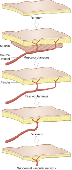
Such simplicity faded after Milton disproved flap length–width ratios, and asserted instead that flap viability was dependent on the vascular supply of the given flap territory. This led to a renaissance in flap construction, based on an improved understanding of the anatomic basis of the circulation to the skin, initiated by McGregor and Morgan, when they discovered that some regions of the body had discrete and relatively large subcutaneous vessels that had pierced the deep fascia to follow a predictable course. Comparatively large cutaneous flaps, if oriented along the axis of that vascular pathway, consistently maintained complete viability, and appropriately were called axial flaps (e.g., deltopectoral flap, groin flap).
Orticochea reported that the inclusion of muscle in the flap could result in larger skin flap survival. McCraw et al. more precisely explained the reasoning behind this association, and called the vessels coursing from the muscle to the skin “musculocutaneous” arteries. Interestingly, Tanzini in 1906 had published the first report of such a musculocutaneous flap. Tanzini et al., were well aware of the existence of branches from the muscle necessary to form these compound flaps. Nevertheless, it was not until the late 1970s that musculocutaneous flaps became a standard reconstructive option.
The zeal to adopt cutaneous flaps relying on musculocutaneous vessels perhaps delayed the recognition of other important contributions to the “fascial plexus” that would eventually allow cutaneous flaps to stand alone. Pontén deserves credit for reintroducing fasciocutaneous flaps, although he was not quite sure why the inclusion of the deep fascia with his “superflaps” resulted in a longer flap survival length than could be predicted for random flaps of comparable width. Note that Esser and Schwerer, and Gillies in the early part of the last century, had already suggested that it was advantageous to include the deep fascia with skin flaps, so that perhaps all along, this, too, was common knowledge.
The “fascial plexus” essential for survival of these fasciocutaneous flaps is not a discrete structure per se, but represents a confluence of subfascial, intrafascial, and suprafascial vascular plexuses within the dermal, subdermal, superficial adipofascial (above Scarpa’s fascia), and deep adipofascial layers, where each is a component part of a magnificent array of interconnected vessels. The medical student Manchot, more than 100 years ago, was also well aware that “larger cutaneous arteries … appear from the fissure between … muscles …” as intermuscular or septocutaneous perforators of the deep fascia. These, along with the direct perforators of axial flaps and musculocutaneous perforators, are today the major contributors to the “fascial plexus” from the underlying source vessels of a given angiosome.
As microsurgical tissue transfer became widely adopted, there was an exponential growth in flap options. Virtually every tissue could then be transferred including muscle, bone, nerve, viscera, etc. As well, modifications and combinations such as perforator flaps, conjoined and chimeric flaps were reported leading to an explosive development in choices of flaps. The challenge was to classify all of these flaps into a system that allows teaching, research, and communication regarding flaps.
Skin Flaps and Their Classification
In the last few decades there have been many attempts to provide a modern classification schema for skin flaps. The simplest system is to describe all skin flaps as having direct cutaneous, septocutaneous, or musculocutaneous perforators. However, many authors have published other concepts of the vascular supply to skin flaps, which has led to some degree of confusion about the terms. In this section, we aim to clarify the terms used in the most well-known classifications.
Nakajima et al. (1986) separated skin flaps into five types: random pattern, fasciocutaneous, adipofascial, septocutaneous, and musculocutaneous flaps.
Cormack and Lamberty (1984) emphasized that the term “fasciocutaneous” implied retention of a system of vascularization within the given flap, and does not refer to any specific tissue constituents per se. Thus, even if the skin (e.g., adipofascial flap) and/or deep fascia (e.g., subcutaneous flap) are excluded from a flap that is dependent on the remaining “fascial plexus,” such a flap would still be a “fasciocutaneous” flap. A fasciocutaneous flap can be composed of any or all of the tissue layers found between the skin and deep fascia. By this definition, Cormack and Lamberty were further able to contract all skin flaps into a tripartite system that included direct cutaneous ( sic . axial), musculocutaneous , and fasciocutaneous flaps.
Cormack and Lamberty Classification
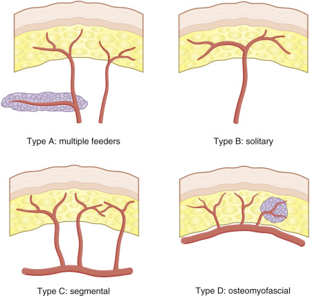
Cormack and Lamberty classified fasciocutaneous flaps into four major types ( Fig. 2.2 ), differentiated by the origin of the circulation to their “fascial plexus.” Their Type A flap had multiple “fascial feeders” or perforators that did not require specific identification, reminiscent of the random skin flap. Type B flaps contained a large, solitary septocutaneous perforator. The Type C flap relied on multiple and usually diminutive segmental septocutaneous branches, so that elevation of these flaps almost always necessitated inclusion of the source vessel with the flap, in order to maintain their complete integrity. Type D is similar to Type C in that it is based on multiple small perforators; however, it is raised as an osteomyofasciocutaneous flap.
Mathes and Nahai Classification
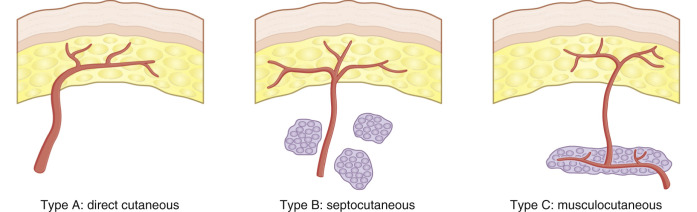
Mathes and Nahai’s (1997) classification of fasciocutaneous flaps based on the type of deep fascial perforator ( Fig. 2.3 ) is similar to Cormack and Lamberty’s classification of flaps: their Type A is a direct cutaneous flap, in which the vascular pedicle travels deep to the fascia for a variable distance then pierces the fascia to supply the skin (e.g., groin flap, temporoparietal fascia flap). Type B is a septocutaneous flap, which has a vascular pedicle that courses within an intermuscular septum (e.g., lateral arm flap, radial forearm flap). Type C is a musculocutaneous flap and is based on a vascular pedicle that is traveling within the muscle substance (e.g., DIEP flap).
Nakajima Classification
Nakajima et al. expanded the subtypes of fasciocutaneous flaps into six forms (Types I–VI, or A–F), each based on a distinctly different perforator of the deep fascia ( Fig. 2.4 ). Most had been previously described, e.g., their Type I “direct cutaneous flaps” were identical to the axial flaps of McGregor and Morgan. The Type II “direct septocutaneous flaps” were identical to Cormack and Lamberty’s type B fasciocutaneous flaps, and the type V “septocutaneous perforator flaps” would be the same as the latter’s Type C. The Type VI “musculocutaneous perforator flaps” resembled traditional myocutaneous flaps.
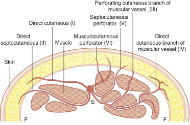
The remaining two types of flaps hypothesized by Nakajima et al., based on a “direct cutaneous branch of muscular vessel” (IV) and “perforating cutaneous branch of a muscular vessel” (III), were without question different and that is their most important contribution. Later, using computer graphics imaging, Nakajima et al ., in a 3-dimensional view of the “fascial plexus,” demonstrated that the axiality, vessel size, and suprafascial course of these vessels proved that the function of these “muscular vessels” was to provide nutrition to the skin, and only secondarily to the involved muscle. The “direct cutaneous branch of muscular vessel” remains an enigma due to its variable presence and has been little described. However, the “perforating cutaneous branch of muscular vessel” has become recognized as the basis for the perforator flaps. Some have argued that these are the only “true” perforator flaps.
The fasciocutaneous flap classification schemas of Cormack and Lamberty, Nakajima et al., and then Mathes and Nahai (see below) are very similar, as each uses subtle differences in circulation patterns to distinguish their flap subtypes. This is also a pragmatic approach, as the vascular supply, if nothing else, must be correctly identified and protected to ensure flap viability. Yet even further consolidation of cutaneous flap types is possible.
Taylor’s “Angiosome” Concept-Based Classification
Angiosomes refer to specific 3-dimensional blocks of tissue, including skin and deeper tissues layers that are supplied by specific source arteries. In 1893, Spalteholz performed injection studies using pigment mixed in gelatin to demonstrate a pure (intermuscular) or impure (i.e., primarily supplied deeper tissues, mostly muscle) course of cutaneous perforators. All arteries to the skin could most simply be considered to be either “direct” or “indirect” branches from an underlying source vessel. Taylor has repeatedly argued that the direct vessels are the primary cutaneous supply, and it is irrelevant if they have first traversed intermuscular or intramuscular septa, as their main destination always is to the skin. The indirect vessels emerge above the deep fascia as terminal, spent branches whose main purpose was to supply the deeper tissues, so they are in reality only a secondary means of cutaneous blood supply. It can be further argued that all deep fascia perforators would be “direct” if they coursed from the source vessel to perforate the fascia without first passing through some other tissue intermediary, or otherwise they would be “indirect” perforators ( Fig. 2.5 ). All corresponding cutaneous flaps would then be either direct perforator flaps or indirect perforator flaps.
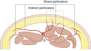
Because the perforator supplying a muscle perforator flap, by definition, must have first traveled through the substance of that muscle, in this classification system, these would be indirect perforator flaps. Neurocutaneous flaps would be an excellent example of indirect non-muscle perforator flaps. These flaps rely on an intrinsic and extrinsic neurocutaneous or venocutaneous vascular supply that accompanies a peripheral cutaneous nerve. The extrinsic vascular supply can often be a true artery, and, depending on the nerve, both structures can simultaneously pierce the deep fascia before proceeding within the subcutaneous tissues. The major purpose of this accompanying vascular system is to provide circulation to the nerve, and only secondarily (or indirectly) are cutaneous branches given off that will support an overlying cutaneous flap. Niranjan et al. pointed out that perforators similar to those accompanying the cutaneous nerves can also arise independently from fascioperiosteal or tenosynovial branches, which in turn also can supply indirect non-muscle perforator flaps. Thus, indirect perforator flaps, more than any other cutaneous flap, deserve a separate categorization to ensure the appropriate dissection of whatever are the intermediary structures, while protecting the requisite vascular supply.
Muscle Flaps
In the 1980s, muscle flaps became very popular, since it was a reliable method to transfer large skin flaps. Over the decades, the use of muscle flaps has evolved so that muscle function is rarely sacrificed simply to supply skin blood supply but rather, muscle flaps are used when a large volume of flap is required to fill dead space or muscle function is required, as, for example, with facial reanimation. It remains important that the pattern of vascularity of muscles will determine muscle flap survival and must be respected.
Mathes and Nahai Muscle Flap Classification
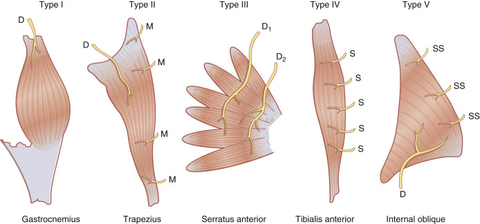
Mathes and Nahai (1981) categorized muscles into Types I–V, based on their vascular supply ( Fig. 2.6 ). On the other hand, it is not just a coincidence that the majority of musculocutaneous perforators arise near where the dominant pedicle enters the hilum of that muscle. The astute student of muscle perforator flaps must be fully aware of the most reliable muscle types, which pedicle is dominant, and where they are typically located to better predict the presence of musculocutaneous perforators. This is essential for skilful elevation of these flaps, even though the muscle itself is not to be included.
Taylor Classification
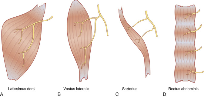
Taylor et al. have also divided muscles into four groups (Types I–IV) that differ by their mode of motor innervation ( Fig. 2.7 ). This classification becomes important when a dynamic muscle transfer is considered, to ensure capture of an appropriately functioning muscle unit.
Other Flaps
Vascularized bone flaps are used less frequently than soft tissue flaps. Again, as for all other flaps, a good system would categorize these primarily on the basis of their circulatory patterns.
Serafin Classification
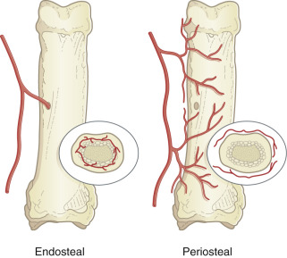
Serafin has divided osseous flaps according to whether the flap has a direct (endosteal) or indirect (periosteal) circulation ( Fig. 2.8 ). Vascularized joint transfers could similarly be subclassified according to the source of vascularization of their bony constituents.
Combined Flaps
In extraordinary circumstances, the creative combination of flaps from a single donor site is used to repair challenging defects where multiple tissue islands are required. The sporadic introduction of variations of combined flaps has created much confusion in terminology, since minor modifications of technique often have been called different names or given eponyms, but the basic flap composition has remained unaltered. There are two major subtypes of combined flaps that are distinctly different in regards to the physical relationship of their component parts, yet both are similar in that each anatomic territory that is part of that combination always retains an independent blood supply.
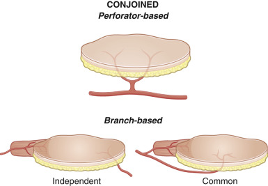
Conjoined Flaps
Harii et al. first introduced the concept of combined flaps when they described a “combined myocutaneous flap and microvascular free flap.” This captured the skin territories of the latissimus dorsi musculocutaneous flap and groin flap, where both remained connected together to essentially form a bipedicled flap with the thoracodorsal and superficial circumflex iliac vessels remaining as pedicles at opposite ends, respectively. Such a conjoined flap or Siamese flap (named after the conjoined Siamese twins, Chang and Eng, 1811–1874) will have multiple flap territories that remain dependent due to some common physical junction, yet each territory retains its intrinsic and independent vascular supply ( Fig. 2.9 ).