Chapter 7 Filler Injection of the Eyebrows


10.1055/b-0040-178125

Chapter 7 Filler Injection of the Eyebrows


Introduction


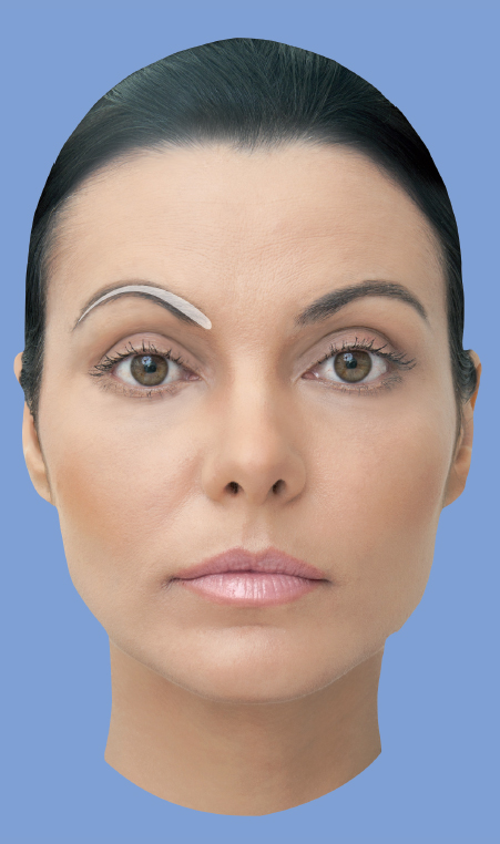
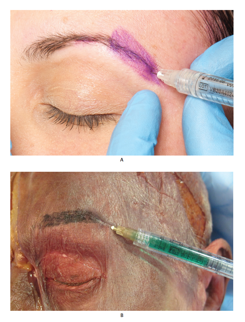
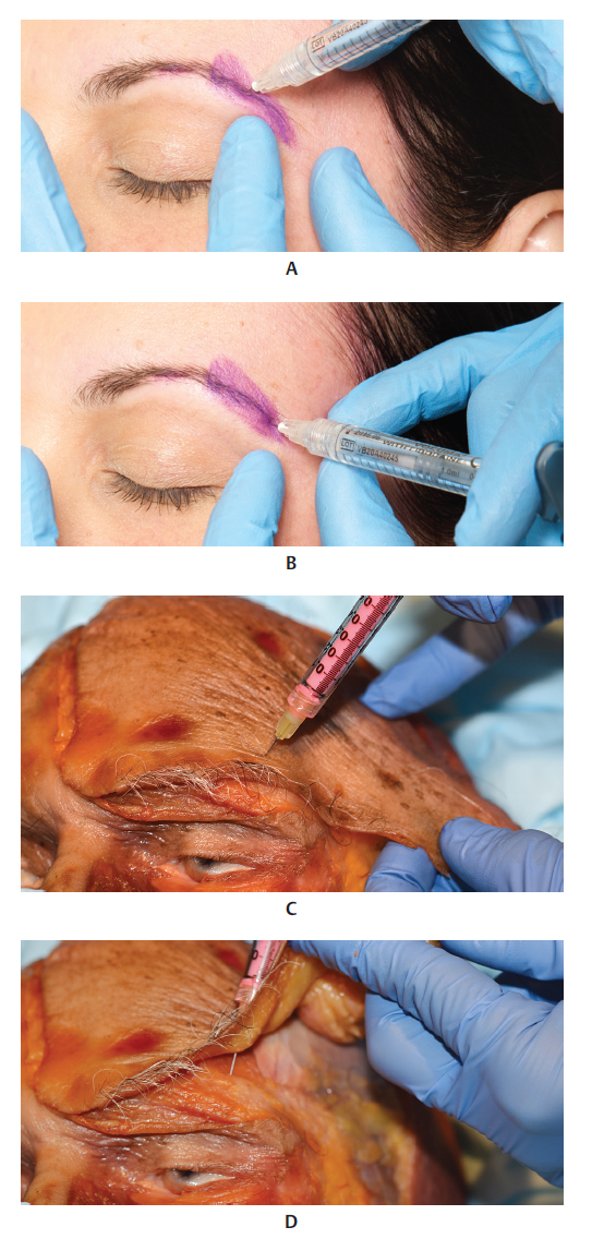
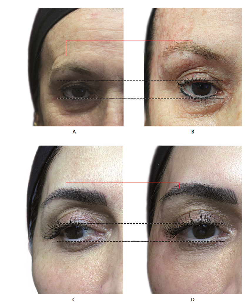
If the eyes are the windows to the soul, then the eyebrows are certainly their frame, as they give the face a format and define the eyes and forehead. The eyebrows are one of the most versatile characteristics of the human face. They influence the perception of beauty, and play a fundamental role in sexual dimorphism, facial recognition, and nonverbal communication, 1 since subtle changes in their position convey different emotions, from anger to surprise. 2 In 1974, Westmore X described the ideal female eyebrow as being slightly curved, with its apex approximately aligned with the lateral corneal limbus. The medial extremity should start at the vertical line of the ala of the nose and the medial palpebral commissure, as shown inFig. 7.1 A. The lateral extremity should end at the oblique lined traced between the lateral ala of the nose and the lateral palpebral commissure. Male eyebrows have a similar medial and lateral alignment but are generally lower and straighter. 2


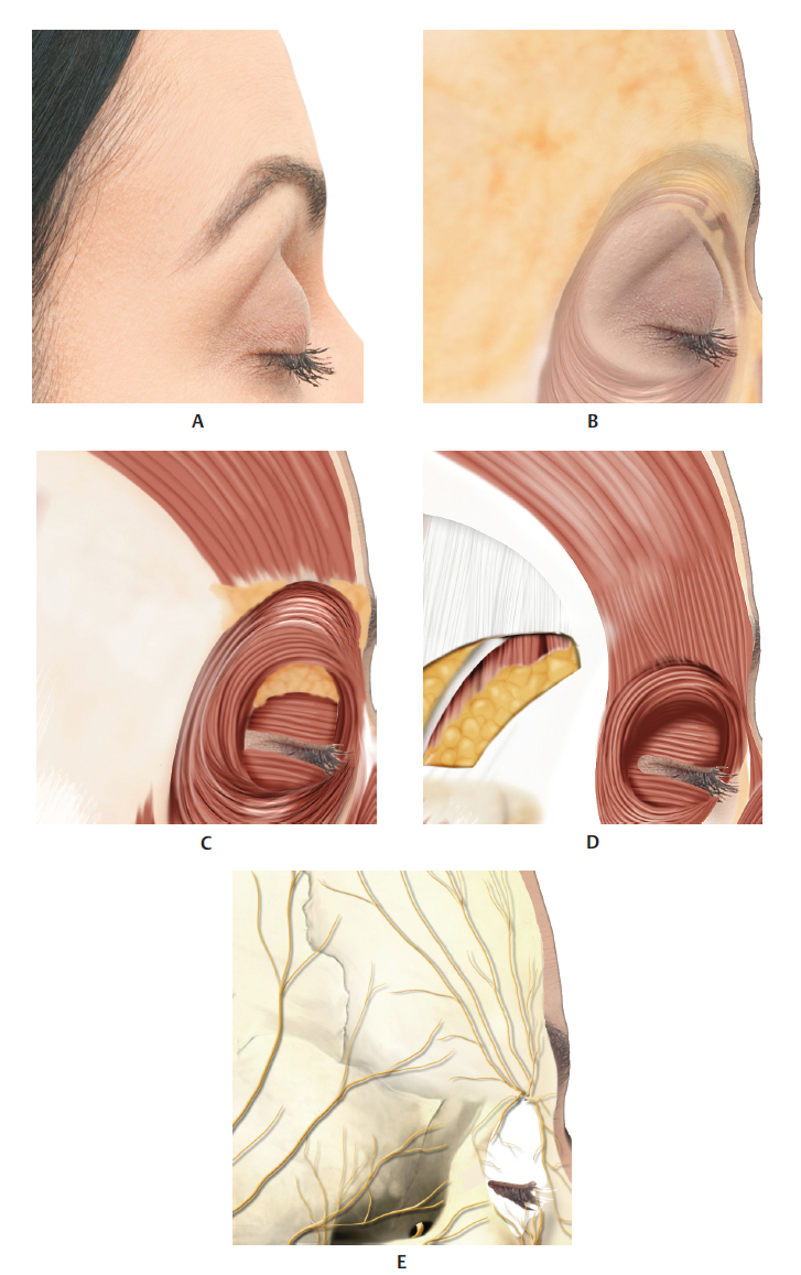
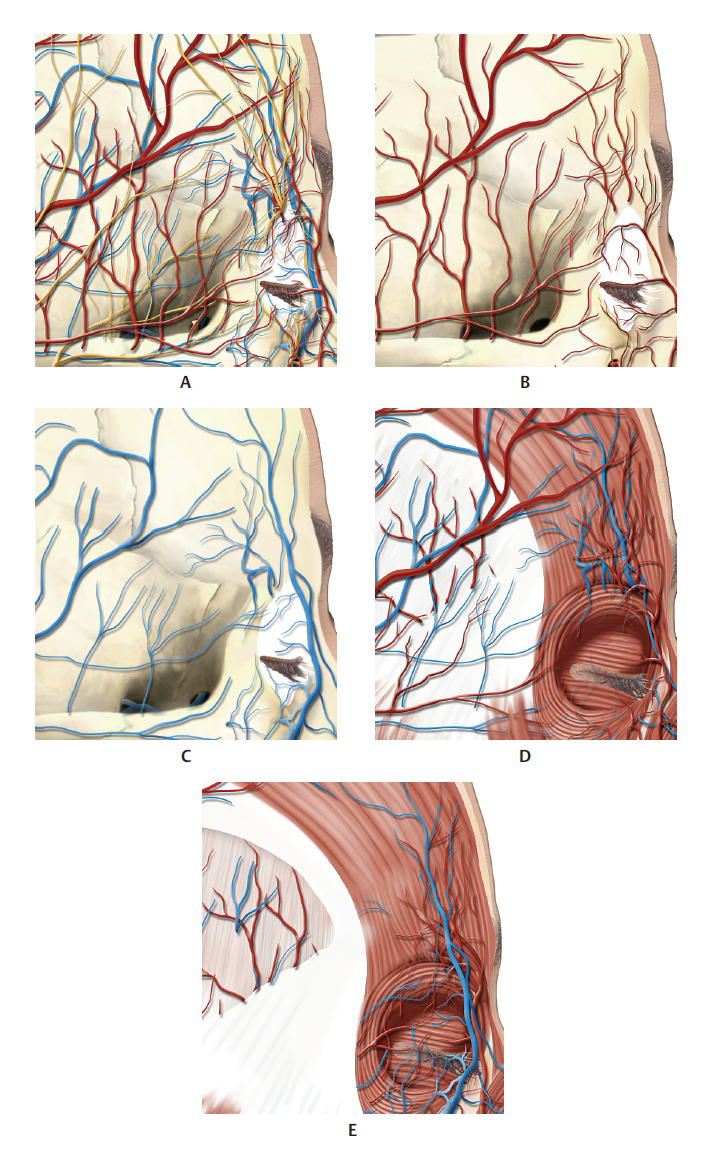
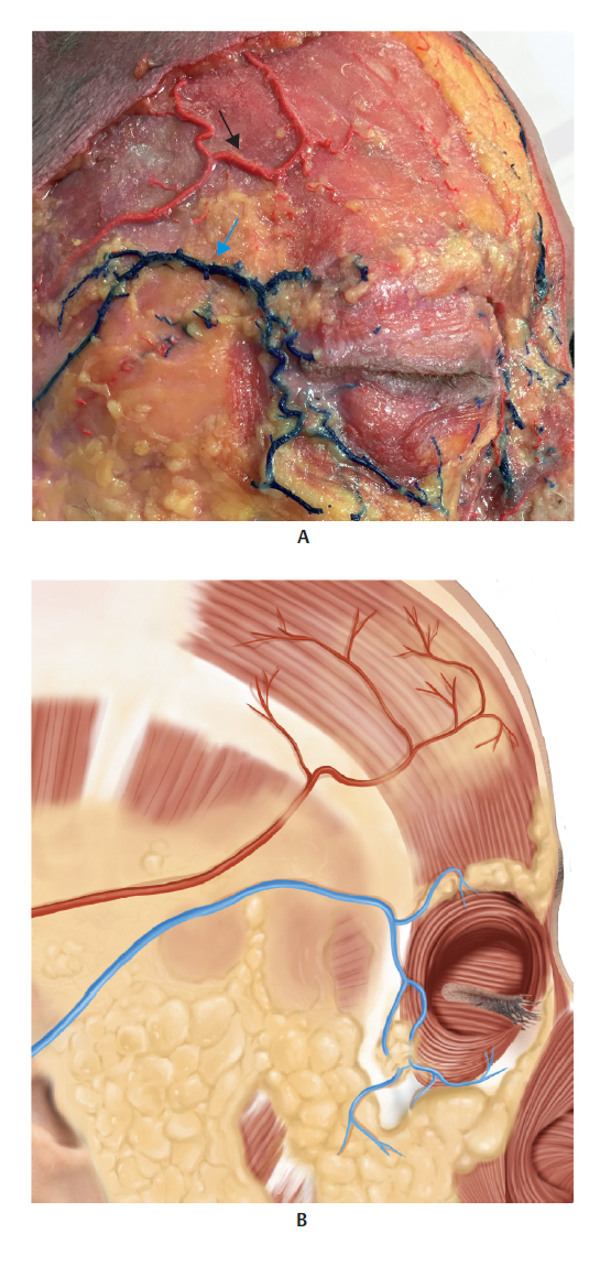
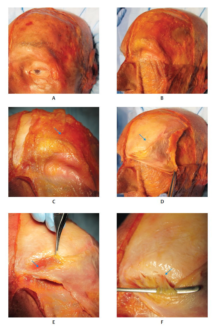
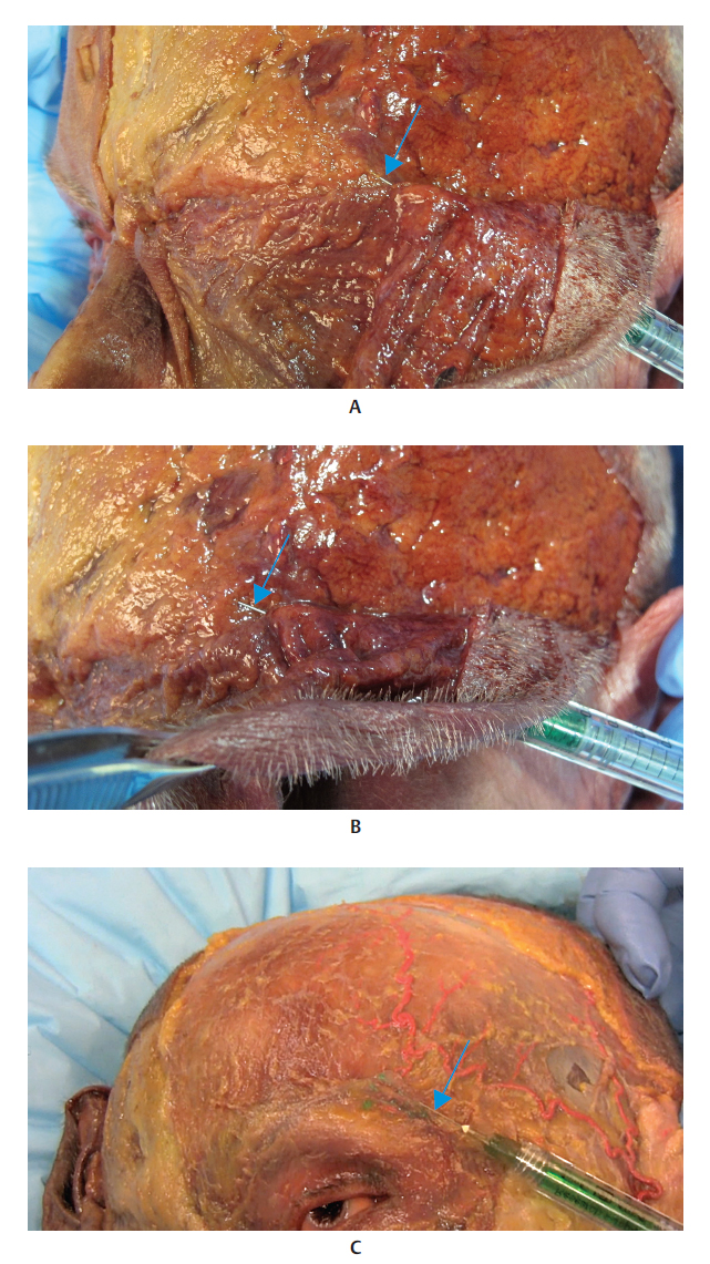
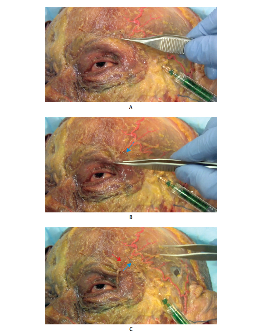
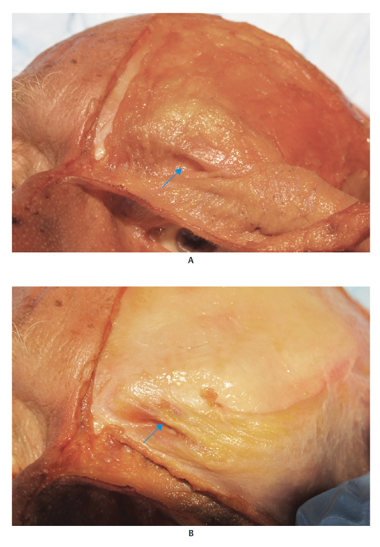
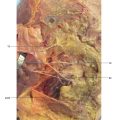
The eyebrow region presents four layers, from the outermost to innermost regions: skin containing sebaceous glands, apocrine glands, and hair follicles; subcutaneous tissue that connects the skin to the aponeurosis of the overlying muscles; superficial layer of the frontal and orbicularis oculi muscles (OMs), and the deep layer, the corrugator supercilii; and the periosteum. 3 Retroorbicularis oculi fat (ROOF) is the deep fat pad located below the orbicular muscle, originating medially at the level of the supraorbital nerve and extending laterally to the superior orbital rim (Fig. 7.2).


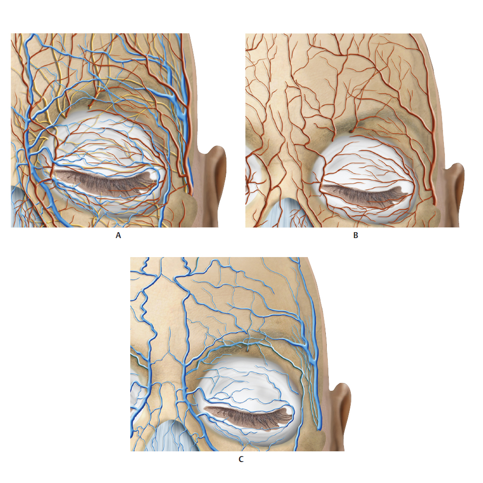
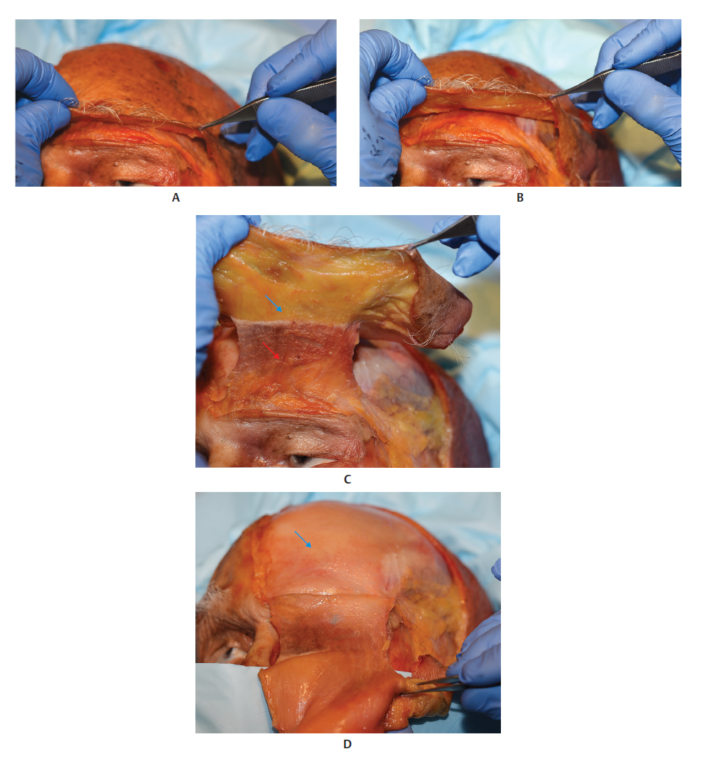
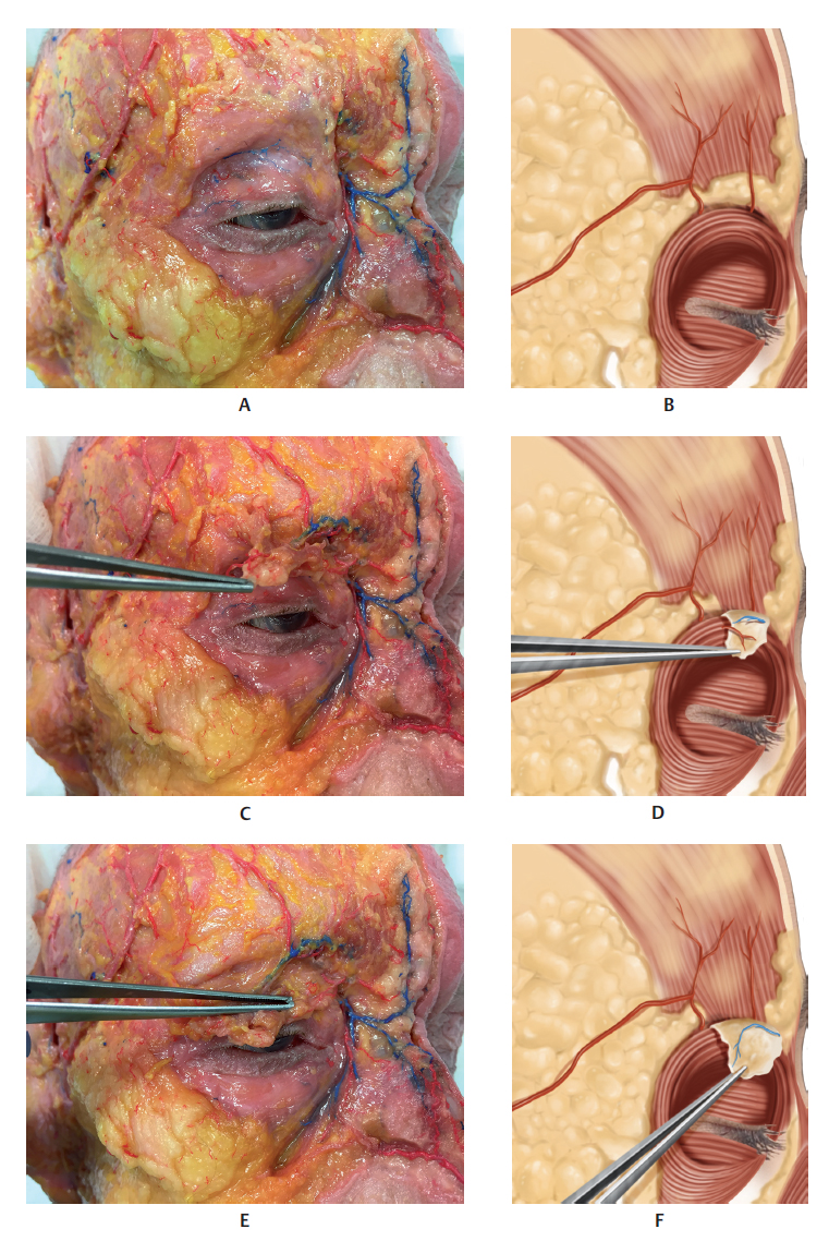
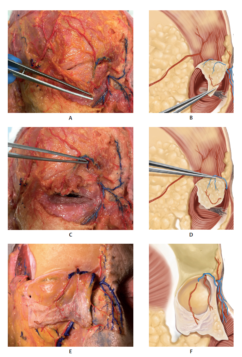
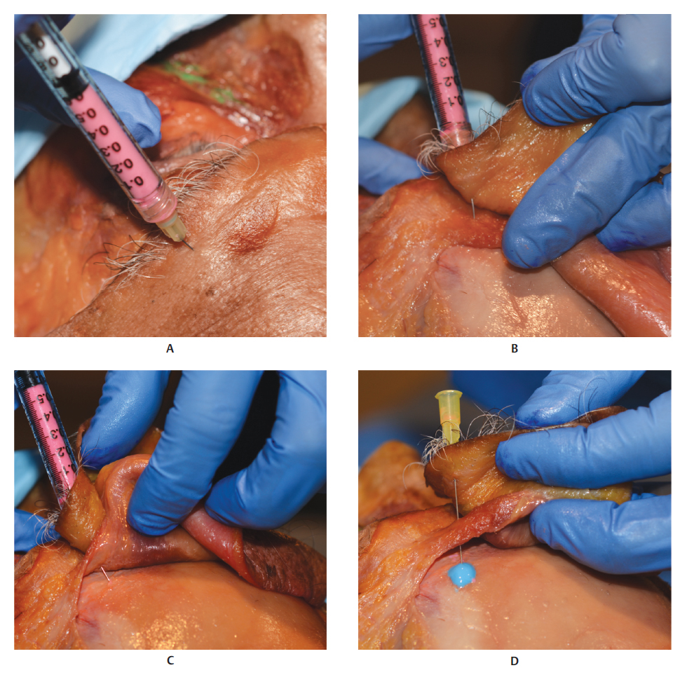
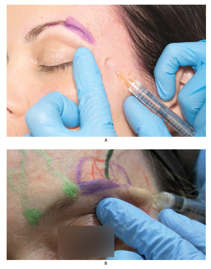
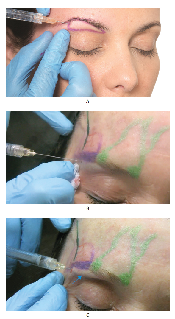
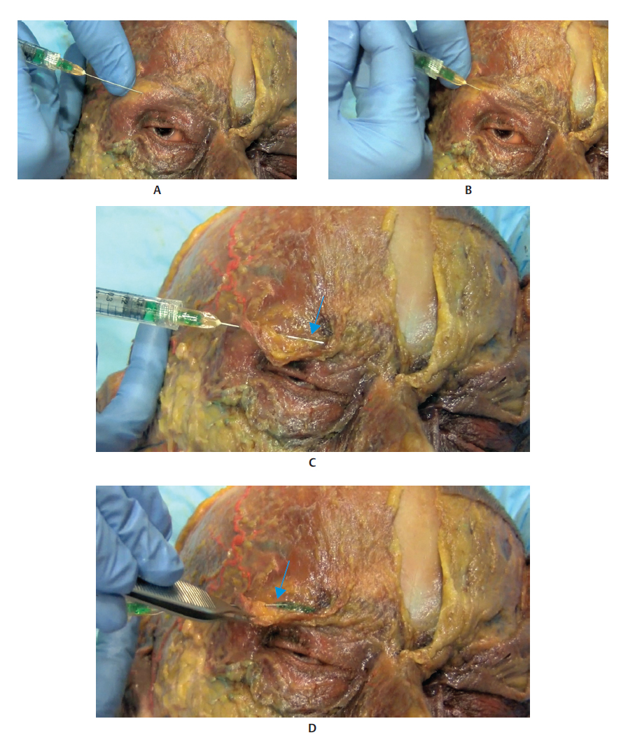
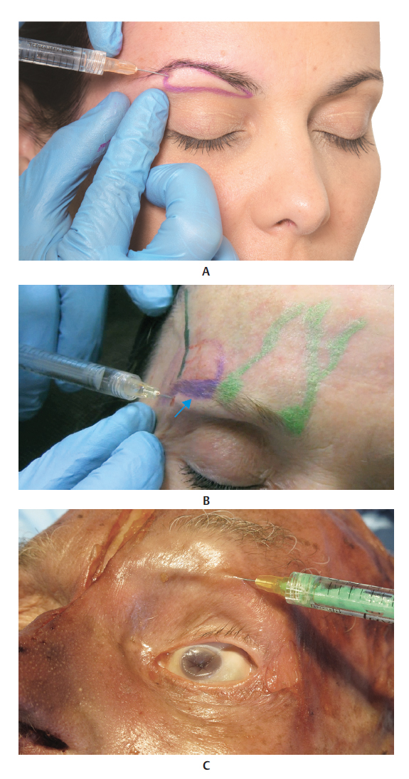
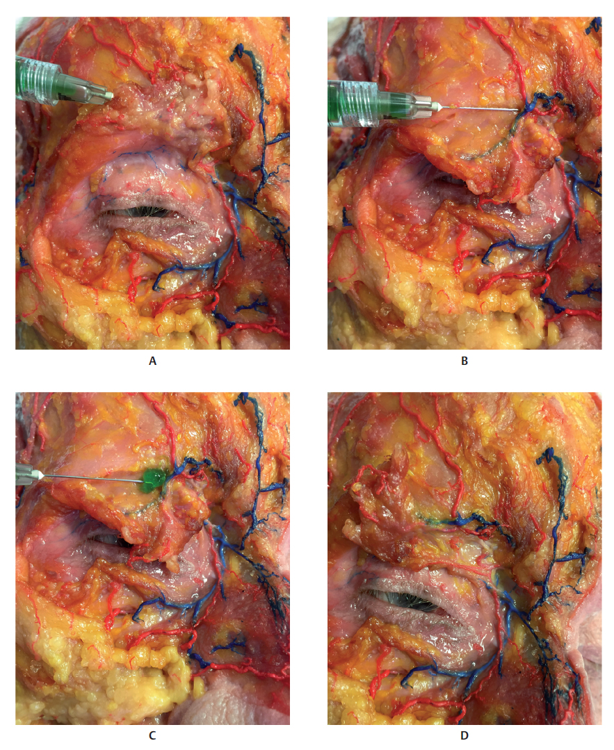
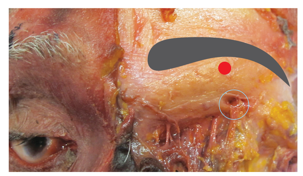
The supraorbital and supratrochlear notches (or foramens in some cases) are located approximately 2.7 and 1.7 cm, respectively, from the midline of the glabella, where the respective vessels and nerves emerge. Caution must be exercised when applying filler injections to this region, as an accidental intravascular injection into one of the distal branches of the ophthalmic artery can lead to a rare but very severe complication consisting of embolization of the central retinal artery, leading to amaurosis. Therefore, in-depth knowledge of the irrigation of the orbital region and its ramifications is important (Fig. 7.37.13).


Techniques


Superficial Technique Using a Needle


SeeFig. 7.14 and7.15.


Deep Technique Using a Needle


SeeFig. 7.167.18.


Cannula Technique


SeeFig. 7.19 and7.20.


Region Below the Eyebrow


SeeFig. 7.217.26.


References

1 Carruthers J, Carruthers A. Social significance of the eyebrows and periorbital complex. J Drugs Dermatol 2014; 13(1, Suppl):s7–s11 2 Lam VB, Czyz CN, Wulc AE. The brow-eyelid continuum: an anatomic perspective. Clin Plast Surg 2013;40(1):1–19 3 Andre P, Azib N, Berros P, et al. Anatomy and volumizing injections. Paris: E2e Medical Publishing
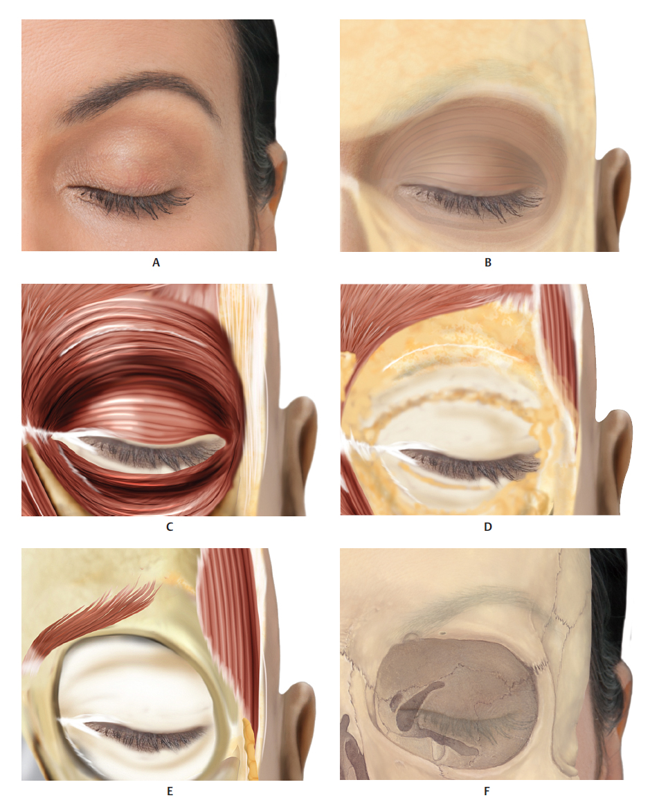
Fig. 7.1 A. Frontal view of the brow region.B. Same region after removal of the skin. The superficial fat pads (SFPs) are visible.C. Same region after removal of the skin and SFP. The orbicularis oculi muscle (OM) is visible.D. Same region after removal of the skin, SFP, and OM. Retroorbicularis oculi fat (ROOF) is visible.E. Same region showing the orbit and corrugator muscle.F. Bone structure of the aforementioned region.
Fig. 7.2 Frontal view of the brow region.A. Corresponding innervation and vascularization.B. Corresponding arterial vascularization.C. Corresponding venous vascularization.
Fig. 7.3 A. Lateral view of the brow region.B. Same region after removal of the skin, showing superficial fat pads (SFPs).C. Same region after removal of the skin and the SFP. The frontal muscle and the palpebral and orbital portions of the orbicularis oculi muscle (OM) are visible over the retroorbicularis oculi fat (ROOF).D. Same region after removal of: the superficial temporal fascia, orbital portion of the OM, and ROOF. The temporal part of the buccal fat is visible below the deep temporal fascia and above the temporal muscle.E. Bone structure of the same region in which the corresponding innervation is visible.
Fig. 7.4 Right profile view showing details of the brow region.A. Corresponding vascularization and innervation.B. Corresponding arterial vascularization.C. Corresponding venous vascularization.D. Arterial and venous vascularization integrated in the frontal muscle and orbicularis oculi muscle (OM).E. Corresponding vascularization above the muscles of the upper third of the face. The superficial temporal fascia has been removed, revealing the vascularization above the deep fascia.
Fig. 7.5 A andB. Frontal view of a 3D digital model.C. Profile view of the 3D digital model highlighting the brow region.
Fig. 7.6 Profile view of a fresh cadaver with skin being folded down.
Fig. 7.7 A. Profile view of a fresh cadaver being dissected.B. Skin folded down to expose the superficial fat pad (SFP) of the eyebrow, forehead, and temples.
Fig. 7.8 A. A fresh cadaver in frontal position with skin being folded down exposing the superficial fat pad (SFP) of the eyebrow.B. Close-up ofA.
Fig. 7.9 A. Anterior or frontal branch of the superficial temporal artery (STA) (black arrow) and middle temporal vein (MTV) (blue arrow), which are drained by the superciliary vein of the brow region and upper eyelid and by the zygomaticofacial and orbitomalar veins of the malar region and lower eyelid.B. Illustration ofA showing the STA and MTV above the temporal muscle.
Figs. 7.10 A andB. A dissection of a fresh cadaver in frontal position with skin being folded back in the frontal region, in an intraciliary incision.C. Exposure of the superficial fat pad (SFP) of the forehead (blue arrow) and the frontal muscle (red arrow) .D. Exposure of the epicranial aponeurosis and the frontal bone (blue arrow) located below the frontal muscle that was folded back.
Fig. 7.11 A. A fresh cadaver with skin and superficial fat pad (SFP) of the eyebrow folded back. The orbicularis oculi muscle (OM) is visible above the frontal muscle.B. Illustration corresponding toA.C. A fresh cadaver with orbital portion of the OM being folded anteriorly, revealing the supraorbital vein and artery.D. Illustration corresponding toC.E. Same cadaver with orbital portion of the OM totally folded anteriorly, revealing the supraorbital vein and artery above the frontal muscle.F. Illustration corresponding toE.
Fig. 7.12 A. A cadaver with orbital portion of the orbicularis oculi muscle (OM) totally folded down, revealing the supraorbital vein and artery above the frontal muscle. Note the periorbital bony rim.B. Illustration corresponding toA.C. Sample as in Fig.A, with the supraorbital artery and vein being pinched, with focus on the supraorbital foramen.D. Illustration corresponding toC.E. Same cadaver with the frontal muscle totally folded back, making it possible to see the supraorbital vein and artery emerging from the corresponding foramen.F. Illustration corresponding toE.
Fig. 7.13 A andB. Profile view of a fresh cadaver with skin folded down exposing the superficial fat pad (SFP) of the eyebrow.C. Skin of the frontal region being folded down with an intraciliary incision.D. Frontal muscle being folded back exposing the epicranial aponeurosis and the frontal bone (blue arrow).E. Note the corrugator muscle (blue arrow) below the frontal muscle.F. Note the supraorbital foramen and exposure of the supraorbital nerve, vein, and artery (blue arrow) below the corrugator muscle.
Fig. 7.14 A. Profile view of a model with highlighted tail of the eyebrow and 30 GX 13 mm needle inserted into the deep dermis and/or superficial subcutaneous plane, where the product is injected with a retrograde injection technique.B. Profile view of a fresh cadaver with a 30 GX 13 mm needle inserted into the deep dermis and/or superficial subcutaneous plane, where the product is injected with a retrograde injection technique.
Fig. 7.15 A. A fresh cadaver with skin folded down, revealing the superficial fat pad (SFP) of the eyebrow and a 30 Gx 13 mm needle(blue arrow) simulating the application of the product to the deep dermis and/or the superficial subcutaneous plane.B. Close-up ofA.C. Skin and SFP of the eyebrow removed. Simulation of application of the product with a 30 Gx 13 mm needle(blue arrow) above the orbital portion of the orbicularis oculi muscle (OM), showing the ideal region for superficial filler injections between the deep dermis and/or the superficial subcutaneous tissue.
Fig. 7.16 A andB. Profile view of a model with highlighted tail of the eyebrow and a 27 GX 13 mm needle inserted into the supraperiosteal plane in two different positions. At these sites, the product is injected using a bolus technique.C. Profile view of a fresh cadaver profile with a 27 GX 13 mm needle inserted into the supraperiosteal plane.D. Skin folded back to expose the fat pad of the eyebrow. 27 GX 13 mm needle inserted into the supraperiosteal plane, where the product is injected with a bolus technique.
Fig. 7.17 A. A fresh cadaver in inverted craniocaudal position with 27 GX 13 mm needle inserted into the supraperiosteal plane.B. Skin has been folded up, exposing the fat pad of the eyebrow.C. Skin and fat pad of the eyebrow have been folded up, exposing the epicranial aponeurosis and the frontal bone.D. Skin and fat pad of the eyebrow have been folded up, exposing the epicranial aponeurosis and the frontal bone, and application of the product (turquoise-colored) using a bolus technique.
Fig. 7.18 A. Middle and upper thirds of the right side of the face of a patient with indication for eyebrow lifting.B. Patient underwent a hyaluronic acid (HA) filler injection using a needle to lift the eyebrow. Note that the position of the eyebrow is higher after treatment. The patient also received filler injections in the temporal and frontal regions of the face.C. Another patient with indication for filler injection to lift the eyebrow.D. The patient underwent a HA filler injection using a cannula to lift the eyebrow. Note that the position of the eyebrow is higher after treatment.
Fig. 7.19 A. Profile view of a model with highlight of the tail of the eyebrow and a 25 GX 13 mm cannula inserted into the submuscular plane (orbital portion of the orbicularis oculi muscle [OM]), where the product is injected using a retrograde injection technique.B. Purple diagram marking the tail of the eyebrow and a 25 GX 13 mm cannula inserted into the submuscular plane (orbital portion of the orbicular muscle of the eyes), where the product is injected using a retrograde injection technique. Note the raised outline in the skin indicating the position of the 25 GX 13 mm cannula.
Fig. 7.20 A. Profile view of a fresh cadaver with skin and fat pad of the eyebrow folded back. 25 GX 13 mm cannula inserted below the orbital portion of the orbicularis oculi muscle (OM), which has been pinched.B. Orbital portion of the OM being folded anteriorly and the tip of the 25 GX 13 mm cannula(blue arrow) below the muscle is visible.C. Orbital portion of the OM totally folded anteriorly and the tip of the 25 GX 13 mm cannula(blue arrow) below the muscle is visible. The tip of thered arrow points to the orbicular branch emerging from the frontal branch of the superficial temporal artery (STA).
Fig. 7.21 A. Profile of a model with markings highlighting the infraorbital region (below the eyebrow) and a 25 GX 13 mm cannula inserted into the submuscular plane (orbital portion of the orbicularis oculi muscle [OM]), where the product is injected using a retrograde injection technique.B. Profile of another model with indication for a filler injection of the infraorbital region (below the eyebrow), with a 25 GX 13 mm cannula pointing toward the site where it should be inserted.C. 25 GX 13 mm cannula inserted into the infraorbital region (below the eyebrow) in the submuscular plane (orbital portion of the OM), where the product is injected using a retrograde injection technique. The raised outline in the skin(blue arrow) indicates the position of the tip of the 25 GX 13 mm cannula.
Fig. 7.22 A. 25 GX 13 mm cannula being inserted into the infraorbital region (below the eyebrow) in the submuscular plane below the orbital portion of the orbicularis oculi muscle (OM).B. 25 GX 13 mm cannula already inserted into the infraorbital region (below the eyebrow) in the submuscular plane below the orbital portion of the OM.C. Upper part of the orbital portion of the OM has been folded down, making it possible to see one-third of the 25 GX 13 mm cannula(blue arrow). D. Green-colored product being injected through the 25 GX 13 mm cannula into the submuscular plane below the upper part of the orbital portion of the OM, which was pinched and folded down(blue arrow).
Fig. 7.23 A. Profile view of a model on whom the infraorbital region (below the eyebrow) has been marked, and a 25 GX 13 mm cannula inserted into the submuscular plane (orbital portion of the orbicularis oculi muscle [OM]), where the product is injected using a retrograde injection technique.B. Profile of another model with a 25 GX 13 mm cannula inserted into the infraorbital region (below the eyebrow) in the submuscular plane (orbital portion of the OM), where the product is injected using a retrograde injection technique. The raised outline in the skin(blue arrow) indicates the position of the tip of the 25 GX 13 mm cannula.C. Profile view of a fresh cadaver with a 25 G x 13 mm cannula inserted into the infraorbital region (below the eyebrow) in the submuscular plane below the orbital portion of the OM.
Fig. 7.24 A. Left profile view of a fresh cadaver with skin folded down exposing the fat pad of the eyebrow. 25 Gx 13 mm cannula inserted into the infraorbital region (below the eyebrow) in the submuscular plane below the orbital portion of the orbicularis oculi muscle (OM) of the eyes. Theblue arrow shows the raised outline of the cannula.B. Skin and frontal muscle have been folded down, exposing the supraorbital neurovascular bundle(blue arrow) below the corrugator muscle.
Fig. 7.25 A. Right half-profile view of a fresh cadaver with skin and superficial fat pad (SFP) removed. 25 Gx 13 mm cannula inserted into the infraorbital region (below the eyebrow) in the submuscular plane below the orbital portion of the orbicularis oculi muscle (OM).B. Orbital portion of the OM has been folded down, revealing a 25 Gx 13 mm cannula already inserted into the infraorbital region (below the eyebrow) anterior to the frontal muscle.C. Orbital portion of the OM has been folded down, making it possible to see the 25 Gx 13 mm cannula and the green-colored product being injected anterior to the frontal muscle of the orbital rim. Note the proximity to the supraorbital vein and artery emerging from the same corresponding foramen.D. Same image with focus on the supraorbital vein and artery emerging from the same foramen. Note the supratrochlear vein and artery, more medially.
Fig. 7.26. Left side of the face of a fresh cadaver exposing the epicranial aponeurosis, frontal bone, and supraorbital foramen; the supraorbital foramen (blue circle) is visible in a peculiar position. Thered dot shows the normal location of the foramen. In view of such anatomical variations, it is highly advisable to always use the submuscular plane, not touch the periosteum, and never use a needle for procedures in this region.

Only gold members can continue reading. Log In or Register to continue

Stay updated, free articles. Join our Telegram channel

Sep 28, 2020 | Posted by in Aesthetic plastic surgery | Comments Off on Chapter 7 Filler Injection of the Eyebrows

Full access? Get Clinical Tree

Get Clinical Tree app for offline access