Chapter 4 Anatomy of the Aging Process
Introduction
In order to rejuvenate the face effectively, it is necessary to understand the aging process and its consequences on facial structures.
Up until a few years ago, only lines and folds were treated, meaning that the approach to facial rejuvenation was only two-dimensional. However, studies of the human face using three-dimensional computed tomography (CT), magnetic resonance (MR), and the dissection of cadavers have now made it possible to understand that lines and folds are simply the final markers of an overall process that involves not only the skin, but also the bone structure, fat pads, and muscles.
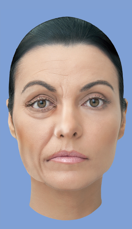
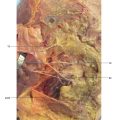
Aging causes reduction in the support and elasticity of the skin; resorption and remodeling of bone structure; atrophy and downward shift of the fat pads; and increase in facial muscle tension. The convex areas of the face and the characteristic arches of youth turn into flattened or concave areas, which stop reflecting light, and create shadows (Fig. 4.1). 1
Bone Alterations— Remodeling of the Cranial Skeleton
The cranial skeleton is essential for the three-dimensional contour of the face because it provides the support on which the skin envelope and the fat pads lie. If the morphology of this supporting base changes, the overlying soft tissues will protrude differently.
The craniofacial skeleton tends to expand continuously during an individual’s lifetime with aging. 2 Over 40 years ago, Enlow followed the lengthwise growth of the facial skeleton from childhood through to adulthood, using serial cephalometrograms. He observed that the entire face becomes longer (except in cases of tooth loss), deeper in the anteroposterior plane, and broader. 3 More recently, Pessa 3 used three-dimensional stereolithography and Shaw et al 4 used three-dimensional CT scans to study individuals from different age groups. They confirmed the changes in the shape of the skeleton over the years and provided additional details (Fig. 4.2).
It is important to note that despite an overall increase in size, some areas suffer resorption with age. The maxilla, including the piriform aperture of the nose, the superomedial and inferolateral portions of the orbital rim, and the area preceding the “bulldog” fat of the mandible are all more susceptible to this resorption. This is probably caused by differential growth of the craniofacial skeleton, which makes it possible for a child’s cranium to take on the proportions of an adult cranium. Contrary to what was thought previously, it is impossible to prevent remodeling of the facial skeleton, regardless of the condition of the teeth, and despite toothless patients presenting accelerated resorption of the maxilla and mandible.
Craniofacial skeleton = centrifugal growth with selective resorption

During the aging process, contraction, shrinking, and bone loss only occur if there are other supervening factors, such as tooth loss, osteoporosis, or calcium metabolism deficiencies. 3
Bone changes in each part of the face and their respective clinical consequences will be discussed below.
Periorbital Region
The orbital aperture grows with age, both in area and in width. However, resorption or expansion occur irregularly, and more intensely in the superomedial and inferolateral portions of the orbital rim. 4 Using three-dimensional stereolithography, Pessa 3 demonstrated the distortion of the inferior orbital rim over the years. As shown inFig. 4.3A, the skeleton of a young man exhibits a thick inferior orbital rim, which is well defined and symmetrical. The arrow shows how the lateral orbital bone structure is robust. However, inFig. 4.3B, the rim has become asymmetrical, deep, and fragile over the years, with loss of lateral support(arrow). 6 This distortion can be classified into three types from the clinical point of view (Fig. 4.4).
Middle Third of the Face
The skeleton of the middle third of the face is composed of the maxilla in the medial and intermediate thirds, and the zygomatic body and arch in the lateral third. The maxilla is the bone of the face that suffers the most resorption in the course of a lifetime. 2 In different studies, Pessa 3 and Shaw et al 4 measured the maxillary angle of young and elderly patients and observed a significant reduction in this angle, indicating extensive bone resorption and loss of maxillary projection, as shown inFig. 4.5. Consequently, they confirmed Lambros’s theory that continuous remodeling of the facial skeleton can be summarized as a clockwise rotation of the maxilla relative to the base of the cranium. In other words, the maxilla rotates in an inferior and posterior direction.
Related posts:

Stay updated, free articles. Join our Telegram channel

Full access? Get Clinical Tree


