Chapter 15 Filler Injection of the Preauricular Region and the Earlobe
Introduction
The ear is a complex structure, which has peculiar anatomical characteristics starting from the embryonic stage through to adult stage. Patient complaints in relation to ears vary according to age: In childhood, being flap-eared is the main complaint; as patients grow older, neoplastic lesions, fissured lobes, and aging of the region take over as dominant complaints. 1
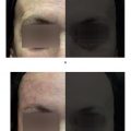
The application of hyaluronic acid (HA) is indicated for patients who present with aging of the earlobe, flaccidity, folds, and loss of volume and support in the region (clinically, elongated or pendulous earlobes). In addition to the aged appearance, patients complain that earrings are no longer supported in the earlobes, and this gives rise to fissures of the orifices. This technique is ideal for those who wish to have their earrings firmly supported in place (Fig. 15.3). 1 –. 3
Anatomy
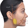
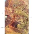
The anatomy of the ear is composed of the keratinized squamous epithelium and the dermis adhering to the perichondrium. Subcutaneous tissue is scarce or absent. Only the lobe contains adipose tissue, layered with dense conjunctive tissue. The sebaceous and apocrine glands are located mainly in the concha of the ear and the scaphoid fossa. The ear is vascularized by branches of the external carotid artery. The posterior auricular artery emerges below the ear, continues toward the cranium, supplying the posterior part of the ear, while the anterior auricular branches of the superficial temporal artery supply the anterior region (in front of the tragus). The superficial temporal artery flows superficially in the subcutaneous part of this region, and its pulses can be easily felt (Fig. 15.1,15.2, and15.4–15.7).
Technique
HA is used for the middermis. The material is deposited in the inferior portion of the lobe, around the orifice of the earring, in the middermis. Both the fanning injection and retrograde injec tion techniques can be used.
On average, 0.3 to 0.4 ml is used for each lobe, injecting slowly, until all the depressions are filled, paying attention to avoid overcorrection. The area should be massaged gently, so the product molds and is distributed uniformly throughout the earlobe. In case of bleeding, the area should be compressed for 3 to 5 minutes in order avoid the formation of hematomas and ecchymosis. The use of heavy earrings should be avoided for approximately 7 to 10 days. Treatment of this region is usually long lasting because of the absence of movement and the low local metabolic activity.
Related posts:
Stay updated, free articles. Join our Telegram channel

Full access? Get Clinical Tree


