Chapter 13 Filler Injection of the Submalar Triangle
10.1055/b-0040-178131
Chapter 13 Filler Injection of the Submalar Triangle
Introduction
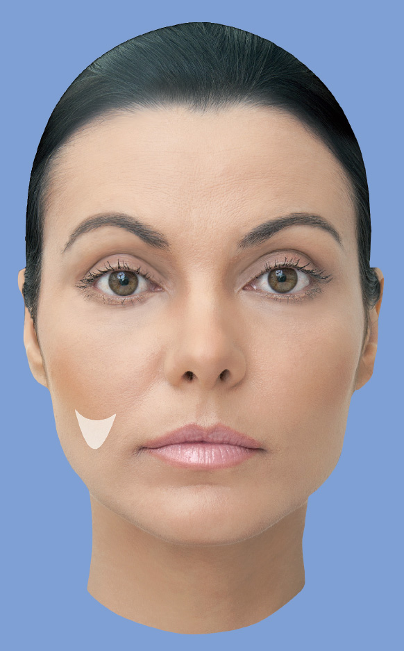
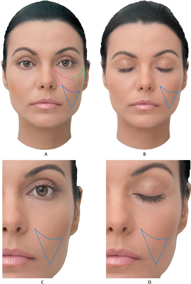
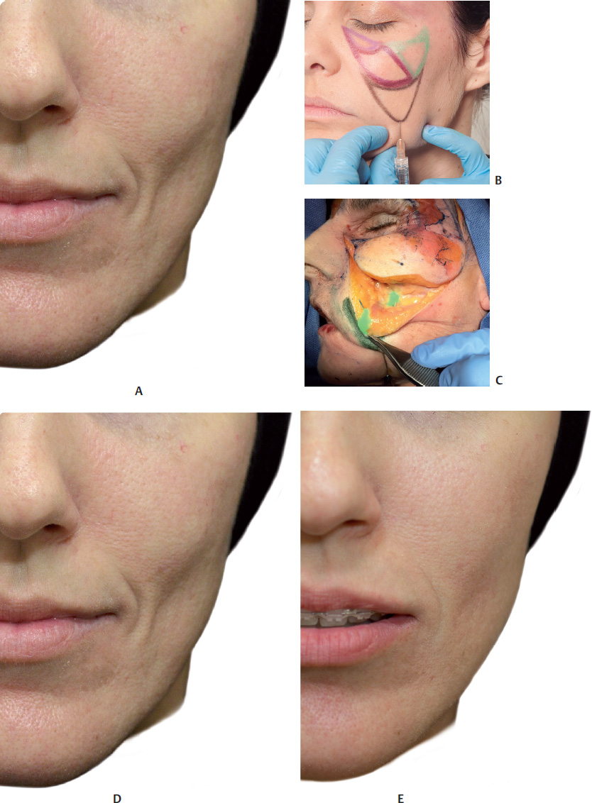
Loss of volume in the submalar region manifests clinically in the form of an inverted triangle, which is delimited medially by the nasolabial fold, superiorly by the body of the zygomatic bone, and laterally by the masseter muscle. The buccal fat pad (Bichat’s fat pad) gives volume to the submalar triangle. It is a fundamental component in this system, and is exceptionally sensitive to weight loss, trauma, cachexia, and, in particular, aging process. In cases of advanced atrophy, the patient has a skeletonized appearance.
Anatomy
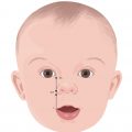
The buccal fat pad (Bichat’s fat pad) is of glandular origin and was first described by Heister in 1732. However, Marie François Xavier Bichat, a French anatomist and physiologist, was the one who, in 1802, recognized its fatty nature. The buccal fat pad is directly related to the chewing muscles, and in childhood, it helps in sucking. This explains the cherubic appearance of infant faces.1,.2
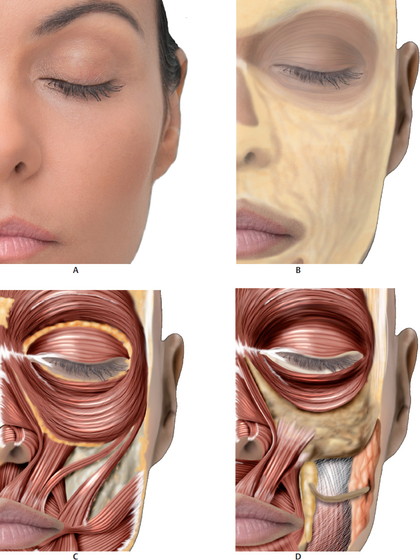
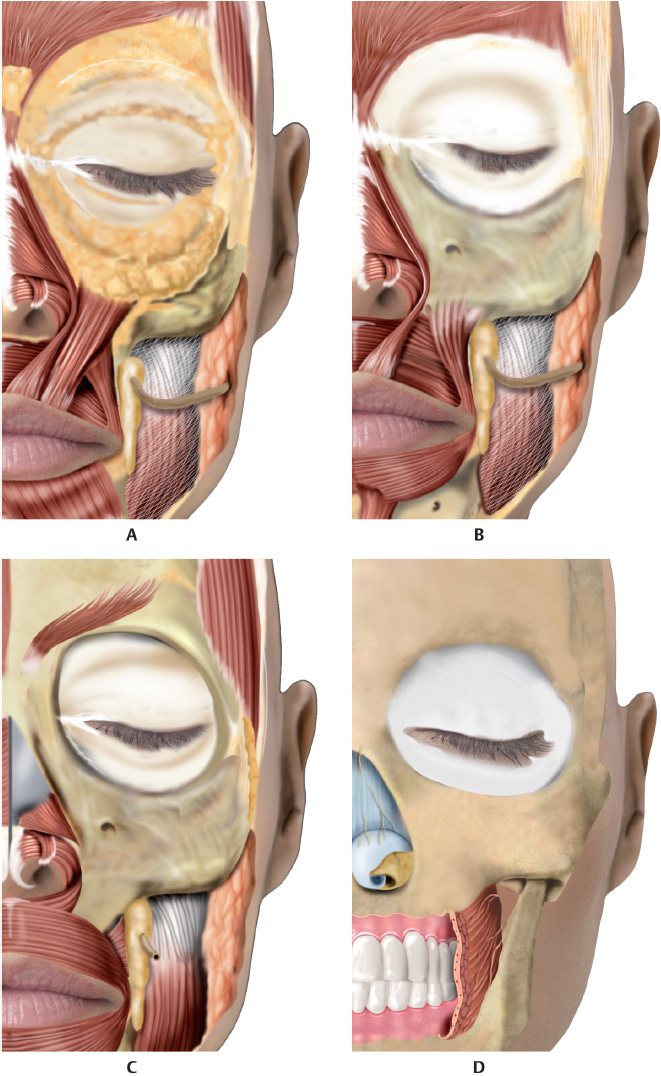
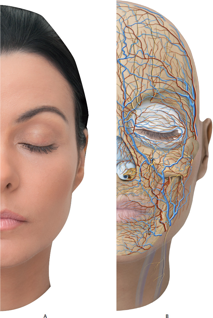
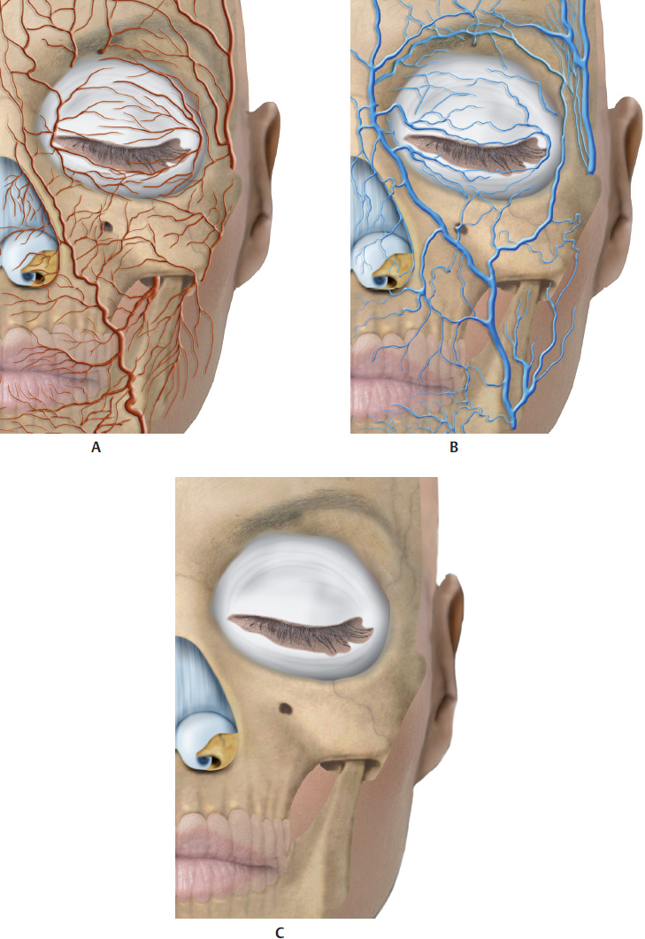
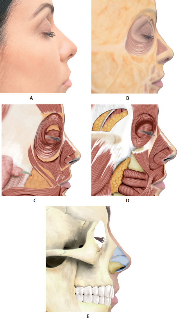
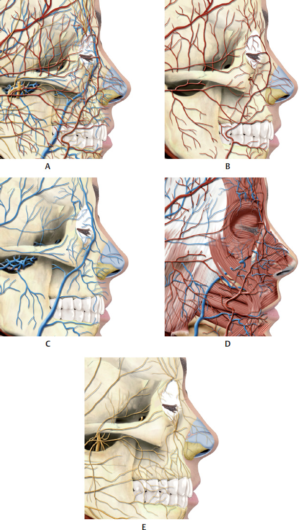
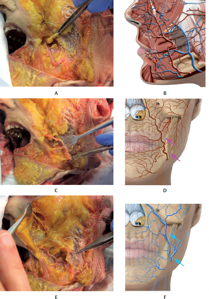
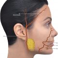
It is a deep fat pad (DFP) in the center of the submalar triangle. It has a triangular shape and extensions into the masseter, temporal, and pterygoid muscles. It is located over a fissure above the buccinator muscle and below the superficial muscular aponeurotic system (SMAS). The facial artery and vein are located anteriorly to Bichat’s fat pad. The transverse facial vessels irrigate its superior portion, above the parotid duct, and branches of the internal maxillary artery also contribute to vascularization of the area.
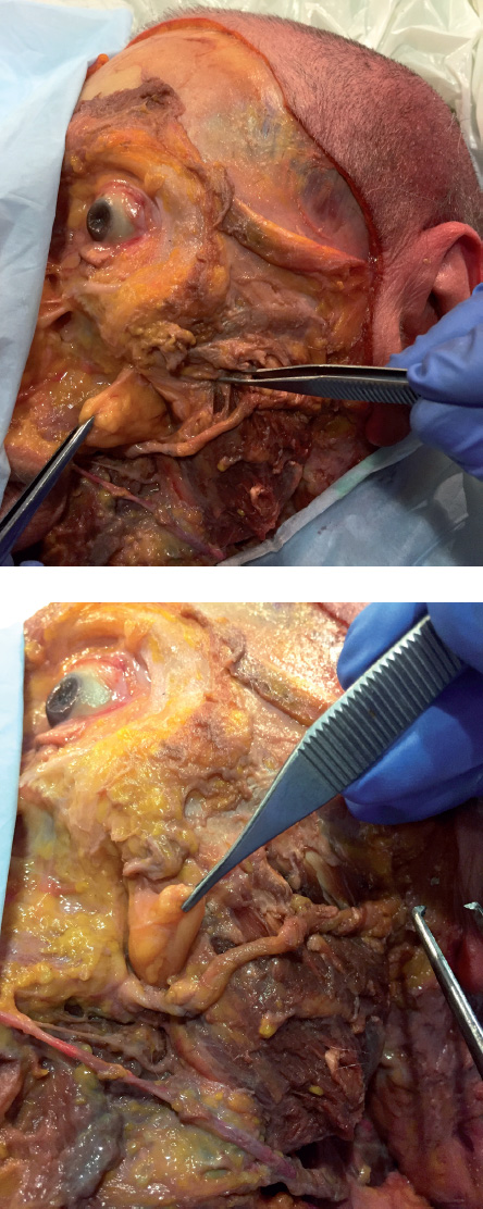
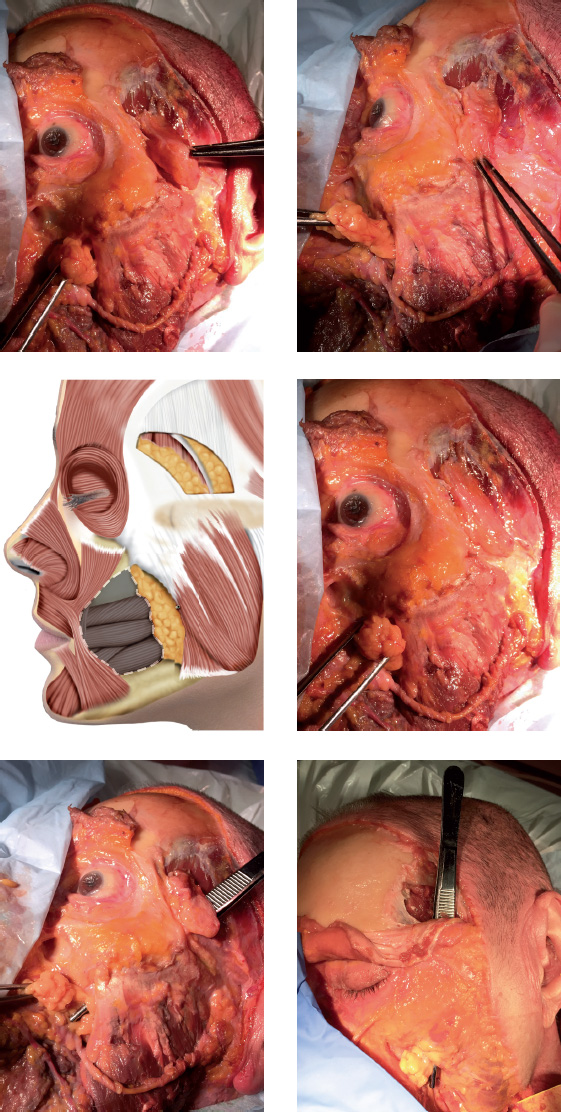
The parotid duct and the zygomatic and buccal branches of the facial nerve are intimately related to the buccal fat pad (Fig. 13.1–13.6).
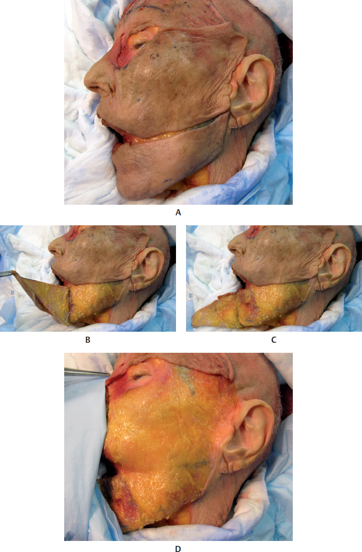
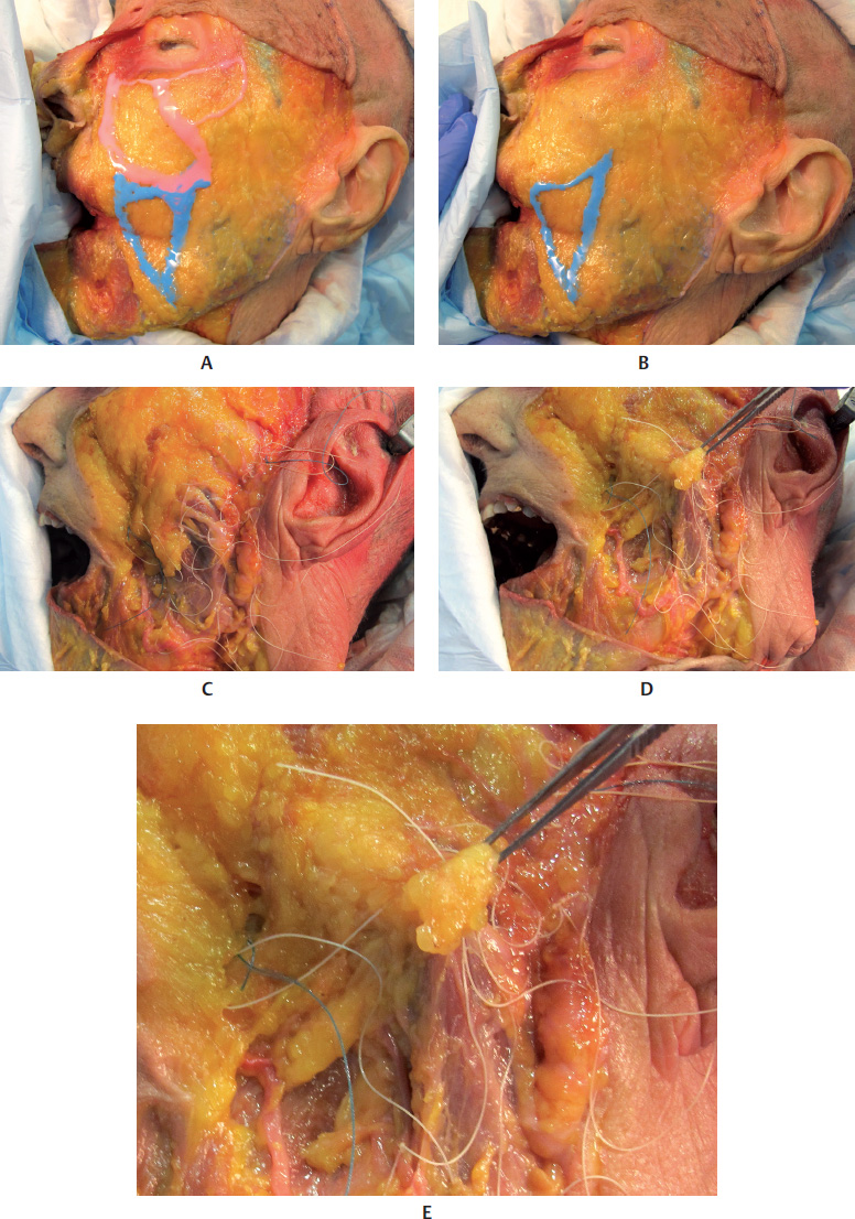
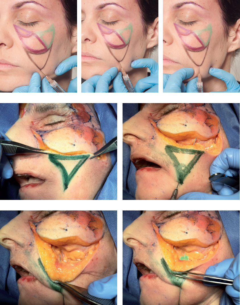
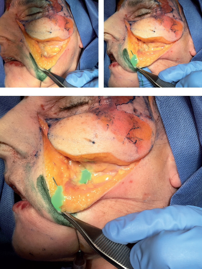
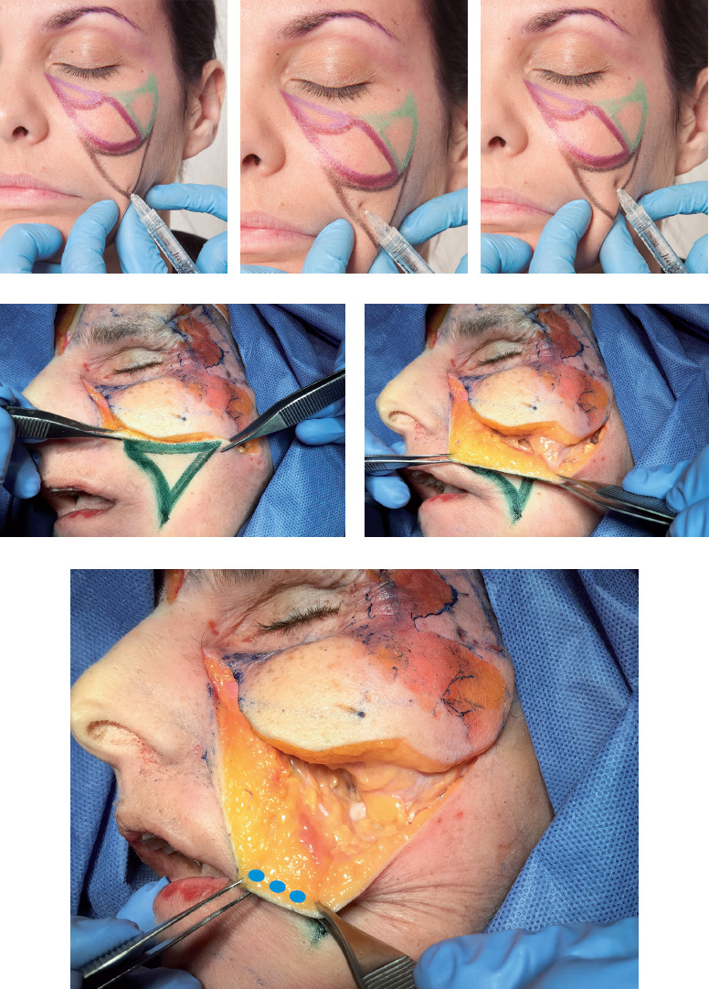
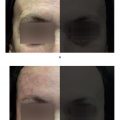
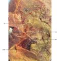
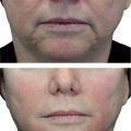
1 Tobias GW, Binder WJ. The submalar triangle: Its anatomy and clinical significance. Facial Plast Surg Cl. Facial Contouring and alloplastic implants. 1994;2(3):255–632 Stuzin JM, Wagstrom L, Kawamoto HK, Baker TJ, Wolfe SA. The anatomy and clinical applications of the buccal fat pad. Plast Reconstr Surg 1990;85(1):29–37Fig. 13.1 A. Anterior view of the anatomy of the submalar region.B. Same region after removal of the skin to expose the superficial fat pads (SFPs) and focus on the buccal fat pad.C. Same region after removal of the skin and the SFPs.D. Same region after removal of the skin, SFPs, orbital portion of the orbicularis oculi muscle (OM), and zygomatic muscles. Parts of the buccal fat pad, parotid duct (above the masseter), and parotid gland are visible.Fig. 13.2 A. Anterior view of the anatomy of the submalar region after removal of the skin, superficial fat pads (SFPs), palpebral and orbital portions of the orbicularis oculi muscle (OM), and zygomatic muscles.B. Same. region after removal of the skin, the SFPs, the palpebral and orbital portions of the OM, the medial and lateral suborbicularis oculi fat (SOOF), the levator muscle of the upper lip, and the zygomatic major and minor muscles.C. Same. region after removal of the levator muscle of the upper lip and the ala of the nose, and the levator muscle of the angle of the mouth. Parts of the buccal fat pad, dissected parotid duct, masseter muscle, and parotid gland are visible.D. Bone. structure of the submalar region and a part of the buccinator muscle.Fig. 13.3 A. Anterior view of the left side of the face of a 3D digital model andB. Vessels, arteries, and nerves of the submalar region.Fig. 1.2 Anterior view of the face focusing on the submalar region.A. Corresponding arterial vascularization.B. Corresponding venous vascularization.C. Bone structure of this region.Fig. 13.5 A. Lateral view of the right side of the face of a 3D digital model.B. Same region after removal of the skin, showing superficial fat pads (SFPs).C. Same region after removal of the skin and the SFPs. Palpebral and orbital portions of the orbicularis oculi muscle (OM) can be seen, with the latter portion over the medial and lateral suborbicularis oculi fat (SOOF) pads. The levator muscle of the upper lip and ala of the nose can be seen, as well as the levator muscle of the angle of the mouth, and the zygomatic major and minor muscles above the deep fat pads (DFPs).D. Same region after removal of the orbital portion of the OM, medial and lateral SOOF pads, SFPs, and DFPs. A part of the buccal fat pad (Bichat’s fat pad) can be seen above the buccinator muscle.E. Bone structure of the submalar region.Fig. 1.2 Right profile view with focus on the submalar region.A. Corresponding vascularization and innervation.B. Corresponding arterial vascularization.C. Corresponding. venous vascularization.D. Arterial. and venous vascularization integrated in the frontal muscle, orbicularis oculi muscle (OM), nasal muscles, greater zygomatic muscle, levator muscle of the lip, buccinator muscle, and masseter muscle.E. Corresponding. innervation.Fig. 13.7 A andB. Anterior view of the delimitation of the submalar triangle.C andD. Close-ups ofA andB.Fig. 13.8 A. Left side of the face of a fresh cadaver specimen.B andC. Skin has been folded back to show the superficial portion of the buccal fat pad (Bichat’s fat pad).D. Buccal fat pad in detail.Fig. 13.9 A. Left side of the face of a fresh cadaver specimen with skin folded back showing delimitation of the malar and zygomatic superficial fat pads (SFPs) (marked in pink).B. Delimitation of the superficial portion of the buccal fat pad (Bichat’s fat pad) (marked in blue).C. The specimen after removal of the superficial portion of the buccal fat pad.D. Deep portion of the buccal fat pad being pinched.E. Deep portion of the deep buccal fat pad (Bichat’s fat pad) in detail.Fig. 13.10 A. Left side of the face of a fresh cadaver specimen with buccal fat pad being pinched; the facial artery can be seen at the front.B. Illustration showing the buccal fat pad between the facial artery and vein.C. Pinching. of the facial artery.D. Illustration of the facial artery.E. Pinching of the facial vein.F. Illustration of the facial vein.Fig. 13.11 Left side of the face of a fresh cadaver specimen after removal of the superficial fat pads (SFPs) and pinching ofthe buccal fat pad (Bichat’s fat pad).Fig. 13.12 Left side of the face of a fresh cadaver specimen and illustration after removal of the superficial fat pads (SFPs). Note the deep portion of the buccal fat pad (Bichat’s fat pad) and its extension up to the temporal region.Fig. 13.13 A, B, andC. Left side of the face of a fresh cadaver specimen showing delimitation of the submalar triangle (markings in green) and the superficial portion of the buccal fat pad.D. Superficial portion of the buccal fat pad being folded back to show the deep buccal fat pad, located below the superficial muscular aponeurotic system (SMAS).Fig. 13.14 Demonstration of a filler injection technique in the submalar triangle with a cannula in the deep buccal fat pad, below the superficial muscular aponeurotic system (SMAS). Note thegreen product. in the deep plane, below the levator muscles of the upper lip.Fig. 13.15 Demonstration of a filler injection technique in the submalar triangle with a cannula in the superficial buccal fat pad, above the superficial muscular aponeurotic system (SMAS). Note thegreen product in the subcutaneous plane, above the zygomatic muscle.Fig. 13.16 Demonstration of a filler injection technique in the submalar triangle with a needle in the superficial buccal fat pad, above the superficial muscular aponeurotic system (SMAS). The procedure can be performed with a linear antegrade or retrograde injection.Fig. 13.17 A. A patient with loss of volume in the region of the submalar triangle.B. Delimitation of the area to be filled.C. Filler injection in the area using the cannula technique in the superficial and deep planes. Note thegreen product above and below the greater zygomatic muscle.D andE. The patient before and after a filler injection in the submalar triangle, using a superficial and deep cannula technique.
Only gold members can continue reading. Log In or Register to continue