Chapter 43 Calf and thigh implants
• Calf and thigh implants in the femoral region are described.
• Leg perimeter augmentation is described.
• The indications are for patients with thin legs, sequelae to poliomyelitis, and for burn and accident sequelae.
• Montellano implants were conceived by the author.
• There is a real augmentation of 2.5 to 3.0 of the leg perimeter.
Introduction
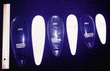
Plastic surgery has allowed us to improve numerous body contouring problems along the years. Amongst those, new techniques have emerged in the treatment of deformities of the lower extremities secondary to poliomyelitis, burns, trauma and the so-called thin legs, in order to increase the perimeter of the legs and obtain a certain harmony. Implant use in calf and thigh surgery has gained great interest as it has shown great results. Silicone gel implants mimicking the legs’ musculature have been designed by the author, allowing anatomical and natural postoperative results (Fig. 43.1). Retractors were also created in order to facilitate the insertion of these implants.
Background
In 1979 Carlsen1 showed the use of hard silicone implants for calf augmentation, but also emphasized the fact that they were not the best implants for this surgery, as there were many complications. In the same year, Glicenstein3 published an article where he used cigar-shaped silicone gel implants for calf augmentation, through a skin incision in the middle third of the leg. Then in 1985, Montellano5 presented his first cases of calf augmentation using anatomical silicone gel implants at the 22nd National Brazilian Meeting of Plastic Surgery.
Anatomy
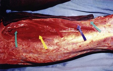
The calf is composed of three major muscles:
1. The soleus, which contains important vascular structures, namely the posterior tibial artery, vein and nerve, and inferiorly the external saphenous vein and nerve.
2. The gastrocnemius, which is approximately 18 cm long, with its inferior part located at ±5 cm from the internal malleolus.
3. The lateral gastrocnemius, approximately 12 cm long, with its inferior part located at ±10 cm from the external malleolus.
These three muscles converge inferiorly at the heel, where they form the Achilles tendon, also known as triceps surae (Fig. 43.2).
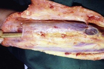
The implant is placed in the medial-posterior region of the thigh, under the following muscles: internal rectus, adductor magnum, semimembranous and vastus medialis muscles (Fig. 43.3).
Materials and Methods
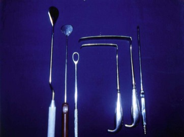
Implants used for calf augmentation vary in size and measure approximately 14, 18 or 22 cm long, 4 to 6 cm wide and 2 to 3 cm in height. Those used for thigh augmentation have a similar triangular shape, but are slightly larger and present an additional guide at their distal extremity, which facilitates their insertion up to the bottom of the previously created pocket. Dissectors used for these procedures, also designed by the author, have a wide and blunt tip. Their sizes vary from 25 to 30 cm in length. Also, retractors such as “L” wide and Finochietto2 types are used for these procedures (Fig. 43.4).
Surgical Technique
Calf Implants
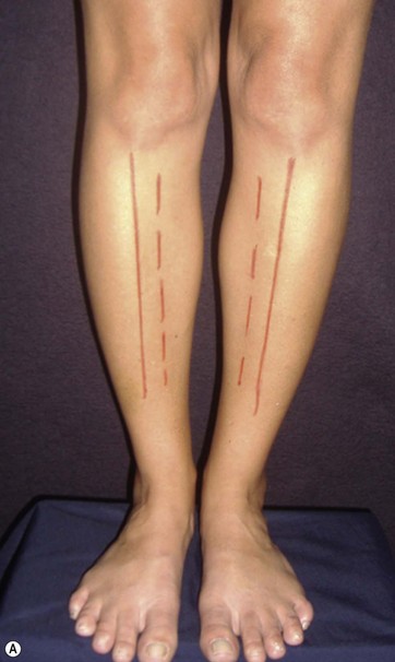
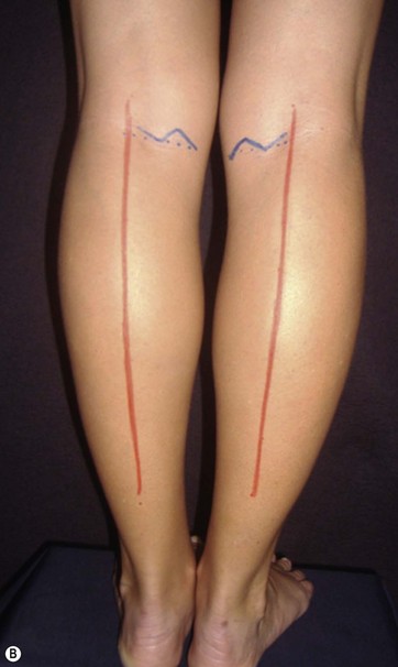
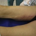
Demarcation of the incision lines and the undermining regions is done preoperatively with each patient in the standing, sitting and supine positions, in order to ensure the correct placement of the implants. The lateral borders of the gastrocnemius muscle are delimited; a distance of 10 cm above the internal malleolus and superficially a 3 cm zig-zag incision above the popliteal fossa are also marked (Fig. 43.5). The surgery is performed with the patient in the supine position under epidural or general anesthesia, and in some cases under local anesthesia with mild sedation of the patient, in the presence of an anesthesiologist.
< div class='tao-gold-member'>
Related posts:
Stay updated, free articles. Join our Telegram channel

Full access? Get Clinical Tree


