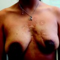
Fig. 43.1
Capsular contracture and skin alteration after an augmented breast treated with lumpectomy and radiotherapy
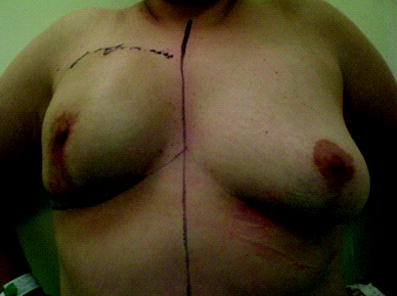
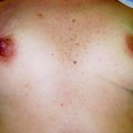
Fig. 43.2
Capsular contracture and asymmetry after mammoplasty with an implant and radiotherapy
Breast-conserving therapy with implant removal is a less desirable option. Women having breast augmentation often have scarce breast tissue, which is actually why many undergo this procedure. In addition, the presence of an implant results in thinning of the stretched overlying breast tissue over time. One study reported that native breast tissue comprised 50 % of overall breast volume [17, 18]. Therefore, this is a suitable option only for a very small group of patients who have a considerable amount of remaining tissue. In these cases, mammoplasty techniques should be used as a T Wise pattern or vertical scar technique (Lejour’s technique).
Patients who are candidates for partial breast irradiation, especially those who are candidates for intraoperative radiotherapy could benefit from lumpectomy and implant maintenance [19]. Despite the paucity of evidence, this could be a good option for resection alone and local glandular flap partial reconstruction (Fig. 43.3). Figure 43.4 shows a flowchart for decision-making regarding augmented breast surgery and oncologic surgery.
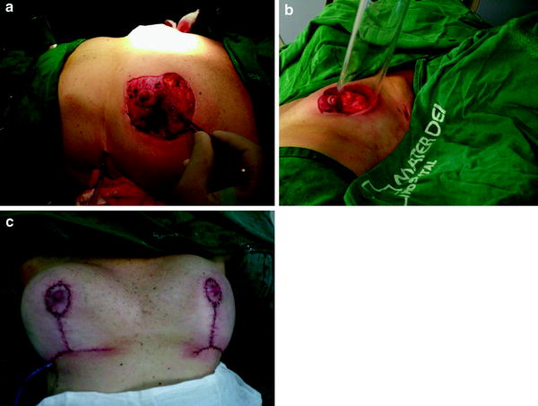
Fig. 43.3
a Lumpectomy after implant removal and inferior pedicle mammoplasty. b Intraoperative radiotherapy at the tumor bed c Final result after lumpectomy and implant reinsertion (new implant)
Fig. 43.4
Flowchart for the surgical decision for an augmented breast and lumpectomy. IORT intraoperative radiotherapy
43.1.2 Total Breast Reconstruction
As previously discussed, mastectomy and immediate reconstruction seems to be the best treatment for breast cancer patients with preexisting breast augmentation [20–22]. Decisions on the type of reconstruction should be made according to local conditions following mastectomy. If a large amount of skin needs to be removed, reconstruction with autologous tissue is more suitable, e.g., a transverse rectus abdominis myocutaneous flap or deep inferior epigastric perforator flap. A latissimus dorsi flap with an implant is also a good option for these cases. An extended latissimus dorsi flap without an implant would probably not be a good option, since patients usually hope for a reconstructed breast that is the same size as before. With use of this technique, it is difficult to achieve the desired result. The choice of technique can be challenging, because many patients with augmentation surgery are thin and the donor site can be insufficient.
In contrast, if native skin can be preserved, a skin-sparing mastectomy (SSM) or nipple-sparing mastectomy (NSM) is performed. Reconstruction can easily be performed with a single-stage implant (implant or definitive breast expander) or a two-stage implant (tissue expander plus implant exchange). When implant-based reconstruction is chosen, it is critically important to evaluate both the quality of the skin and muscles (pectoralis major muscle and serratus muscle). As shown in previous chapters, adequate implant reconstruction is performed with a good muscular pocket that partially or completely covers the implant. In a partially covered implant where the skin is compromised (vascular suffering, infection, necrosis), the implant can be exposed and should be removed.
Definitive implant reconstruction is desirable in patients in whom a minimal amount of skin needs to be removed. The reason is that it is a faster technique with no donor site complications [23, 24]. In a previously augmented breast, skin coverage is rarely a problem and good cosmetic results can be achieved.
The need for adjuvant radiotherapy may play an important role in the decision for reconstruction. Although some authors strongly contraindicate reconstruction because of a high complication rate (up to 70–90 %) [25], good results have been achieved by many other authors, showing patient satisfaction of up to 80 % [26]. Indeed, we recommend implant reconstruction whenever feasible, even in a scenario of adjuvant radiotherapy. If complication happens a further autologous reconstruction can always be performed. Figures 43.5, 43.6 and 43.7 show the results of breast reconstruction with and without radiotherapy.
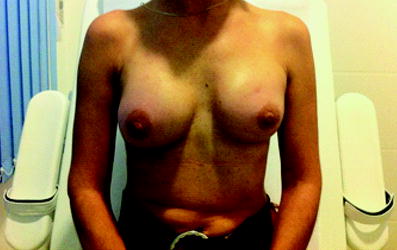
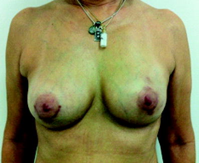
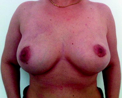
Fig. 43.5
Left breast capsular contracture after mastectomy and implant reconstruction in a breast-augmented patient followed by radiotherapy
Fig. 43.6
Left breast implant-based reconstruction after left nipple-sparing mastectomy (NSM) in an augmented patient with radiotherapy
Fig. 43.7
Bilateral implant-based breast reconstruction after bilateral NSM in an augmented patient with no radiotherapy
Tumor location and skin incision is of major importance to surgical outcome. Skin or nipple–areola complex (NAC) necrosis can translate into reconstruction failure if there is exposure of the implant. There is no study addressing the use of a preexisting augmentation mammoplasty incision to perform mastectomy. When choosing an incision, the surgeon must consider the oncologic outcome and preexisting scarring which can translate into abnormality of the skin and NAC irrigation. Figures 43.8 and 43.9 show a periareolar approach in which a preexisting scar from breast augmentation is used.
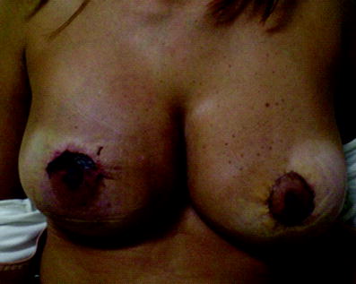
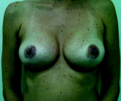
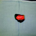
Fig. 43.8
Periareolar mastectomy and reconstruction in previously augmented patient with partial necrosis of the nipple–areola complex (NAC)
Fig. 43.9
Periareolar bilateral mastectomy and reconstruction in a previously augmented patient with no complications
Preexisting breast surgery is a well-known factor related to postoperative complications. Skin incisions for augmentation mammoplasty are periareolar (complete or partial) in the inframammary fold or in the axillary line when it is not associated with mastopexy (vertical or inverted-T pattern). SSM is performed with removal of the NAC, so the incision must be made in the central portion of the breast. However, when NSM is indicated, the incision can be made in any part of the breast (periareolar, inframammary fold, etc.). Therefore, the surgeon can attempt to use the preexisting scar to perform NSM.
To predict the surgical outcome relative to surgical access for mastectomy and reconstruction, an analogy was made between studies evaluating NSM incisions according to outcome. Wijayanayagam et al. [27] showed that a radial incision and an inframammary fold incision (in small breasts) are good options with a low risk of NAC or skin necrosis. Algaithy et al. [28] showed a low risk of necrosis with a superolateral radial incision and a high risk of complications with a circumareolar and periareolar incision. Figure 43.10 shows a radial approach to mastectomy and reconstruction in a patient with periareolar breast augmentation.
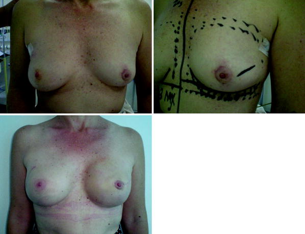
Fig. 43.10
Patient with periareolar breast augmentation and capsular contracture in the preoperative period, and postoperatively after left breast mastectomy and reconstruction using a radial scar and right breast implant exchange
Therefore, a complete periareolar incision or a large circumareolar incision should be discouraged. Inframammary fold incisions should be performed in selected cases and only in patients with small breasts. A periareolar 180° incision can be performed, although the risk of wound dehiscence and skin necrosis is higher owing to direct skin traction during surgery. Table 43.1 shows the risk of skin and NAC necrosis according to the location of the incision and breast size.
Incision | Large breast | Medium-sized/small breast |
|---|---|---|
Complete periareolar | High risk | High risk |
Periareolar 180° | Moderate risk | Moderate risk |
Circumareolar | High risk | High risk |
Radial | Low risk | Low risk |
Inframammary fold | High risk | Low risk |
Another issue that should be discussed is whether the implant should be exchanged during surgery or whether the old implant should be maintained. Many authors consider that implant exchange is mandatory when the implant is located in the subglandular space because it must be removed for adequate patient treatment. Other considerations that are clearly in favor of implant exchange are implant rupture, capsular contracture, infection, and poor cosmetic result [20, 21]. Few publications have advocated the possibility of maintaining a preexisting implant in the case of a new-generation implant located in the submuscular space [29]. Actually, this should be an exception rather than the rule, applied only to strictly selected cases. Figure 43.11 shows a flowchart of decisions on augmented breast and total breast reconstruction.
Fig. 43.11
Flowchart of indications for breast reconstruction after mastectomy in augmented patients
43.2 Breast Reconstruction After Breast Reduction Mammoplasty
As previously discussed, breast reduction mammoplasty is the fifth commonest cosmetic intervention in the USA. A considerable number of patients undergoing this procedure will develop breast cancer at some time in their lives. The procedure per se reduces the risk of breast cancer. Some studies have shown up to 50 % reduction in breast cancer risk [30].
Related posts:

Stay updated, free articles. Join our Telegram channel

Full access? Get Clinical Tree


