Key points
- •
Inconspicuous incision location.
- •
Adequate incision length for accurate and meticulous pocket dissection.
- •
Large enough incision so as not to damage the breast implant.
- •
Accurate anatomic realignment of the tissues.
- •
Tension free skin closure.
Introduction
The chosen incision technique should be based upon the type of breast surgery to be performed, the patient’s anatomy, the type and size of the breast implant and the preferences of the patient and surgeon. If a mastopexy is planned as a part of the procedure then, ideally, one of the mastopexy incisions should be utilized so as to avoid additional unnecessary scarring on a woman’s breast. The same is true if a scar already exists on a breast and the procedure is not otherwise compromised. Patients often choose an incision based upon it leaving the least conspicuous scar. The surgeon should share this goal, leaving as little surgical evidence as possible.
Basic principles should include placing incisions inconspicuously and along relaxed skin tension lines regardless of location. The incisions should be as short as possible, but as long as necessary so as not to compromise accurate and meticulous pocket dissection and insertion of the breast implant without damage. Even though some implants can be placed through an incision as short as 2 cm, the quality of the scar is more dependent upon minimizing tissue trauma from retraction injury than upon the length of the scar. In general, it is important that tissues disrupted by surgery be accurately and anatomically realigned. Reapproximating the fascia, when available, provides a strong closure and minimizes tension on the edges of the wound. Deeper layers should be sutured to eliminate any dead space. Closure of the deep dermis should create eversion of the wound edges so the skin suture can perfectly align the skin edges without any tension.
Suture selection should be based upon utilizing the least amount of suture material necessary to effectively close the wound. It should resorb as quickly as possible after adequate tissue strength in the healing wound has been accomplished. Typically, deeper layers of tissue require larger absorbable sutures. The scar is minimized if the skin suture is placed below the skin as a permanent running or pullout subcuticular suture.
Incision selection
Inframammary incision
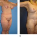
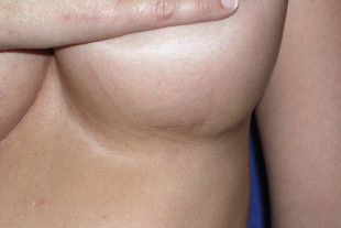
Historically, the inframammary incision has been the most commonly used incision since the inception of breast implantation surgery (Cronin 1964). Women who have a well-defined inframammary fold and slight glandular breast ptosis are the best candidates for this incision. Incisions that are planned correctly and lie precisely in the new inframammary fold heal very well and are difficult to see not only from a standing position, but also while lying down ( Figure 2.1 ). An incision in this location (a relaxed skin tension line) has very little tension on it from the stretch of the new implant, but it must be placed accurately ( Figure 2.2 ). A scar misplaced superiorly on the breast mound becomes apparent when the patient lies down whereas a scar placed too low is evident in any position. Sometimes even a well planned incision becomes too high as an implant descends, a phenomenon less likely with the tissue adhesion from aggressively textured implants. Some surgeons shy away from the inframammary incision because of this inherent unpredictability. Discussions will follow as to how to more precisely predict the location of the inframammary scar.
The inframammary incision is perhaps the simplest and quickest of all breast augmentation incisions. It allows immediate and direct access to any desired dissection plane with very little disruption of breast tissue. This incision is often necessary in patients who receive larger implants or those requiring textured implants where longer length incisions are frequently necessary. It is an ideal incision for anatomic shaped implants where the orientation of the implant is crucial to the overall success of the surgery. This is also the incision of choice for secondary or revision breast implant surgery. The inframammary incision is not a good choice in patients with a non-existent inframammary fold or a high inframammary fold which is to be lowered as a part of the surgical plan ( ). Usually a 4 cm incision provides sufficient exposure to facilitate accurate pocket dissection for most saline breast implants and smaller silicone breast implants. Textured surface implants, larger smooth surface silicone implants and cohesive gel implants may require incisions up to 6 cm in length for proper insertion while avoiding damage to the implant.
Operative technique
The most important consideration in the placement of the inframammary incision is predicting the final position of the inframammary fold. There are two methods for determining the post-operative inframammary fold location, one simple and the other more complex. Keep in mind that regardless of the method chosen, other variables must be accounted for such as the type of breast implant selected for the patient, the pocket location and the fibrous connections from the deeper tissues to the skin.
The type of implant selected makes a significant difference in the final location of the inframammary fold. Smooth implants are known to ‘drop’ in the early post-operative course. How far they drop is dependent upon the volume (weight) of the implant and how tight the attachments are from the superficial tissues to the deeper tissues; stronger in young nulliparous women and weaker in older patients especially if they have had children. Placement is another consideration. Subpectoral implants in a dual plane pocket tend to drop more than implants placed in a submammary position or complete submuscular position. Lastly, breast implants with a more aggressive textured surface tend to be more predictable in terms of their final position because of adherence from the implant to the surrounding tissues. Thus, the ultimate inframammary location becomes more predictable since these implants are less likely to drop.
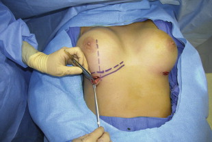
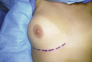
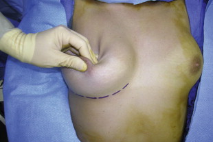
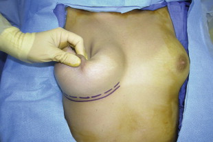
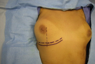
The simplest method for determining the post-operative inframammary fold location is by marking the patient’s existing inframammary fold while the patient is in the standing position ( Figure 2.3 ). Markings for the new inframammary fold are made with the patient on the operating room table in the supine position. The covered nipple is held with the thumb and index finger pulling it away from the chest wall. The remaining fingers are used to push the breast gland inferiorly ( Figure 2.4 ). The new curvilinear inframammary fold is marked along the crease created by this maneuver ( Figure 2.5 ).
A more comprehensive method for determining the inframammary fold location utilizes the tissue based planning principles in the High Five System ( ). This method analyzes variables such as breast implant volume, implant base width; the patient’s measured breast width, anterior pull skin stretch, nipple to IMF distance on stretch and breast parenchyma to stretched breast envelope fill. Once an implant has been selected based upon these variables, a reference chart is used to determine the recommended new IMF fold location. This selection is based in part upon the base width and volume of the breast implant, the recommended nipple to inframammary distance based upon the author’s experience and the patient’s pre-operative nipple to inframammary fold distance under maximal stretch. The inframammary fold location is then determined based upon the difference in the recommended and measured IMF fold position. This new fold position is calculated prior to surgery, noted in the patient’s chart and physically marked in the preoperative holding area or in the operating room.
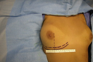
Ultimately, the goal in planning the location of the inframammary fold is to have the inferior border of the implant lay at the fold with the point of maximum projection of the implant located directly behind the nipple. Therefore, the radius of a round implant or the distance from the point of maximum projection to the lower edge of an anatomic implant should equal the distance from the nipple to inframammary fold. Once the inframammary fold position has been determined, a vertically oriented paramedian line is drawn from the medial aspect of the areola inferiorly to bisect the newly drawn inframammary fold ( Figure 2.6 ). This marks the medial extent of the planned inframammary incision. The marking is then carried laterally along the new inframammary fold for 4–6 cm depending upon the procedure to be performed and the implant type and size ( Figure 2.7 ).
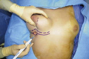
The incision is performed with a 10 or 15 blade down through skin into the mid dermis. Further dissection is carried out while holding the breast between the fingers and thumb lifting it away from the chest wall. Dissection proceeds superiorly at about a 20 degree angle down to the chest wall with an electrocautery device ( Figure 2.8 ). The electrocautery device should be set on the lowest possible setting to avoid collateral thermal damage to the tissues. The dissection passes through the subcutaneous tissue and Scarpa’s fascia to the retroglandular fatty tissue. From this point, the lateral diagonal border of the pectoralis major muscle and its fascia as well as the breast gland are visible making it easy to proceed with the desired pocket dissection.
Closure of the inframammary fold should include Scarpa’s fascia with simple interrupted 3-0 or 4-0 absorbable sutures placed approximately 1 cm apart ( Figure 2.9 ). There should be closure of the subcutaneous tissue and deep dermis with simple interrupted 4-0 absorbable suture with eversion of the wound edges so the skin edges are touching one another without tension. Finally, there should be perfect alignment of the epidermis with a permanent running or pullout subcuticular suture or skin glue ( Figure 2.10 ). Depending upon the thickness of the tissues, Scarpa’s fascia, the subcutaneous tissue and the deep dermis may all be included in a single suture.