Bioprosthetic Materials for Plastic Surgery of the Breast
G. Patrick Maxwell
Allen Gabriel
Introduction
Over the last century breast surgery has evolved from a rarely performed surgical venture to a daily occurrence that has become an important part of the rehabilitation process following aesthetic and reconstructive surgeries. The aesthetic quality of breast surgery, fostered by technical advances, has emerged from that of amorphous blobs appearing as breast mounds to nearly normal appearing breasts. Symmetry, which previously was hardly possible and seldom achieved, is now the standard for which we strive.
History
Controversy regarding the safety of silicone gel–filled breast implants, which were usually placed in a subglandular pocket, resulted in a 1992 moratorium on their use for aesthetic breast augmentation. This forced U.S. surgeons to use saline implants, which were placed under the muscle, thereby gaining extra tissue coverage to conceal the untoward contour irregularities of these implants (1,2,3). At the same time, the majority of reconstructive surgeons converted to reconstructing breast mounds with saline implants. During the 14 years of the moratorium, surgeons were trained to use saline implants for both reconstructive and aesthetic procedures.
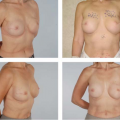
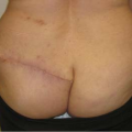
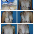
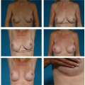
Revisionary breast surgery (secondary or tertiary), which is often performed for the late complications of implant-based breast surgeries, poses a continual challenge to plastic surgeons. These procedures are complex, challenging, and unpredictable. Over the years we have had to deal with thinned breast tissues from large implants that have been placed either in subglandular or subpectoral space or reconstructed with saline implants, leading to long-term complications such as implant extrusion, rupture or deflation, capsular contracture, palpability, rippling, “double-bubble” appearance, “Snoopy breasts,” symmastia, and implant malposition (1,2,3).
For many years capsular contracture has plagued plastic surgery as the most common complication of aesthetic and reconstructive breast surgery (4,5). The majority of revisionary surgeries are performed due to capsular contracture (5,6). Many etiologies have been proposed for this process, and it is clear that its prevention in primary cases includes sound techniques, including precise, atraumatic, bloodless dissection; appropriate triple-antibiotic breast pocket irrigation; and minimization of any points of contamination during the procedure (7,8). Treatment of an established capsule can be more challenging, and multiple techniques have been used for this. The bottom line to any pathophysiologic process is to understand the disease at the cellular level. In this case, it is clear that at the cellular level, capsular contracture is most likely caused by any process that will produce increased inflammation, leading to formation of deleterious cytokines within the periprosthetic pocket. Historically, options for revision and improvement have included replacing saline implants with silicone implants, using capsular flaps to gain additional stability and coverage, or performing a site change operation, which does not allow for complete resolution of some of the implant issues (1,9). Capsular flaps are available, but some patients have extremely thin tissues, and these flaps only allow for subtle improvements. Consequently, in addition to all of the techniques for treating and preventing capsular contracture proposed by others (1,4,7,10,11,12,13,14,15,16), we believe that acellular dermal matrix (ADM) is another modality for fighting the evolution of the capsule. ADMs can counteract the inflammatory process, adding additional availability of tissue in-growth and controlling the interface of the pocket.
Acellular Dermal Matrix
Use of ADMs has been popularized in both breast and abdominal wall reconstructions and has been reported in a range of clinical settings, including hernia repair, facial and eyelid surgery, cleft palate repair, soft tissue augmentation, tendon repair, ulcer repair, and vaginal sling repair (17,18,19,20,21,22,23,24,25,26,27). In reconstructive cases, they have been used to replace tissue, extend existing tissue, or act as a supplement. In aesthetic cases, they have been used to correct implant rippling and displacement, including symmastia (28,29,30).
Immediate breast reconstruction using tissue expanders or implants has become one of the most commonly used surgical techniques, which has made visible rippling and contour deformity a more frequently encountered problem (31). The recent use of allogeneic tissue supplements avoids the problems of autologous tissue coverage and provides camouflage, thus decreasing rippling and increasing soft tissue padding (31,43).
The adjunctive use of ADMs in breast reconstruction has been shown to shorten the tissue expansion/implant reconstructive process, avoid mastectomy flap contraction during the latency period of expansion, provide an additional layer of tissue between the skin and implant, and offer an additional option for immediate, single-stage breast implant reconstruction (18,19,44,45,46).
The rising demand has spurred tremendous growth in the number of available ADMs. Published research regarding the use and efficacy of ADMs in immediate breast reconstruction is growing but has not kept pace with the market explosion. The many features and indications of all ADMs can confound the decision-making process for surgeons who want to incorporate ADMs in their treatment armamentarium.
Acellular dermal matrices can be categorized as either xenograft or allograft in origin. A surgical graft of tissue from
one species to an unlike species (or genus or family) is termed a xenograft, whereas the transplant of an organ or tissue from one individual to another of the same species with a different genotype is termed an allograft. All are produced with a similar objective of removing cellular and antigenic components that can cause rejection and infection. The lack of immunogenic epitopes enables the evasion of rejection, absorption, and extrusion (18,19,27). Production processes allow basement membrane and cellular matrix to remain intact. This scaffold is left in place to allow in-growth of the host’s fibroblasts and capillaries to eventually incorporate as its own. Much of this scaffold matrix consists of intact collagen fibers and bundles to support tissue in-growth, proteins, intact elastin, hyaluronic acid, fibronectin, fibrillar collagen, collagen VI, vascular channels, and proteoglycan–-all of which allow the body to mount its own tissue regeneration process (18,19,26,27,47,48,49,50).
one species to an unlike species (or genus or family) is termed a xenograft, whereas the transplant of an organ or tissue from one individual to another of the same species with a different genotype is termed an allograft. All are produced with a similar objective of removing cellular and antigenic components that can cause rejection and infection. The lack of immunogenic epitopes enables the evasion of rejection, absorption, and extrusion (18,19,27). Production processes allow basement membrane and cellular matrix to remain intact. This scaffold is left in place to allow in-growth of the host’s fibroblasts and capillaries to eventually incorporate as its own. Much of this scaffold matrix consists of intact collagen fibers and bundles to support tissue in-growth, proteins, intact elastin, hyaluronic acid, fibronectin, fibrillar collagen, collagen VI, vascular channels, and proteoglycan–-all of which allow the body to mount its own tissue regeneration process (18,19,26,27,47,48,49,50).
Related posts:
 Follow-Up After Surgery for Primary Breast Cancer: Breast-Conserving Therapy and Mastectomy
 Breast Implants: Materials and Manufacturing Past, Present, and Future
 Reconstruction of the Irradiated Breast
 Perforator Flaps in Breast Reconstruction
 Lipomodeling of the Reconstructed Breast
 The Inframammary Approach to Augmentation Mammaplasty
Stay updated, free articles. Join our Telegram channel

Full access? Get Clinical Tree


