General Principles
Living cells respond to physical and biochemical cues, e.g., hormones, cell–cell signaling molecules, and with the physical environment through the extracellular matrix (ECM), enabling them to adapt to changes in the physiological environment. Regulation of cell function (growth, migration, proliferation, and differentiation) is dependent upon contact or adhesion between cells and the substrate (ECM). ECM has three key components: collagen – the structural framework of ECM – proteoglycans, and adhesive matrix proteins (e.g., fibronectin, vitronectin). Initial contact of cells with a solid substrate is mediated by adsorbed adhesive proteins. Cell surface receptors subsequently form bonds leading to cytoskeletal reorganization and progressive spreading of the cell on the substrate. These receptors transduce biochemical signals to the nucleus by activating the same intracellular signaling pathways that are used by growth factor receptors. In addition to biochemical cues, mechanical forces play an important role in cell adhesion, growth, and possibly differentiation. These responses are mediated through internal cell receptors that respond to mechanical forces. Cell binding to ECM through specific cell–substrate contacts is critical to cell-growth control through mechanical forces resulting in alterations in cell shape and cytoskeletal tension.
A biomaterial is a nonpharmaceutical, nonviable material in intimate contact with living tissue in order to treat, augment, or replace any tissue, organ or function of the body. Ideal properties of a biomaterial include: biocompatibility, noncarcinogenicity, lack of allergenicity, cost-effectiveness, and ease of handling. Biomaterials or tissue engineering scaffolds contain chemical and structural information that may control tissue formation in a manner similar to cell–cell communication and cell–ECM interaction ( Fig. 3.1 ). An appropriate 3D scaffold should facilitate normal cellular organization and behavior, define and maintain the desired tissue volume, whilst also promoting host integration and implant vascularization.
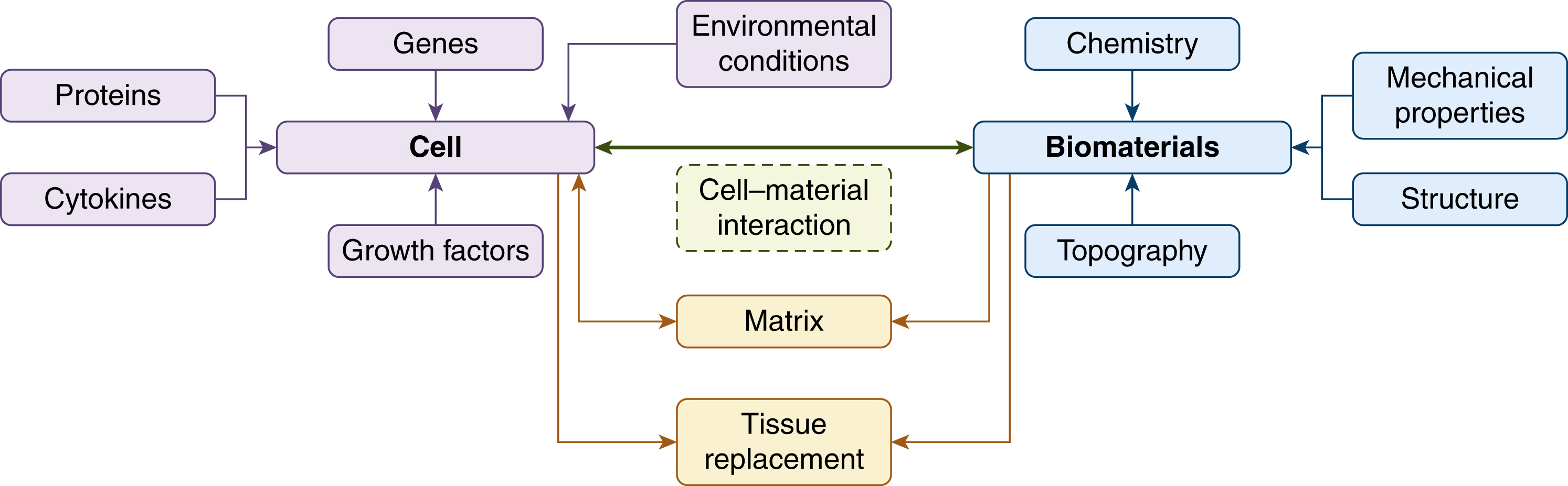
Ultimately, the scaffold should undergo nontoxic degradation as it is replaced by healthy host tissue, rendering a biodegradable scaffold ideal for use in tissue engineering. The cell–biomaterial interface also plays a significant role in the choice of scaffold due to the direct impact on cell adhesion, which is an important step in the survival of anchorage-dependent cells. Both cell proliferation and differentiation can be controlled by functionalization of the biomaterial surface; these interactions between cells and biomaterials have been shown to occur at the nanoscale.
With the advent of nanotechnology, nanoparticles are being merged with synthetic scaffolds to develop nanostructured biomaterials, enhance interaction of proteins that control cell adhesion and thus, tissue formation. Nanocomposites can be defined as multiphase solid materials where one of the phases has a dimension of less than 100 nm. Nanofibrous composite scaffolds are found to decrease immunogenicity, improve the capacity for cell interaction, and to harbor increased concentrations of fibronectin and vitronectin, the adsorption proteins that reduce apoptosis of transplanted cells. Biomaterials can be classified as bioinert, bioresorbable, or bioactive. A bioinert material does not induce a toxic response from the body on implantation and is usually associated with fibrous encapsulation, whereas a bioresorbable material undergoes degradation in the host by hydrolysis, enzymes or osteoclasts. Bioactive materials produce a biological response from the body that results in the formation of a bond between the material and host tissue. The type and site of biomaterial implantation governs a tissue response: hard tissues (bone, enamel, and dentin), soft tissues (breast and ocular implants), vascular implants (stents, heart valves, and vascular grafts) and connective tissue (cartilage, tendons, and ligaments).
A variety of possible scaffold biomaterials are available. Among these are biological polymers (e.g., collagen, elastin, chitosan, silk), biodegradable synthetic polymers (e.g., PGA, PLA, PDLA), synthetic biomimetic compounds (e.g., hydroxyapatite, calcium phosphates), allografts and xenografts or combinations of the above. Geometric variables such as porosity, pore size, and pore morphology are crucial. A porous scaffold provides a large surface area for neovascularization, nutrient and waste exchange, cell migration, and matrix deposition. Scaffolds can be made porous using several different methods. Porogens (i.e., salt or paraffin microspheres) can be incorporated in the polymer and subsequently leached out. Freeze drying of homogenized polymer solvents is another option, in addition to gel casting of organic scaffolds and sol-gel foaming. Surface properties, such as surface topography and chemistry, guide cellular behavior and control protein adhesion. If a biodegradable material is chosen for implantation, the rate of resorption should match the speed at which the body replaces the missing tissue and its degradation byproducts should be nontoxic. Finally, the biomaterial of choice ought to be economically viable and conform to the regulatory bodies.
Autologous Grafts
The word autologous means “related to self” and refers to tissue that has been derived or transferred from the same organism. Autologous grafts or “autografts” can be harvested and transferred from several different tissues in the body, e.g., skin, bone, tendon, cartilage. The focus of this section is on the autologous transfer of skin, adipose tissue, bone, and cartilage.
Skin Autografts
From superficial to deep, skin is composed of epidermis, dermis, and skin appendages. It has several functions, ranging from physical protection, prevention of fluid loss, protection against ultraviolet radiation, regulation of body temperature, sensation, immunological surveillance, to protection from microbiological organisms. Injured skin can heal itself through different phases of wound healing and epithelialization, whereby epithelium is reestablished across a wound. The different phases of wound healing consist of hemostasis, inflammation, proliferation, and remodeling, whereas epithelial repair consists of mobilization and migration of epithelial cells, followed by mitosis and differentiation into stratified squamous epithelial cells. If the injury to the skin is grave or the area of skin loss large, this process may take several months and result in significant scarring. In this instance the use of a skin autograft can aid the healing process. Skin grafts can either be split- or full-thickness. Split-thickness skin grafts (STSG) contain a variable amount of dermis and can be harvested from several sites (i.e., thigh, buttocks, and scalp). The donor site heals through epithelial repair and does not require closure. Full-thickness skin grafts (FTSG) contain the entire dermis and are usually taken from areas of the body with sufficient skin laxity to allow direct closure (i.e., groin, forearm, arm, postauricular and supraclavicular areas).
Skin grafts heal in four phases:
- 1.
Adherence of the graft to its bed via immediately formed fibrin bonds.
- 2.
Serum imbibition. Skin grafts swell in the first 2–4 days after application due to absorption of fluid.
- 3.
Revascularization commences around the 4th day and is a consequence of new vessel formation along new and existing vascular channels in the graft.
- 4.
Remodeling is the process whereby the histological structure of the graft returns to that of the normal skin.
Skin grafts can fail to “take” due to several reasons, e.g., hematoma, infection, excessive shearing, an inappropriate bed, or technical errors such as placing the graft upside down on its bed or allowing it to dry before application.
The main limitation of skin grafts is their tendency to contract. Contraction of grafts is more pronounced with split-thickness skin grafts compared to full-thickness skin grafts.
Adipose Tissue Autografting
Although autologous fat transfer has gained popularity over the past two decades, Neuber first described the technique in the medical literature in 1890s. The injectable fat transfer was first described in the 1920s. In the 1950s Peer reported that fat grafts lose approximately 45% of their weight and mass per year. He also emphasized the importance of a good vascular recipient bed combined with meticulous hemostasis to promote optimal graft survival. Autologous fat transfer can be regarded as the ideal filler. It induces neither host reaction nor a carcinogenic response. Being minimally invasive renders the procedure safe, and fat is also readily available and inexpensive. Limitations include the unpredictable and variable rate of absorption, the need for overcorrection, and the requirement for a surgical procedure.
Donor Site
The most common site is the abdomen due to availability and ease of access when the patient lies supine. Other sites include the flanks, extremities (inner thighs, inner knees, inner arms, trochanteric area) and gluteal area. A study investigating the viability of lipoaspirates in vitro using flow cytometry from three different anatomical sites – abdomen, flank, and thighs – found no difference in fat viability 12 weeks post transplantation. Currently there is no consensus on the best donor site for autologous fat harvest.
Extraction Techniques
Vacuum extraction, syringe aspiration, and direct fat excision are well-described techniques in harvesting fat tissue. Although syringe aspiration is the most popular technique, there exists no current objective evidence to corroborate the benefit of this technique relative to other methods in terms of weight or volume of the fat grafts isolated. However, high negative pressure exerted on adipocytes in conventional liposuction has been reported to cause up to 90% adipocyte rupture. Coleman popularized autologous fat transfer employing the syringe aspiration technique. A 3-mm cannula is connected to a 10-mL syringe and aspiration is achieved through manual suction by withdrawing the plunger.
Infiltration with Local Anesthetic Agents
Some clinicians do not use local anesthetic during fat harvest but the majority inject the donor site with short-acting local anesthetic agents with or without epinephrine. The most commonly used agent is lignocaine with concentration doses ranging from 0.5% to 2% and doses of epinephrine ranging from 1:80,000 to 1:200,000. Coleman recommends using 0.5% lignocaine with 1:200,000 adrenaline. The main clinical advantage of infiltration with local anesthetic combined with epinephrine are reduction of potential donor site pain, blood loss and bruising.
It is uncertain whether the use of lidocaine adversely impacts fat graft survival. In a study by Moore et al an inhibitory effect of lidocaine on adipocyte growth in cell culture was observed. In contrast, Keck et al demonstrated that exposure to a variety of anesthetic solutions increased adipocyte viability in contrast to a dry technique. Furthermore, animal models pretreated with saline or lidocaine and epinephrine have demonstrated no significant effects on fat graft volumes or histological architecture.
Preparation
Centrifugation is a popular method of fat graft preparation. The process separates the nonviable components, including blood, oil, and lidocaine, leaving behind the adipocyte-rich segment for transfer. Coleman advocates 3 minutes of centrifugation of aspirated fat at 3000 rpm. This process separates the fat into a bottom layer of tumescent lidocaine and blood, a middle layer of usable fat graft and a top layer of oil from ruptured adipocytes. Other clinicians advocate the use of centrifugation at different spinning rates and duration.
Rohrich et al have recently challenged the process of centrifugation and found no quantitative difference between the viability of processed and unprocessed fat graft viability. Similarly, Ramon et al utilized a mouse model to demonstrate that fat cell survival after 16 weeks between centrifuged and noncentrifuged fat grafts in terms of weight and volume were comparable, whilst histological analysis showed noncentrifuged fat grafts exhibited less fibrosis.
Washing harvested fat grafts as a form of preparation has also been described, using lactate Ringer’s solution to improve viability. The rational is to decrease levels of inflammatory mediators from the graft and minimize the immune response at the recipient site. However, there is a paucity of high-level evidence in the literature to affirm that washing harvested fat graft improves overall graft survival.
Application
To maximize graft survival the adipocytes must be in close vicinity to a blood supply. Studies have suggested that fat placed within 2 mm of an arterial supply will survive and greater distances risk necrosis and fibrosis. Thus the surface area of the fat graft in contact with a vascular recipient bed is essential to optimize graft survival. The “fanning out” technique is one method of injection that ensures small aliquots of fat graft are spread over a large surface area. Other commonly used injection methods, such as linear threading, serial puncture, and cross-hatching, can also be used to achieve the desired outcome. Slower injection speeds of 0.5–1 mL/s have resulted in greater fat graft survival rates compared to faster injection speeds of 3–5 mL/s. A curved microcannula 2–3 mm in diameter or blunt-tip needle 14–19 gauge is commonly used for autologous fat transfer.
Complications
In general, autologous fat transfer is a safe procedure with a low risk profile. However, patients must be warned about bruising, swelling, contour irregularity, and infection, in addition to fat necrosis and calcification. Rare cases of severe complications such as fat embolism of cerebral artery and retinal artery leading to stroke and blindness, respectively, have been reported in the literature during soft tissue augmentation of nasolabial fold and periorbital area. ,
During the consent process it is important to ensure that the common and rare risks of fat transfer are made evident. Moreover, it important to discuss the limitations of the procedure, with emphasis on the unpredictability of the graft take in regard to partial and complete absorption over time.
The rise in popularity of autologous fat transfer in addressing contour deformity and volume restoration over the past few decades has propelled this technique to the forefront of reconstructive and aesthetic surgery. Despite its widespread adoption, there remains no consensus on the optimal donor site or most effective method of harvest and preparation. Further objective randomized clinical trials are required to guide current variations in practice. However, autologous fat transfer functions as a powerful tool in the armamentarium of plastic surgeons and practitioners must keep abreast of developments in order to ensure optimal graft outcome.
Bone Autografts
The success of bone autografts in the treatment of nonunions is well described. In the tibia, union rates of more than 90% have been reported using iliac crest bone graft at a mechanically stable site. Bone grafting has also been effective in treating recalcitrant and infected nonunions as well as aiding the healing at the docking site of nonunions treated with distraction osteogenesis. In addition to the mechanical properties and volume effect, the biological properties of bone autografts are also advantageous.
Autologous bone grafting has been considered as the gold standard in management of bone defects and nonunions. However, the introduction of a new generation of bone substitutes has challenged this notion, largely due to the morbidity associated with the harvesting procedure with reports of donor site pain, infection, and hematoma of varying incidences between 9% and 55%.
Bone grafts heal by the following mechanisms: (1) adherence to the surrounding tissue, i.e., incorporation, (2) creeping substitution, also referred to as osseoconduction, a process whereby the bone graft acts as a scaffold along which angiogenesis occurs and new bone is formed by progenitor cells, and (3) osseoinduction, the differentiation of mesenchymal stem cells into osteocytes.
Autologous bone grafts possess biological advantages over allografts and synthetic bone grafts as they confer greater histocompatibility as well as an excellent combination of osteogenic, osteoinductive, and osteoconductive properties.
Autologous Cancellous Bone Grafts
The trabecular structure of cancellous bone generates a large surface area. This permits a greater incorporation of cellular components (mesenchymal stem cells and osteoblasts) and provides excellent osteogenic and osteoinductive properties. Additionally, the sizable trabecular surface area facilitates revascularization and integration of the graft at the host site. Conversely, a significant limitation of cancellous bone is its poor mechanical strength during the initial phase. However, increased stability is achieved within months due to the biological capacity of trabecular bone graft to induce new bone formation, once incorporated.
Autologous Cortical Bone Grafts
Cortical bone autografts have a different biological profile compared to cancellous bone autografts. In contrast to the low density of cancellous bone, the dense and highly organized structure of cortical bone provides initial mechanical strength and stability. However this structural integrity compromises the available surface area and constitutes a barrier to vascular in-growth and remodeling during graft incorporation. Furthermore, the subsequent lack of cellularity and growth factors diminishes the osteogenic potential of cortical bone grafts. The integration of cortical bone is mediated through osteoclastic activity resulting in resorption of the cortices and loss of bone. This results in transient weakness with reduction of mechanical strength of up to 75%.
Autologous Vascularized Bone Grafts
Autologous vascularized cortical bone grafts have favorable biological attributes relative to standard autologous nonvascularized autografts. In addition, they are mechanically superior during the initial 6–12 months after the grafting procedure and offer a greater provision of nutrients to deep structures of the graft. Despite their many advantages, they carry an increased risk of donor site morbidity and can be more technically challenging during harvest and implantation, requiring both orthopedic input and microvascular skills. It has been proposed that more than 90% of the osteocytes may survive transplantation if patent vascular anastomoses and stability of the graft are achieved. Osteoneogenesis by graft and host can lead to expeditious incorporation of the graft, whilst the residual weakness of the construct remains minimal.
Due to the advantageous and cost-effective combination of biological and mechanical properties, bone grafts continue to be important tools in the management of bone defects and nonunions.
Cartilage Autografts
Cartilage does not possess the regenerative capacity of bone or other connective tissues due to its lack of vascularity. Cartilage is composed of chondroitin sulphate, a gelatinous ground substance, into which collagen and elastic protein fibers are incorporated. It is flexible, very durable, and resistant to compressive forces. There are three types of cartilage: hyaline, elastic, and fibrocartilage. The most abundant type is hyaline, found in the nose, ear, trachea, larynx, smaller respiratory tracts, costal cartilages, and the articular surfaces of synovial joints. It is the precursor to bone in most of the embryonic skeleton. Injury to or congenital deformity of cartilage can result in pain, loss of mobility, and significant deformity. The management of moderate to severe cartilage defects is challenging due to the lack of intrinsic ability to self-repair.
Autologous Cartilage Grafts
Autologous cartilage grafts are commonly used in the reconstruction of congenital ear deformities, augmentation or revision rhinoplasty, and nipple reconstruction post mastectomy. They can be harvested from the costal cartilages, septum of the nose, and the ear. Autologous cartilage grafts can provide excellent results, but are associated with significant donor site morbidity and the final aesthetic result can be dependent on the carving skills of the operating surgeon.
Osteochondral Autograft Transfer
When reconstructing injured joint surface cartilage, transplants of cylindrical osteochondral plugs can be harvested from non-weight-bearing portions of the joint and transferred to the site of the defect. Osteochondral autograft transfer (OAT) offers the prospect of restoring the defect with true hyaline articular cartilage with greater wear characteristics and joint function. Furthermore, this technique can be performed in a single procedure using reusable equipment without the requirement of external laboratory assistance.
A recent 10-year follow-up study showed superior clinical results with OAT compared with microfracture in young athletes with focal osteochondral defects. Several studies have corroborated similar improvements in both pain and function in patients with osteochondral defects of the knee.
One of the primary limitations is defect size, as the technique is best suited to small and medium-sized focal chondral defects . Much larger lesions up to 9 cm 2 require multiple osteochondral plugs and carry the risk of greater donor site morbidity. Other disadvantages include limited donor site availability and a size mismatch between the donor and recipient site.
Autologous Chondrocyte Implantation
Autologous chondrocyte implantation (ACI) aims to provide hyaline cartilage for repair of injured articular cartilage. This cell-based technique takes place in three stages: (1) 200–300 mg of cartilage is harvested arthroscopically from a lower weight-bearing area (intercondylar notch, superior ridge of the femoral condyles); (2) chondrocytes are isolated after enzymatic removal of the matrix, grown in vitro for 4–6 weeks, and then (3) applied to damaged areas during a second procedure.
ACI holds the advantage of treating larger lesions (up to 10 cm 2 ) with articular hyaline cartilage with minimal donor requirements. A clinical study of ACI demonstrated long-term improvement of symptoms in patients with isolated femoral condyle lesions and osteochondritis dissecans at 5–11 years after treatment. Similar results were reported 10–20 years after ACI, where 74% of 224 patients described symptomatic improvement.
Despite promising reports, ACI is technically more challenging and carries associated risks of graft failure, hypertrophy, and subsequent high reoperation rates of between 9% and 20%. It is also dependent on in vitro expansion of chondrocytes, relying on external laboratories and higher costs. This theoretically also increases the chance of introducing infection, whilst also necessitating two operations.
Allografts and Xenografts
Allografts can be defined as graft tissue obtained from a donor of the same species as the recipient, but comprised of a different genetic make-up. The absence of donor site morbidity, general successful outcomes, and decreased surgical time render allografts a popular alternative to autologous grafts. In recent years, natural ECM have been procured, processed, and marketed for clinical use as patches to reinforce tissue repair.
Bone Allografts
Bone allografts can be applied to aid healing of bone injured either by trauma or secondary to iatrogenic causes. Survival of bone grafts depend on systemic factors that also affect wound healing, e.g., age, nutritional status, immunosuppression, diabetes, obesity. Scarring, infection, and previous radiotherapy at the site of the recipient bed can also adversely affect graft survival.
Allograft preparation requires treatment with ethanol in order to remove cellular material, followed by gamma irradiation for bacterial, fungal, and viral eradication. However, allografts lose some of their biological properties through the sterilization process and synthetic bone substitutes have only osteoconductive properties if combined with human growth factors or autograft.
Fascia Allografts
An engineered fascial device may be utilized for ligament repair, extension of rotational muscle transfers, grafting lacerated muscles, periosteal coverage, and wound healing. Fascia lata or fascia from the iliotibial tract are most commonly used and have several advantages: biocompatibility, ability to be replaced by the host tissue, good mechanical properties, and ease of handling.
An in vitro study comparing the histological features of allograft iliotibial bands with tibialis anterior tendons reported that they shared a similar structure composed of aligned collagen fibers. Moreover, they observed that allograft iliotibial bands were superior in regard to creep, stress-relaxation, and viscoelastic properties. In addition, allograft iliotibial bands also demonstrated low cytotoxicity with a higher propensity to favor cell attachment and infiltration over time.
Dermal Matrices
Integra
Integra (Integra LifeSciences Corp., Plainsboro, NJ, USA) is a dermal regeneration matrix consisting of bovine collagen, chondroitin-6-sulphate and a silastic membrane. It has gained widespread use in the clinical treatment of deep partial-thickness and full-thickness burn wounds, full-thickness skin defects, and chronic wounds. The bovine collagen dermal component integrates with the host and the temporary epidermal silicone is peeled away as the dermis regenerates. A very thin autograft is then grafted onto the neodermis.
Integra has been successfully used in reconstructive surgery to augment soft tissue coverage over structures such as bone or tendon. However, it requires a two-stage procedure prior to sufficient defect coverage to allow for adequate vascularization of the template. Integra provides immediate donor site coverage independent of wound size. Additionally, it creates a smooth gliding surface between underlying structures, e.g., paratenon or nerves and STSG. Long-term outcomes after radial forearm flap donor site coverage with Integra and STSG show successful functional and aesthetic defect coverage. A similar long-term study also reported improved scar cosmesis and skin elasticity after comparing Integra combined with STSG relative to STSG alone in acute burn treatment. However, disadvantages to its use include cost, infection risk, and foreign body reaction.
AlloDerm
AlloDerm (LifeCell Corporation, Branchburg, NJ, USA) is fabricated by decellularizing cadaveric skin. After processing, the skin is reduced to a basement membrane and correctly aligned dermal collagen matrix that will theoretically not be rejected. During host integration, it is invaded by fibroblasts, and undergoes neovascularization and neoepithelialization, without evidence of rejection or adverse reaction to the implant from the host.
AlloDerm has been shown in clinical trials to be well tolerated. It has been used extensively in burns surgery to provide biological coverage of full-thickness burns and as a framework for split-thickness skin grafts and for intraoral mucosa resurfacing after fistula repair. Its ability to support neovascularization and tissue ingrowth permits persistent volume correction. This gives AlloDerm an advantage over other injectable forms of collagen. A significant drawback of AlloDerm sheets are the requirement for an incision for placement. An injectable form of AlloDerm (Micronized AlloDerm) has been created by homogenizing AlloDerm sheets cut into strips. The product comprises of injectable-sized particles of AlloDerm that maintain the ultrastructure of the dermis and can be introduced easily through a 26-gauge needle.
Several large published series of patients undergoing breast reconstruction utilizing acellular dermal matrices have reported low rates of capsular contracture. , In addition, animal studies have corroborated the absence of a capsule around implants where human acellular dermal matrix was placed, which was both observed clinically at the time of implant exchange and also confirmed histologically from biopsies.
Strattice
Strattice (LifeCell Corporation, Branchburg, NJ, USA) is an acellular reconstructive tissue matrix derived from porcine dermis. It undergoes proprietary processing that removes cells and reduces the risk of xenogeneic rejection response. The presence of the α-Gal epitope on porcine tissues has been associated with rapid rejection. The enzymatic processing of Strattice significantly reduces the amount of α-Gal epitope in the absence of damage to the matrix integrity (collagen and elastin architecture).
Since its introduction in 2008, Strattice has been utilized in wide array of reconstructive procedures including primary breast reconstruction, breast revision surgery, and in abdominal wall reconstruction and hernia repair.
Performance of Strattice has been evaluated by implantation into the abdominal wall of a primate excisional repair model. The animals tolerated the implants well and exhibited no clinical signs of inflammation, laxity, hernia or visceral tissue adhesion. Immunological evaluation of cell infiltration demonstrated an early, mixed cellular inflammatory response at 2 weeks, which was transient and diminished to baseline levels by 3 months. A study comparing three non-cross-linked, commercially available extracellular matrix scaffolds (Strattice, Veritas, and XenMatrix) showed that the intrinsic denaturation temperature for Strattice, Veritas, and XenMatrix was 51°C, 38°C, and 44°C, respectively. In addition, the susceptibility to collagenase degradation was increased in Veritas and XenMatrix. These findings suggest a greater number of extracellular matrix modifications in the Veritas and XenMatrix scaffolds. In an in vivo primate model, Strattice permitted cell repopulation and was remodeled over 6 months. In contrast, Veritas was unstable at body temperature, absorbed rapidly and associated with moderate inflammation whilst XenMatrix caused severe inflammation.
The use of Strattice for repair of incisional hernias in Yucatan pigs compared to primary closure demonstrated that primary closure resulted in greater scarring and bone hyperplasia along the incision line. In contrast, Strattice elastic modulus and tensile stress were similar to fascia and it facilitated a more complete wound healing response relative to primary closure.
Clinically, Strattice may enhance soft tissue support in patients with poor quality mammary soft tissue support, especially in implant-based breast reconstruction post-mastectomy patients and in those undergoing augmentation mammoplasty. Furthermore, it has been associated with fewer implant-related complications, including capsular contracture, rippling, palpability, and malposition.
Alloplastic Materials
Metals
An ideal metal implant possesses the characteristics of biocompatibility, high tensile strength and resistance to corrosion. Advantages of metallic biomaterials include lack of donor site morbidity and widespread availability. Disadvantages include eliciting a host reaction and their considerable cost. Metals are routinely used in fracture fixation (plates, screws, and wires), cranial plates, and artificial joints. Most metals used as biomaterials are alloys. An alloy is a mixture of two or more metals into a single material. Commonly used alloys include the following.
Stainless Steel
Stainless steel is an alloy composed mainly of iron, chromium, and nickel. It is highly corrosion-resistant in the physiological environment compared to other formulations. Chromium forms a strongly adherent surface oxide layer which renders it resistant to corrosion. Despite the favorable anticorrosive properties of stainless steel, the leaching of metallic ions can lead to inflammatory reactions in surrounding tissues and cause pain.
Cobalt Alloys
Vitallium is an example of an alloy composed of cobalt, chromium, and molybdenum (Co–Cr–Mo). This alloy has similar strength to stainless steel but is lighter and less resistant to corrosion.
Titanium (Ti)
Titanium has the capacity to form direct chemical bonds with mineralized bone matrix. This “osseointegration” allows for robust incorporation between soft tissue as well as between implant and bone, and confers significant utility in prosthetic reconstruction. In the first stage the titanium implant is placed into bone and covered by periosteum or bone for a minimum period of 3 months. Subsequently, during the integration process the histological appearance shows calcified matrix in direct apposition to the metal surface without intervening fibrous tissue. It is this interaction which provides unparalleled stability and endows great mechanical strength to titanium implants.
Maxillofacial prosthetics largely rely on the support of titanium osseointegrated implants and titanium microplates are routinely use for bony internal fixation. Combined with aluminum and vanadium alloy, titanium becomes less malleable. There is also less scatter artifact on radiographic images compared to cobalt alloys and stainless steel.
Gold
Gold (Au) has limited applications in the field of plastic surgery. Although it is resistant to corrosion, it lacks sufficient mechanical properties and is costly. It still serves a purpose in facial palsy patients where it is utilized to create iatrogenic ptosis through gold loads placed on the upper eyelid to address lagophthalmos. However, a recent review reported fewer complications of infection and capsule formation with the use of platinum due to its higher biocompatibility relative to gold.
Ceramics
Ceramics are solid materials composed of inorganic, nonmetallic substances. Chemically, they are formed through a combination of hybrid covalent and ionic bonds. The atomic repeating order can be short-range (e.g., glasses) or long-order (e.g., crystals). Advantages of ceramics are that they can be chemically inert in numerous environments or designed to be bioactive in the body with a composition that can mimic the mineral component of bone. They are excellent heat and electric insulators whilst also being corrosion-resistant. In addition, they are hard and brittle (fracture without deformation), strong in compression, but weak in shearing and tension. Examples of ceramic materials are porcelain, cements, pottery, and glasses (amporphous).
Bioinert Ceramics
These include oxide ceramics (Al 2 O 3 , ZrO 2 ), silica ceramics, carbon fiber, and synthetic diamond. Alumina (Al 2 O 3 ) and Zirconia (ZrO 2 ) are the two most commonly used structural bioceramics. They are primarily used as modular heads on femoral stem hip components, but can also be used for dental restorations including in crowns and bridges. They undergo less wear than metal components, and the wear particles generated are generally better tolerated. In addition, they have good mechanical and aesthetic properties.
Bioactive Ceramics
These include tricalcium phosphates, synthetic hydroxyapatite substitutes and bioactive glass (e.g., 45S5 Bioglass). Bioactive glass has osteoproductive characteristics and encourages bone cells to produce matrix. When implanted it binds to soft tissue and hard tissue to form a porous matrix that facilitates activation and infiltration of osteoblasts.
Bioglass (composition = 46.1% SiO 2 , 24.4% NaO, 26.9% CaO, 2.6% P 2 O 5 (mol %)) was the first material observed to form an interfacial bond with the host tissue. This bone-binding property is underpinned by accumulation of silicon ions upon implantation and the subsequent formation of a hydroxyapatite coating on the surface of Bioglass. The strength of the bond between Bioglass and cortical bone has been shown to mimic the strength of native host bone.
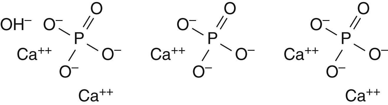
Hydroxyapatite (HA) and tricalcium phosphate are the most commonly used ceramics in plastic surgery. HA, the coral of genus Porites , has a calcium carbonate exoskeleton similar to bone. Through a hydrothermal exchange process of carbonate for phosphate, it adopts a structure identical to bone. The material is available in the form of blocks or granules and can bond to bone without evoking an inflammatory response. It provides a scaffold for bony ingrowth and is not resorbed ( Fig. 3.2 ). A recent study evaluated a hybrid tissue-engineered bone construct of nanocrystalline HA. The authors demonstrated that when the construct was seeded with osteoblasts, the scaffold with HA enhanced cell proliferation and promoted production of mineralized extracellular matrix greater than the plain scaffold.