Introduction
Flap surgery is the commonest procedure in plastic surgery and is the essence of the discipline. Critical to its success is a sound understanding of soft tissue’s blood supply, its compliance, and its mobility. As all flaps “rob Peter to pay Paul,” it is also about conceptualizing the secondary defect and minimizing its consequences. The art and craft of plastic surgery necessarily requires an esthetic sense and experience.
Evolution of Flap Surgery
For almost a century, Manchot (1889) , and Salmon’s (1936) detailed studies of the skin’s vascular design were unknown to clinicians. With limited understanding and the simplistic belief that the skin’s blood supply was based on a random distribution in the horizontal plane, local flap surgery was unpredictable and its progress curtailed by an adherence to dogmatic rules such as length-to-width ratios and the superiority of proximal over distally-based flaps. A generation of surgeons failed to appreciate the simple observation that circumferentially incising large skin lesions in the process of their elevation and removal did not compromise their circulation. The explanation of course was that their blood supply was derived from the depths and not horizontally. Surgeons no doubt were aware of the circulation from below, but the reality was that sufficient numbers of these had to be divided to permit the flaps to transpose or rotate, and ultimately it was the base, fed by the horizontal input, that was the critical lifeline. Not until it would be shown that flaps could be completely islanded and still live was it possible to move flaps based on these deeper vessels. Milton (1970) elegantly highlighted the fallacy of the length-to-breadth ratio using pig studies to demonstrate the existence of arterialized zones of the integument that would survive over extreme lengths, even if completely islanded, provided they retained their arterial source at their base. These exciting findings breathed life back into the clinical study of the soft tissue’s blood supply. True to the adage that history is written by the victors, so too was the blinded acceptance of random pattern proposed by Gillies, derived in part from the neglect of the publications of the Dutchman Johannes Esser (1917), Gillies’ plastic surgery counterpart for the German army in World War I. Instead of the complex tube pedicle (Webster 1937), Esser performed one-stage “arterialized biological island flaps” based on the palpable arteries of the head and neck region. He clearly recognized the fundamental concepts of the flap’s axial blood supply. Furthermore, Gillies was very aware of his work but publicly pronounced it “not up to the standards expected of the British and American people.”
The first clinical application of the new “axial pattern” concept was the groin flap (McGregor 1972), based on the superficial circumflex iliac branch of the femoral artery. The wide arc of rotation of this very long and narrow-based flap of like tissue expanded the single-stage reconstructive options for the regional wounds previously manageable only by multistage transfers requiring protracted immobilization and hospitalization.
What had been anecdotally reported in the early literature and had been empirically adopted in practice, the Indian forehead flap for nasal reconstruction and the epigastric flap in the lower abdomen (Shaw 1920), now made sense. The former flap unwittingly captured the supratrochlear/supraorbital vessels and the latter the superficial epigastrics.
With this new awareness, omentum although not skin, was quickly recognized for its application as an arterialized flap by virtue of it wearing its blood supply on the outside of its surface. The vasculature could be pruned to critical arterial pedicles and tunneled from the abdominal cavity to cover far-flung defects and then skin grafted where needed ( Fig. 2.1 ).
Other fundamental concepts of skin blood supply were soon elucidated. The myocutaneous flaps were described, where the skin relied for its blood supply on the multitude of small vessels perforating through the underlying muscle. Provided the muscle was raised on its blood supply, the overlying skin would survive, even when completely islanded. As muscles typically are vascularized often by a single source at their origin the muscle pedicle added even further length to the arc of flap rotation. The gracilis (Orticochea 1972) and latissimus dorsi myocutaneous flap (Olivara 1976), , reinventing the earlier findings of Tansini (1906), were the first to be described and widely adopted. Muscle flaps without skin followed and the anatomical articulation of their vascular basis further expanded the options for one-stage locoregional reconstructions (Mathes and Nahai 1981) ( Fig. 2.2 ).
Fasciocutaneous flaps recognized for the first time that a significant contribution to skin blood supply was in the plane of the deep fascia and provided this fascia was included within the flap, large areas of previously unreliably vascularized skin would survive (Ponten 1981). This particularly applies to the lower limb. Initially they were designed on the assumption their vascularity was in the plane of the fascia and in the limbs, flaps were based proximally to capture its source. It was soon clear that much of this fasciocutaneous blood supply derived from perforating branches of named deep vessels, emerging vertically between septofascial muscle planes from deep axial vessels and furthermore that these zones could be completely islanded from their proximal connections.
This led to the unifying concept of angiosome blood supply (Taylor and Palmer 1987), where the tissue is considered as a three-dimensional territory or somite structure akin to vertebrate embryological development where not only skin, but whole mesenchymal somites including skin, muscle, and bone, have a vascular zone. This was supported by meticulous cadaver studies and expanded the earlier work of Manchot and Salmon.
Concurrent with this explosion in the understanding of the skin’s blood supply, advances were being made in microvascular surgical techniques and instrumentation initially by the neurosurgeons, (Jacobson and Suarez 1962) and quickly adopted for plastic surgery. Microsurgery was a technical disruptor of its time and had immediate application for replantation. Subsequently the transplantation of toes and territories of skin by anastomosing specific vessels flourished. The first such flap was an omental transfer to the scalp (McLean and Buncke Jr 1972). Skin flaps soon followed (Taylor and Daniel, 1973; O’Brien et al 1973 ). More recently, supermicrosurgery has provided surgeons with greater confidence to explore free, pedicled, and ‘freestyle’ perforator flaps (Koshima 1997), reappraise lymphatic surgery and the impetus to further examine the microvasculature anatomy (Saint-Cyer et al 2009 ; Imanishi et al 2000 ; Narushima et al 2018 ).
Physiology and Anatomy of Skin Blood Supply
Flap surgery involves the transfer of tissue with its vascularity preserved. Necessarily this requires an understanding of the physiology and anatomy of the integument’s blood supply.
The blood supply (15 mL/min per 100 cm³) to the skin, greatly exceeds its metabolic needs because of its homeostatic role in thermoregulation and systemic circulation. This explains the relative robustness of skin flaps.
Perfusion of the skin’s capillary beds is regulated by complex and incompletely understood local and systemic neurohumoral mechanisms. These act on precapillary sphincters and arteriovenous anastomoses influencing the filling and emptying of the dermal plexuses and in turn not just the circulation of the skin and subcutaneous tissue, but also insensible heat loss and venous return to the heart. Practically, it follows that the patient following flap surgery should be kept warm, well perfused, and pain-free to maximize the circulation to the peripheries. Technical factors largely underpin successful flap surgery. Clinical experience suggests that perforator flaps become hyperperfused following circumferential incision of the skin; leeching can temporarily alleviate venous congestion and topical vasodilators may beneficially alter the intrinsic blood supply of a flap. Smoking on the other hand is known to cause vasoconstriction in small vessels.
The circulation of the skin and its underlying structures consists of a non-uniform three-dimensional continuous vascular network. For the purposes of skin flap surgery, conceptualizing this vascular network as consisting of horizontal and vertical components is useful.
Running parallel with the skin, and constituting the horizontal component of the skin’s blood supply, are the numerous vascular plexuses. Most important amongst these are the subdermal plexus and the deeper suprafascial plexus. Where no deep fascia exists an equivalent structure such as the panniculus carnosus (limited to the platysma, palmaris brevis, and the dartos muscles in man) serves a similar purpose. These are the vascular bases of (random) cutaneous and fasciocutaneous flaps.
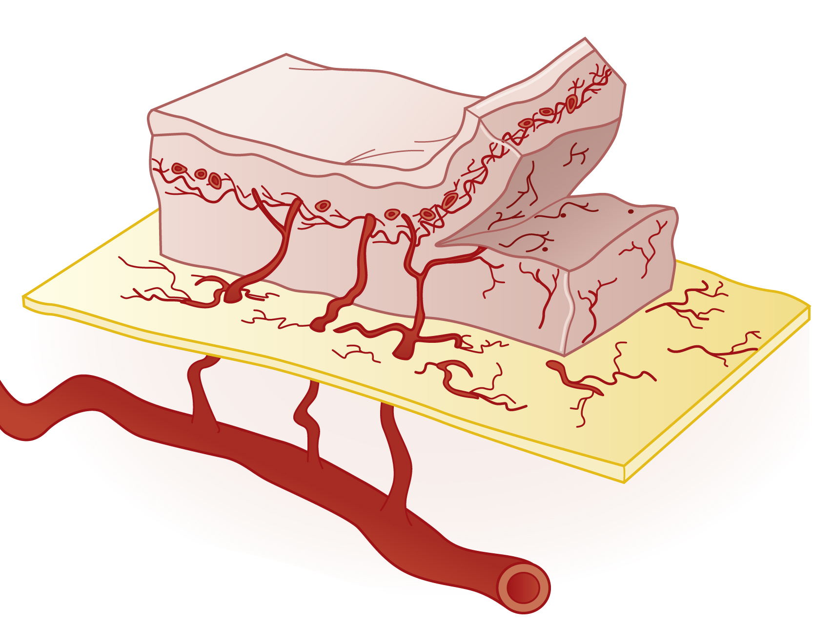
Vessels arising vertically from their source arteries either directly supply the skin, or indirectly supply the skin after nourishing deeper structures such as muscle, tendon, and bone. These are known as “perforators” and they arise from the deep named vessels, axially in the limbs and perpendicularly in the trunk, and follow the dermatomes. They occur in their highest density over the less mobile soft tissue that is adherent to underlying septa; this is particularly evident in the limbs. Perforators anastomose initially with the prefascial plexus before continuing on to the subdermal plexus and are the vascular pedicles on which islands of skin and other soft tissue components may be based . It follows that separate islands of skin and fascia each based on a separate perforator but ultimately deriving from a common arterial axial vessel can be raised on this common axis and permit complex reconstructions with multiple independently oriented flaps.
Renewed interest in the hundreds of cutaneous perforators throughout the body has seen a renaissance in local flap surgery. Based on single perforators, perforator flaps offer greater flexibility to utilize the local tissues and achieve better like-for-like reconstructions all the while minimizing donor morbidity. Crucially, the size of the perforasome remains unknown. Relevant factors include perforator size, direction, density, orientation, and linkages with adjacent perforators and cutaneous nerves, as well as hyperperfusion of the skeletonized perforator , ( Fig. 2.3 ). The blood supply of the skin and its underlying structures can also be divided into vascular territories, or angiosomes , each one being interconnected with its adjacent territory by bi-directional arterioles, the direction interchangeable and determined by the relative pressure in each territory. The angiosome is the vascular basis for composite flaps.
Drainage of the skin is by a reciprocal three-dimensional venous network of avalvular bidirectional veins with a dominant subdermal component. This in turn drains into large caliber subcutaneous veins or venae comitantes that run with perforators. Superficial lymphatics follow subcutaneous veins and deeper lymphatics follow arteries.
Venous flaps are generally transferred as free microvascular flaps where the flap is elevated superficially with only the venous system thereby obtaining a thin flap with little morbidity. The flap is arterialized through its veins by anastomosing them to an artery in varying configurations, the tissue being nourished by retrograde perfusion. There is a small but significant body of work suggesting that these flaps may have a wider application than previously thought. Central to this modification of venous flaps to shunt restricted arteriovenous flaps. There are many classification systems based on efferent, afferent vessels, as well as shunt site.
Indications for Skin Flaps
Skin defects should ideally be repaired by replacement of what is missing and generally this will include fat, as well as skin. In many cases the local laxity of skin will allow direct closure and conversion of the deformity to a linear scar. This should be planned to lie within relaxed skin tension lines (RSTL) of Langer to minimize wound tension, optimize healing, and esthetic outcome. In some instances, although the wound may technically be closed directly, direct closure is undesirable. Excessive tension predisposes to unreliable healing and/or a contour “dish” deformity with dog ears; elliptical wounds tend to lengthen, as they are closed and may distort sensitive adjacent structures. This is a particular issue on the face, where numerous overlapping esthetic and functional subunits lie in close proximity to each other. In other instances, axially oriented linear scars predispose to joint contractures. Here, well designed local flaps from redundant areas can redistribute the tension so as to preserve available tissue, restore normal contour, and minimize the morbidity of the scar.
Though all wounds will eventually heal by secondary intention, infection risk, the need for prolonged wound care, and scar contracture limit this approach to only small convex defects. Defects that cannot be closed directly will need grafting or replacement with flaps. Skin grafts take by engaging with the vasculature of the underlying bed. They are inappropriate in avascular circumstances (exposed bone, tendon, fracture sites, irradiated tissue, and mobile beds) and here flaps are required as they possess an independent blood supply. Grafts often contract and may be a poor color match, and where the underlying fatty tissue is missing, result in contour defects and adherence to deeper tissues. Apart from burns and extensive injuries, where large areas of skin are required, it is generally accepted particularly with the wide range of flap options available, that skin grafts are inferior to flaps and are rarely the first choice. Other exceptions include the dorsum of the hand and foot where thin skin is required and few flaps meet these needs. Full-thickness skin grafts have an important place on the face where the tissues are thin with little subcutaneous fat (eyelids, inner canthus, proximal nose, and sometimes nasal tip). Flaps from adjacent regions to these sites are invariably too thick and may distort adjacent aesthetically sensitive subunits.
Elsewhere, flaps are generally indicated rather than grafts. Small defects are closed by local flaps from the immediate area and find their greatest expression in the head and neck region, particularly in association with skin cancer resection. Flaps may be in-continuity (transposition, rotation) or islanded (advancement) and their execution defines the quality of the plastic surgeon. Because of their near-perfect tissue match, the well-executed local flap may be difficult to spot. Larger defects will require locoregional flaps from a more distant site and are most likely transposed on their narrow base to maximize their reach (arc of rotation). Because their skin texture, color, and thickness may not match that of the original defect, they may require subsequent revision surgery. Such distant flaps may be fasciocutaneous, myocutaneous or muscle flaps with skin grafts. Composites of tissue may be included, muscle or tendon, fascia or bone, and allow functional reconstruction of complex defects. Their vascular basis will be on the vascular pedicle alone or together with their associated skin, muscle or fascial carrier respectively. Usually the secondary defect will closed directly.
Though beyond the scope of this chapter, very large skin or complex defects where a specialized tissue is needed, such as functional muscle or bone, innervated or hair-bearing skin, then free flaps are indicated. They are based on a vascular pedicle and require microvascular anastomosis. Prefabricated (arterialized zone of specialized skin created by the implantation of a vascular pedicle so as to render it transferable as a free flap suitable to match a specific defect) and prelaminated flaps (the neovascularization of composite tissue around a vascular pedicle for subsequent transfer) are more sophisticated free flap indications. Tubed pedicle flaps are now rare, but still find applications where there is an absence of recipient vessels at the defect site. The Indian forehead flap is a surviving example.
Design and Application of Flaps
Two concurrent considerations are critical to the successful planning of local flaps: (1) blood supply and (2) mobility of the adjacent tissue (laxity) ( Table 2.1 ).
Related posts:


Stay updated, free articles. Join our Telegram channel

Full access? Get Clinical Tree


