Fig. 45.1
Patient at admission, presenting a 90% TBSA full-thickness burn injury
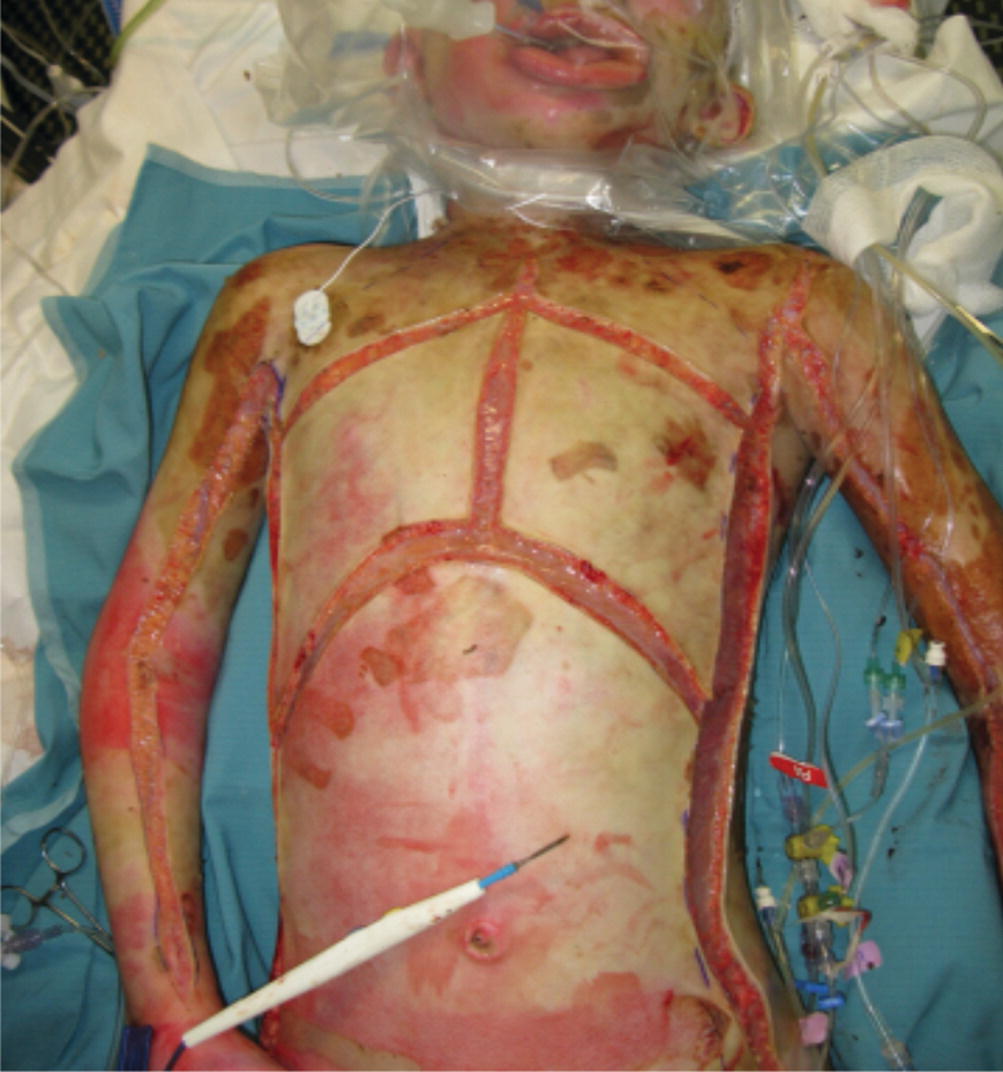
Fig. 45.2
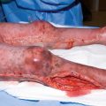
Multiple escharotomies were done upon admission
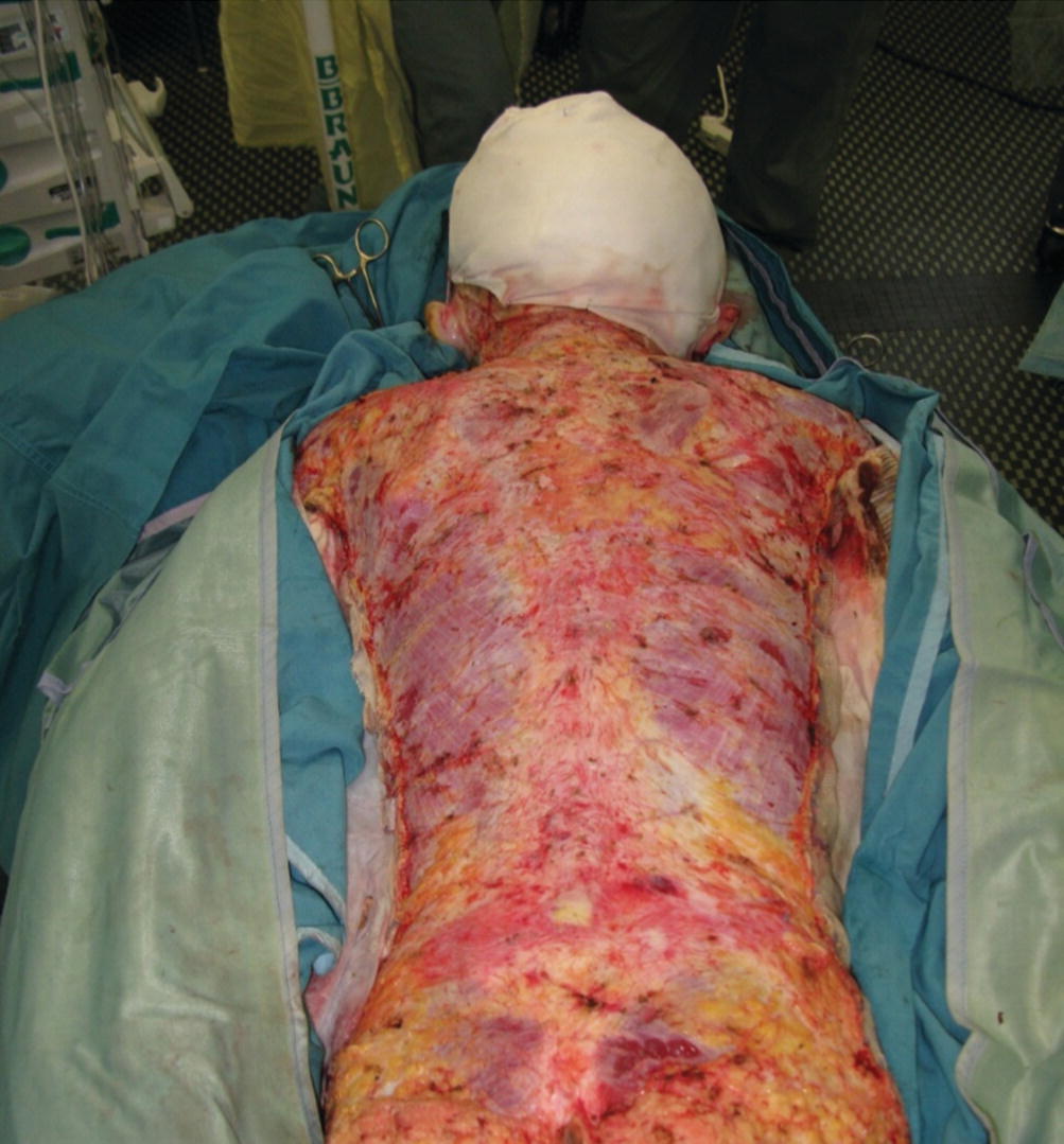
Fig. 45.3
Early debridement of this very deep burn injury down to the fascia
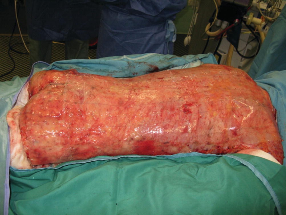
Fig. 45.4
Posterior trunk was first covered with ADM (Integra) meshed in a 1 to 1 ratio (PBD 8)
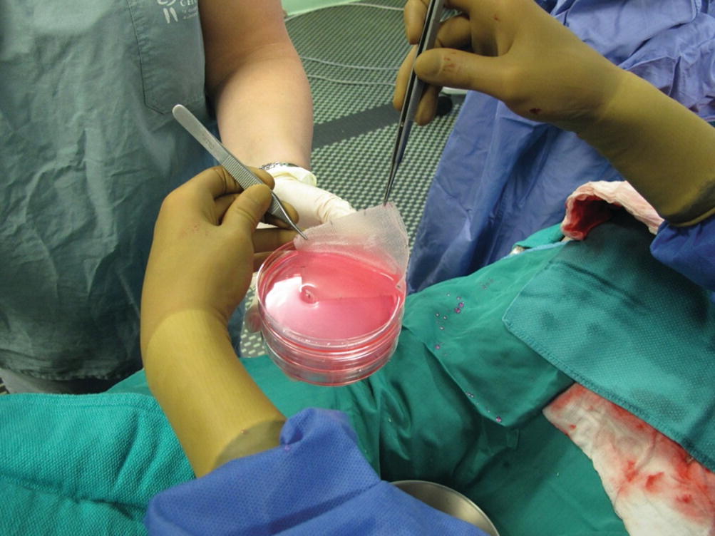
Fig. 45.5
CEA
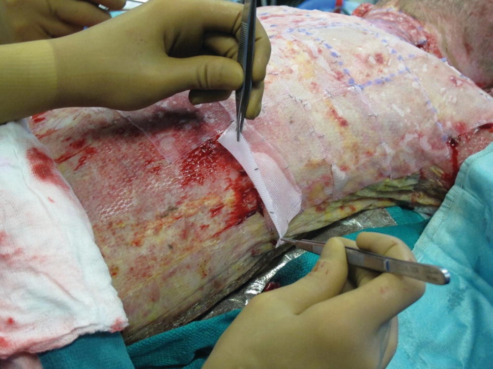
Fig. 45.6
Cultured epithelial cells (CEA) were put in place to substitute the silicone membrane (PBD22)
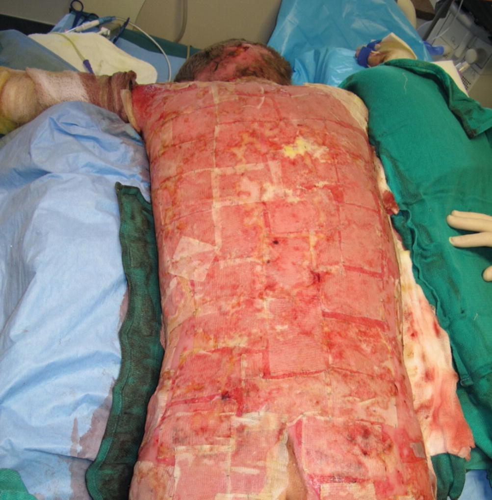
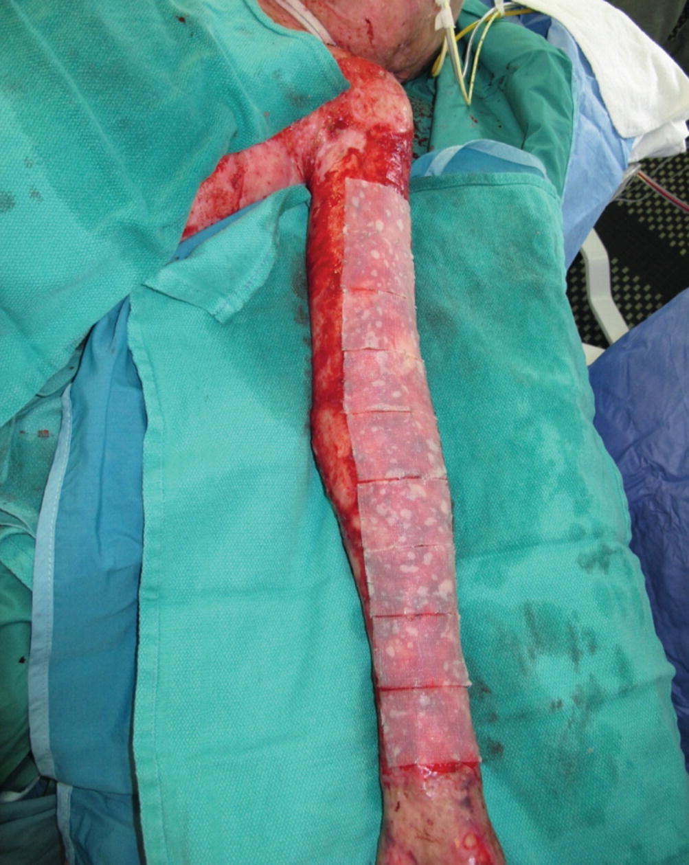
Fig. 45.7
Patches of the ADM-CEA construct showed unstable epithelial coverage in some areas
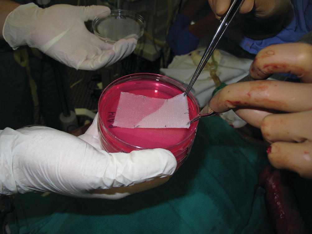
Fig. 45.8
SASSs
Related posts:
 of Burn Patients to the Burn Center Including Burn Wound Evaluation
 Care of the Burned Patient
 Management of Facial Burns, Acute Versus Long-Term, Surgical Versus Non-surgical Face Transplant
 Management of Burn Patients and Fluid Resuscitation
 Improvement in Burn Care
 Necrolysis Spectrum from Basic Theory to Practice Essentials
Stay updated, free articles. Join our Telegram channel

Full access? Get Clinical Tree


