2 Assessment of the Normal and Impaired Lower Extremity
Summary
Before embarking on any lower extremity treatment intervention, not only must an accurate hands-on physical examination be performed but an in-depth history should first be obtained, as often the patient may be the best source of factual information. A complete knowledge of the normal lower limb anatomy and physiology is imperative before attempting to evaluate the impaired lower extremity. Often bone and soft-tissue compromise occurs simultaneously, and thus an orthoplastic approach is preferable to solve not just limb but also life-threatening issues like crush or degloving injuries, as well as the risk of a compartment syndrome. The omnipresent risk of osteomyelitis and the high frequency of diabetic foot wounds today, as with all impaired lower extremities, require a multidisciplinary treatment plan.
Keywords: Gustilo–Anderson classification, orthoplastic approach, compartment syndrome, degloving injury, crush syndrome, Cierny osteomyelitis classification
2.1 Introduction
A comprehensive history as well as a hands-on physical examination is always necessary for the proper assessment of what may be the normal, and definitely for an impaired, lower extremity. Each unit needs to develop a systematic process to achieve this evaluation that is routinely followed to facilitate efficiency and completeness. The impaired lower extremity will, in addition, need to be observed for any acute and chronic complications that may have arisen, whatever the etiology. The complications may include lower extremity fractures, identification of surgical emergencies such as compartment syndromes, the possibility of a crush syndrome, and degloving injuries. A description of the pathology expected with chronic diseases, and in particular osteomyelitis and the diabetic foot, will complete this introduction.
2.2 Assessment of the Normal Lower Extremity
Physicians should have at the least a personal routine for the assessment of the lower extremity to ensure a thorough and focused examination is performed in a timely manner. This should be guided by the history, if available, and requires a complete understanding of lower extremity anatomy, biomechanics, and neurovascular supply as is apropos. In this context, the examination should include assessment of the skin; soft tissues; and the vascular, neurological, and musculoskeletal systems. Selection of appropriate bedside tests as well as more involved investigations to aid with this assessment and an exact diagnosis will be important. An understanding of when and why different imaging modalities should be used such as plain radiographs, computed tomography (CT), magnetic resonance imaging (MRI), ultrasound, and the audible Doppler is imperative. The choices should be made efficiently and timely without redundancy, as extraneous studies in the presence of a critical or life- or limb-threatening injury may lead to a delay in transfer to the operating room which could compromise not only the immediate management of the patient but also ultimate outcome.
2.2.1 Lower Extremity Anatomy
Humans are bipedal; so in the erect position, full weight bearing will be on the lower extremity.1 The fibula and tibia are the bony support of the leg with the latter sustaining 85% of the weight-bearing forces necessary to maintain ambulation.1 This is an important consideration when examining the lower extremity and then selecting potential reconstructive options. The tibia articulates with the femur at the knee joint and joins with the fibula to articulate with the talus to form the ankle joint. The fibula, as does the tibia, serves as a bony surface for multiple muscle and fascial attachments important for integrated locomotion.1
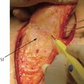
The leg itself is divided into four muscle compartments: anterior, lateral, and posterior—which is again subdivided into a superficial and deep compartment. Knowledge of the location of these compartments and anatomical markers to facilitate their decompression when indicated is essential (▶ Fig. 2.1). The anterior compartment contains the tibialis anterior, extensor hallucis longus, extensor digitorum longus, and peroneus tertius muscles. All four of these muscles are supplied by branches of the anterior tibial artery and innervated by the deep peroneal nerve. The peroneus longus and brevis are found in the lateral compartment of the leg. The peroneus longus receives blood supply from branches of the anterior tibial and peroneal arteries, whereas the peroneus brevis is supplied by only the peroneal artery. Both the peroneus longus and brevis are innervated by the superficial peroneal nerve. The posterior compartment is divided into superficial and deep compartments. The superficial compartment includes both heads of the gastrocnemius, soleus, plantaris, and popliteus muscles. Circulation to the gastrocnemius is supplied by the sural branches of the popliteal artery. The soleus is supplied by the posterior tibial, peroneal, and branches of the popliteal artery. The plantaris muscle is supplied by the sural branches of the popliteal artery. The popliteus is supplied by the genicular branches of the popliteal artery. All muscles of the superficial compartment of the leg are innervated by the tibial nerve. The flexor hallucis longus, flexor digitorum longus, and tibialis posterior muscles are found in the deep compartment. The flexor hallucis longus is supplied by the peroneal artery. The flexor digitorum longus and tibialis posterior are supplied by branches of the posterior tibial artery. All muscles of the deep posterior compartment are innervated by the tibial nerve.
The common vascular anatomy as stated earlier, however, has numerous anomalies thought to be the result of a generalized defect in the mesoderm.2 The venous anatomy of the lower limb can be highly variable as a result of venous malformations occurring during embryonic development in the final phase of embryogenesis.2,3 Park et al4 described four distinct anatomical variations of the femoral and popliteal veins: agenesis, multiplications, variation in anatomical course, and a high union of the tibial veins. Variations in arterial anatomy have also been reported, such as an anterior tibial artery with a high origin; a trifurcation of the anterior tibial artery, peroneal artery, or posterior tibial artery; and a hypoplastic or aplastic posterior tibial artery.5
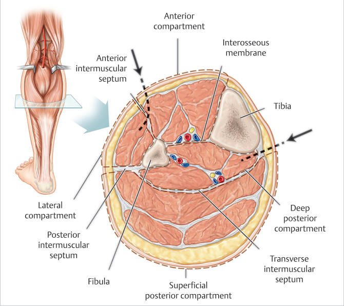
Fig. 2.1 Left mid-leg cross-section showing the four muscle compartments of the leg and the relative location of their investing fascia. The dotted lines emphasized by the arrows should be the direction followed by the anterolateral and posteromedial incisions needed for four-compartment decompression if a compartment syndrome is suspected.
There are also unique factors of the lower limb that can lead to predictable compromise. For example, there is minimal soft tissue overlying the tibia anteriorly. Therefore, even the most minor trauma can lead to exposure of the tibia, and the lack of adjacent soft tissue limits the use of local flaps for its coverage. Adequacy of distal lower extremity blood supply can be complicated by the ubiquitous presence of atherosclerosis. Dependency of the leg and consequently enhanced hydrostatic pressures can contribute to an increased incidence of deep vein thrombosis and venous disease, making reconstruction with any form of soft-tissue flap more susceptible to venous congestion. In addition, following even the slightest insult, patients can be susceptible to venous stasis ulcers which are most difficult to treat.1,6 Finally, unlike other areas of the body, normal sensation to the foot best allows proper ambulation and rehabilitation.
2.3 Assessment of the Impaired Lower Extremity
Treatment of the impaired lower extremity must begin just as for the normal lower extremity with a comprehensive evaluation of not just the defect of the ipsilateral limb but the whole patient. The etiology of these defects may be multifactorial and secondary to trauma, neoplasia, infection, poorly controlled systemic disease, and/or arterial and venous compromise. Regardless of the origin of the defect, the patient should be treated by a multidisciplinary team, and always with a respect for the patient’s autonomy. The patient’s comorbidities, premorbid state, American Society of Anesthesiologists (ASA) grade, smoking status, rehabilitative potential, as well as expectations and motivation should always be taken into consideration before embarking on any treatment course. Following the preservation of life and then limb, the goal of reconstruction will be to restore function. In this context, an acceptable reconstruction will require a stable skeleton capable of supporting the patient’s weight, a robust tissue envelope with satisfactory proprioception and plantar sensibility, as well as having a reasonable appearance.6
2.3.1 Lower Extremity Trauma
Lower extremity trauma should be considered as a potential life-threatening injury due to the risk of high-volume blood loss and the sequelae of inadequate resuscitation, such as hypothermia, hypovolemia or shock, coagulopathy, and adult respiratory distress syndrome.6 Lower extremity injuries should also be considered potentially limb threatening: so obvious after a traumatic amputation, less obvious with a serious vascular injury causing ischemia or a compartment syndrome, but even possible with any open fracture or fracture dislocations resulting in nerve injury.7 Universally, the most common mechanisms of lower extremity trauma still remain motor vehicle accidents, falls, and interpersonal violence.8
2.3.2 History
Open lower extremity fractures are usually a result of high-energy trauma. Treatment can be difficult, as these injuries are usually not solitary but involve multiple-organ systems and each must be assessed and treated simultaneously.9 As well as the mechanism of injury, a thorough history including any past medical or surgical history, allergies, known medications, social history, and premorbid condition must be known. A primary and secondary survey should be performed to look for specific signs indicative of high-energy trauma that would include the presence of other injuries, a large soft-tissue defect, a degloving injury, or a transverse or segmental fracture pattern with comminution.9 Complex bony and soft-tissue injuries may be a clue also of the presence of associated neurovascular compromise.
2.3.3 Examination
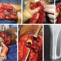
Open tibial fracture is one of the most common lower extremity injuries that requires an orthoplastic approach. The Gustilo–Anderson classification schema (▶ Table 2.1) remains the most widely used system for the classification of open lower extremity fractures (▶ Fig. 2.2).10 The classification is relatively straightforward and offers orthoplastic surgeons a prognostic framework that guides management.
The BOA/BAPRAS guidelines are a robust set of evidenced-based principles also devised to aid in the assessment and management of open tibial fractures.9 They have revolutionized the management of many aspects of lower limb trauma in the United Kingdom and are being increasingly implemented in other European countries. The relevant areas following these guidelines will be summarized as follows.
ATLS principles are employed with airway and spinal management as well as with breathing and circulation support even prior to the arrival of the patient in the emergency department. This should include intravenous access and analgesia. External hemorrhage is stopped via direct pressure or as a last resort by application of a tourniquet. The neurovascular status should be ascertained and documented. Neurovascular examination should include assessment of the capillary refill of the skin, palpation of pedal pulses, as well as the presence or absence of sensation of the medial plantar surface (tibial nerve) and first web space of the foot (deep peroneal nerve).
Repeated neurovascular examinations must be performed particularly for evolving injuries and following any intervention.9 Gross contamination should be removed, and the limb realigned as long as no iatrogenic injury would occur. Photographs ideally need to be taken before the wound is covered with a sterile moist dressing, and this dressing should then not be interfered with until the patient is taken to the operating room. The fracture must be temporarily splinted and radiographs taken (two orthogonal views, as well as the joint above and below). A more thorough examination of the injury should follow in the operating room jointly by a senior plastic and orthopaedic surgeon. This scenario ensures that sterility, illumination, anesthesia, and an expert opinion will be optimal. On-table femoral angiography most simply can also be performed in the operating room if indicated. This initial surgery should be performed at least within 24 hours of the injury. In some cases, however, more immediate surgery without delay should be strongly considered, including the presence of gross contamination, a suspected compartment syndrome, vascular compromise, or other life-threatening injuries.
Table 2.1 The Gustilo–Anderson classification of open fractures10
Gustilo grade definition |
I: Low-energy trauma wound < 1 cm |
II: Wound > 1 cm without extensive soft-tissue damage or avulsions |
III: Extensive soft-tissue damage/loss including any segmental comminuted fractures or fracture open more than 8 h before treatment |
• IIIA: Type III fracture with periosteal coverage of the fracture; extensive soft-tissue laceration or damage |
• IIIB: Type III fracture with periosteal stripping, extensive soft-tissue loss, and bone damage; usually associated with massive contamination |
• IIIC: Type III fracture associated with a vascular injury requiring repair |
Related posts:
 General Wound Preparation and Timing
 The Pertinence of the Reconstructive Ladder and the Reconstructive Elevator
 Supermicrosurgery Approach to the Lower Limb
 Lower Limb Vascularized Composite Allotransplantation
 Using the Flap and Angiosome Concepts to Optimize Functional Lower Leg and Foot Amputations
 Procurement of Thin Flaps as Indicated in the Lower Extremity
Stay updated, free articles. Join our Telegram channel

Full access? Get Clinical Tree


