progressive, and it is important when an infant is noted to have a large segmental hemangioma in the lower body, particularly the lumbosacral region, to refer for evaluation by the appropriate specialties. Early intervention can help prevent and/or manage possible complications.13
TABLE 3.3.1 Disorders and Recommended Treatments | ||||||||||||||||
|---|---|---|---|---|---|---|---|---|---|---|---|---|---|---|---|---|
| ||||||||||||||||
Congenital hemangioma
Kaposiform hemangioendothelioma (KHE)
Tufted angioma (TA)
Pyogenic granuloma (PG)
(VEGF receptors), as well as the genes involved in vascular development (eg, dual specificity phosphatase 5 [DUSP5]), which leads to clonal expansion of endothelial cells. Germline mutations have been found in familial cases and include VEGFR2, TEM8, and mutations on 5q. The placental theory of IHs postulates that the immunohistochemical similarities between IH cells and placental cells (GLUT-1, FcγRII, and Lewis Y antigen expression specifically) infer that IHs are either placental in origin or undergo differentiation toward the placental microvascular phenotype. There is further evidence that the placenta or, possibly, chorioangiomas within the placenta secrete substances that prepare the hemangioma sites for implantation. The approximately 9-month growth phase followed by stabilization and involution also lends credence to this hypothesis. This metastatic niche theory could provide possible future avenues for prevention and therapy.16 Finally, there is discussion regarding the role of hypoxia in the etiology of these lesions. This is supported by their association with hypoxic placental changes, placental insufficiency, low birth weight, regional arterial insufficiency seen in PHACE(S) and LUMBAR syndromes, and retinopathy of prematurity (GLUT-1-positive neovascularization). Estrogen may also play a role as IHs show such a significant predilection for female infants.17,18
TABLE 3.3.2 Reasons to Consider Active Interventions for Infantile Hemangiomas | |
|---|---|
|
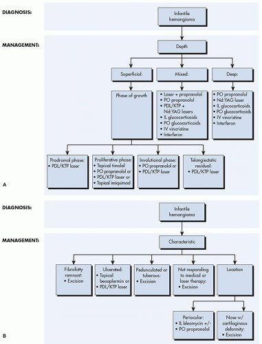 ALGORITHM 3.3.1 Treatment algorithm for infantile hemangiomas. A, Treatment algorithm for infantile hemangiomas based on depth of the tumor. B, Treatment algorithm for infantile hemangiomas based on clinical characteristic of the tumor. IL, intralesional; IV, intravenous; KTP, potassium-titanyl-phosphate; PO, per oral; PDL, pulsed dye laser. |
mitotic spindle by binding to tubulin. Vincristine has been shown to induce rapid and dramatic clearance of IHs, particularly when combined with systemic corticosteroids. Suggested dosage regimens are 0.75 to 1.5 mg/m2 intravenously at weekly intervals for 5 weeks and then slowly tapering by increasing dosing intervals based on treatment response. Caution must be used to prevent extravasation as this can lead to skin necrosis. Additional adverse effects include dose-limiting neurotoxicity, autonomic neuropathy resulting in abdominal pain, constipation, and ileus, and a peripheral mixed sensory motor neuropathy is common. Hematologic toxicity is rarely seen.48,50,54,55,56
577 to 600 nm. Upon absorption of the laser light by hemoglobin, the incoming photon energy is converted to thermal energy which diffuses within the vessels leading to endothelial cell apoptosis, and vessel inflammation and obstruction. If the pulse duration is greater than the thermal relaxation time of the vessels (which is directly proportional to the squared diameter of the target vessels), nonselective thermal damage occurs to the adjacent tissue leading to unwanted destruction and scarring.72,73,74
Related posts:

Stay updated, free articles. Join our Telegram channel

Full access? Get Clinical Tree


