CHAPTER 11
Analysis and Principles of Management
The aim of managing acquired partial defects is to reconstruct the missing contours to replicate the appearance of the normal side. This is achieved by performing the following sequence:
1. Analyzing the defect
2. Choosing the best type of support
3. Deciding whether the reconstruction should be performed in one or two stages
ANALYZING THE DEFECT
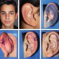
First, the contralateral ear is drawn on a transparent template. Then, it is inverted and placed on the auricular region in its precise location to determine which anatomic subunits are involved in the defect.
Having in mind the three-dimensional architecture of the ear will help to differentiate cases, which at first seem similar.
The following two examples show two small defects of the helix that may seem similar but require different approaches.
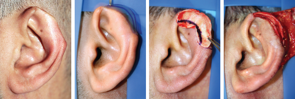
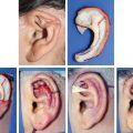
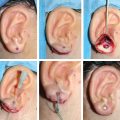
This case demonstrates a deficiency of the upper curved part of the helix. Analysis of the contralateral ear template showed a narrow scaphoid fossa; thus conchal cartilage was used to accurately reproduce the missing helix.
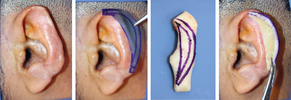
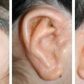
In comparison, this case demonstrates a longer defect of the curved helix, including part of the scapha. Conchal fibrocartilage will not provide enough stability to reproduce correctly the missing contours, and the defect was longer than a conchal graft. Therefore costal cartilage was used. Costal cartilage has the additional benefit of carving a piece that may perfectly reproduce the missing contours of the helix and scaphoid fossa.
Harvesting costal cartilage for a relatively minor defect may seem excessive to some, because this implies a scar on the thorax. But the scar itself is very short, because it is placed directly over the sixthseventh synchondrosis, limiting the dissection and respecting the adjacent ribs.
The following two examples show larger defects requiring different approaches.
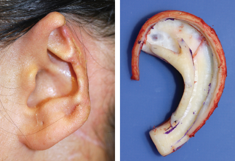
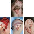
This example demonstrates that when the entire antihelix is unharmed, and the partial defect consists only of the lobule and lower part of the helix, costal cartilage can be used. In this case it was carved from a single segment (sixth).
In contrast, in this seemingly similar defect, the antihelix was damaged, and the posterior wall of the concha was absent. A subtotal framework (with a base and the addition of a new antihelix) was required to reconstruct this defect to increase projection of the middle part of the ear from the frontal view.
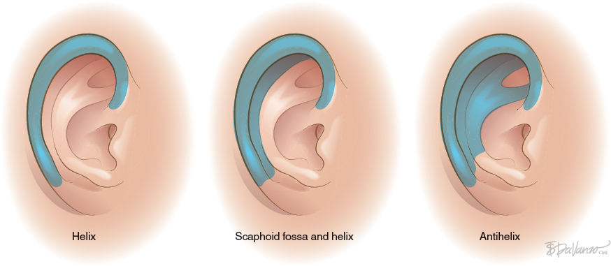
Anatomic Subunits