Dermatitis herpetiformis (DH) is characterized by chronic, itching papules, seropapules, small vesicles and, exceptionally, large blisters. The distribution of these polymorphic symptoms around the elbow, knee, buttock, and back is suggestive of the diagnosis. DH is further confirmed by the accumulation of granulocytes at the papillary dermis, resulting in a subepidermal split formation and by the presence of a unique, granular IgA precipitate in the uppermost dermis. Prognosis is predominantly determined by other autoimmune pathologies, malabsorption, or very rarely by lymphomas. Some of these diseases can be prevented by an early-onset, strict gluten-free diet, which is therefore the suggested treatment option.
Dermatitis herpetiformis (DH) is special among the classic autoimmune blistering skin diseases when considering its dermatologic symptoms, associated diseases, and pathomechanisms. The granular IgA precipitates present at the tips of the papillary dermis of the patients, an observation made by van der Meer in 1969 in Groningen, proved to be pathognomonic for the disease. Contrary to other autoimmune blistering diseases, whereby tissue-bound and serum autoantibodies bind the same target molecule in the skin, no circulating IgA has been detected in DH sera reacting with normal tissue components of the sub-basal membrane zone or any other connective tissue particles within the healthy papillary dermis. The antigenicity of skin-bound IgA remained unknown for 3 decades, until 2002, when epidermal transglutaminase (TG3) was identified by the author’s research group as its main antigen, an enzyme never detected in that area of the normal skin. It has also been confirmed that DH patients have serum IgA autoantibodies to TG3. This article focuses on the clinical data concerning DH, rather than its detailed pathomechanism.
Skin symptoms and disease management
DH can start at any age; rarely, it can be present in toddlers or in the very elderly, but the mean onset is generally in young adulthood or middle age. It is a chronic, very pruritic skin disease, characterized by 1- to 3-mm large papules, seropapules, vesicles, crusted erosions, and excoriations ( Fig. 1 ). Rarely, larger blisters can also develop. The lesions heal with hypopigmentation or hyperpigmentation. In young patients, urticarial plaques might be the predominant skin symptoms ( Fig. 2 ). The severe pruritus and scratching can result in extended lichenification ( Fig. 3 ). In the majority of cases DH is a polymorphic skin disease, and only rarely presents as a bullous dermatosis. Specific symptoms, not always present, are purpura on the fingers and toes, which alone may focus attention on the diagnosis ( Fig. 4 ).
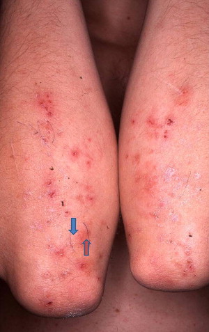
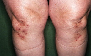
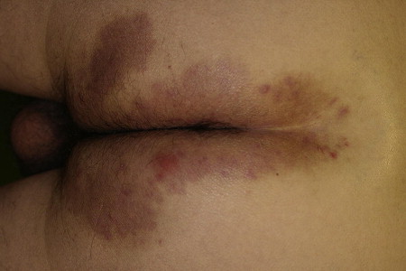
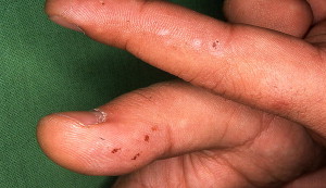
DH has a typical distribution of the skin symptoms; these are, in order of frequency (strongly supported by the author’s personal observations): 1, elbows and knees; 2, buttock; 3, shoulders, middle line of the back, and scapula; 4, scalp; 5 (rarely), purpura on the fingers and toes (see Figs 1–4 ).
Although severe DH is almost a continuous disease with some fluctuation in the severity and itch, it might also present as a relatively mild lichenification, with alternating remissions and relapses: a few patients have skin disease only for a few days between long symptom-free periods. The symptom-free periods may only be in the warmer months when it is sunny. By contrast, in some DH patients sweating may induce disease progression in hot weather. There are patients who, without a gluten-free diet or other medication, remain free of skin symptoms for at least 6 months, in so-called spontaneous remission. One must bear in mind that the underlying celiac disease (CD) and the possibility for further secondary disease development will persist. Spontaneous remissions are rare, but might develop and may last for years, life-long, or just for a short time. Pruritus can precede the skin symptoms and rarely also challenge the otherwise symptom-free time of the patients.
Untreated DH can be a life-long disease with variable severity. Associated uncontrolled diabetes, autoimmune diseases, underlying tumors, and iodine challenges might induce permanent very severe skin symptoms. During pregnancy the disease may improve, but in some cases it is worse. The reason for iodine sensitivity of DH patients is unknown, but is common. A possible relationship with thyroid diseases also remains unraveled. The sensitivity can be proved by patch testing, but iodine-containing drugs also flare up the disease. One of the author’s young patients underwent a thyrotoxic crisis together with a severe DH flare-up after one short visit to a salt cave suggested for her as a “natural therapy.”
Diagnosis confirmation
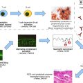
Diagnosis should be made by histologic and direct immunofluorescence (IF) analysis of the skin. The former sample should be taken from the lesional skin, the latter from perilesional, close to symptom-free tissue. On urticaria like lesions the presence of IgA, C3, and eventually IgM and IgG vasculitis is commonly associated with granular IgA and C3 staining of the dermis. In the serum the presence of circulating IgA endomysial antibodies (EMA) or tissue transglutaminase (TG2) autoantibodies should be checked by indirect IF and by enzyme-linked immunosorbent assay (ELISA), respectively. The sensitivity and specificity of these tests are very high for CD, between or above 90% to 95%, whereas for DH it is less: about 75% to 90%. Serum samples from autoimmune diseases as well as sera from normal individuals treated by heat or pH shift might show nonspecific TG2 reactivity. The TG3 antibodies (ELISA) are also gluten dependent and slowly disappear under gluten-free diet (GFD), but later than the EMA or TG2. TG3 antibodies are more commonly present in DH than in CD patients, and in DH patients they have higher avidity and affinity. An upper gastroduodenoscopy and a small bowel histology is strongly advised to visualize and document the upper part of the gastroinstestine and to analyze the initial jejunal histology, due to its strong association with CD.
Enteropathy and Malabsorption
The majority (75%–90%) of DH patients have an associated small bowel disease, a latent or silent CD. It persists and without treatment leads to malabsorption, which may induce secondary diseases: microcytic or macrocytic anemia due to iron, folate, or B12 deficiency, caries, alopecia due to zinc deficiency (see also dental problems), and early or very severe osteoporosis. Weight loss but also weight gain is possible, with unfavorable body mass index (BMI). Most patients have no typical gastrointestinal symptoms, but symptoms may include any of the following: diarrhea, constipation, bloating, abdominal discomfort or pain, and secondary lactose intolerance. Similarly to other gluten-sensitive enteropathy (GSE) patients, in DH patients with severe insulin-dependent diabetes, because of malabsorption the diabetes will be controlled only under a combined, strict GFD and diabetic diet. In childhood short stature, delayed development and puberty, or a high BMI might indicate the underlying GSE. Dental enamel defects, and mineralization disturbances of permanent and decidual teeth may be evident. Half of DH patients have celiac-type permanent-tooth enamel defects, milder than those described for severe celiac disease, and secondary severe caries or early tooth loss is also more common. Although the skin can go into spontaneous remission, the underlying CD and the possibility for further secondary disease development persist.
Diagnosis confirmation
Diagnosis should be made by histologic and direct immunofluorescence (IF) analysis of the skin. The former sample should be taken from the lesional skin, the latter from perilesional, close to symptom-free tissue. On urticaria like lesions the presence of IgA, C3, and eventually IgM and IgG vasculitis is commonly associated with granular IgA and C3 staining of the dermis. In the serum the presence of circulating IgA endomysial antibodies (EMA) or tissue transglutaminase (TG2) autoantibodies should be checked by indirect IF and by enzyme-linked immunosorbent assay (ELISA), respectively. The sensitivity and specificity of these tests are very high for CD, between or above 90% to 95%, whereas for DH it is less: about 75% to 90%. Serum samples from autoimmune diseases as well as sera from normal individuals treated by heat or pH shift might show nonspecific TG2 reactivity. The TG3 antibodies (ELISA) are also gluten dependent and slowly disappear under gluten-free diet (GFD), but later than the EMA or TG2. TG3 antibodies are more commonly present in DH than in CD patients, and in DH patients they have higher avidity and affinity. An upper gastroduodenoscopy and a small bowel histology is strongly advised to visualize and document the upper part of the gastroinstestine and to analyze the initial jejunal histology, due to its strong association with CD.
Enteropathy and Malabsorption
The majority (75%–90%) of DH patients have an associated small bowel disease, a latent or silent CD. It persists and without treatment leads to malabsorption, which may induce secondary diseases: microcytic or macrocytic anemia due to iron, folate, or B12 deficiency, caries, alopecia due to zinc deficiency (see also dental problems), and early or very severe osteoporosis. Weight loss but also weight gain is possible, with unfavorable body mass index (BMI). Most patients have no typical gastrointestinal symptoms, but symptoms may include any of the following: diarrhea, constipation, bloating, abdominal discomfort or pain, and secondary lactose intolerance. Similarly to other gluten-sensitive enteropathy (GSE) patients, in DH patients with severe insulin-dependent diabetes, because of malabsorption the diabetes will be controlled only under a combined, strict GFD and diabetic diet. In childhood short stature, delayed development and puberty, or a high BMI might indicate the underlying GSE. Dental enamel defects, and mineralization disturbances of permanent and decidual teeth may be evident. Half of DH patients have celiac-type permanent-tooth enamel defects, milder than those described for severe celiac disease, and secondary severe caries or early tooth loss is also more common. Although the skin can go into spontaneous remission, the underlying CD and the possibility for further secondary disease development persist.
Related posts:
 A Globally Available Internet-Based Patient Survey of Pemphigus Vulgaris: Epidemiology and Disease Characteristics
 Diagnosis and Clinical Features of Pemphigus Foliaceus
Linear IgA Disease: Clinical Presentation, Diagnosis, and Pathogenesis
Pemphigoid Gestationis: Pathogenesis and Clinical Features
Diagnosis and Clinical Features of Pemphigus Foliaceus
Linear IgA Disease: Clinical Presentation, Diagnosis, and Pathogenesis
Pemphigoid Gestationis: Pathogenesis and Clinical Features
 Pathogenesis of Epidermolysis Bullosa Acquisita
Nail Involvement in Autoimmune Bullous Disorders
Pathogenesis of Epidermolysis Bullosa Acquisita
Nail Involvement in Autoimmune Bullous Disorders
Stay updated, free articles. Join our Telegram channel

Full access? Get Clinical Tree


