Introduction
Advanced practice providers (APPs), such as physician assistants (PAs), advanced practice registered nurses (APRNs), and certified registered nurse anesthetists (CRNAs), are force multipliers for the physician to provide care and reach a broader patient population, especially in subspecialties such as burn reconstruction. The focus of this chapter is an overview of burn reconstruction from the perspective of the APP and practice considerations for the approach and organization of outpatient burn reconstruction that can be useful for any member of multidisciplinary reconstruction teams. APPs facilitate collaboration among a physician-directed multidisciplinary team, allowing for a more comprehensive and patient-focused approach. A brief overview of acute burn management is provided, during which stage the APP may also play an integral role. Any provider participating in acute burn care should strongly consider obtaining Advanced Burn Life Support (ABLS) certification, a targeted educational resource for first responders and other medical personnel focusing on the emergency care of the burn patient in the first 24 hours after burn injury.
Acute burn management
Mechanisms of burn injury
A burn is a complex injury to the body’s tissues caused by contact with heat, chemicals, electrical current, or radiation. The severity of the burn injury is dependent upon the type of offending agent, the duration of contact, and the extent of the body surface area involvement. In the United States, most burn injuries are sustained from thermal or scald mechanisms. Scald injuries account for thousands of patient injuries annually and are the leading cause of injury in children under age 4 years. Although less common, chemical, electrical, and radiation burns are complex injuries that frequently involve deep tissue damage and necrosis, requiring long-term management and reconstruction. Chemical burns are acid or alkali, resulting in skin protein denaturation and coagulation. Acid burns are usually more superficial, resulting in coagulation necrosis and occurring more frequently than alkali burns. Alkali burns are more difficult to manage because they adhere to the tissue, producing a liquefaction necrosis and deeper penetration of the tissues. Electrical burns are devastating injuries that result in coagulation necrosis caused by the intense heat generated by an electrical current. Damage from electrical burns is related to the resistance of the contact point, the amount of voltage, the type of current, and the time of contact. The electrical burn patient may have skin appearing normal but have extensive muscle destruction. Damaged muscle swells beneath the overlying muscle fascia, producing severely elevated tissue pressures and an increased risk for compartment syndrome. Radiation burns range from superficial sunburn to deep tissue injury from prolonged radiation exposure. Severe radiation injuries frequently create chronic, nonhealing wounds that are refractory to traditional treatment modalities and require a specialized reconstruction approach.
Initial burn management
Understanding fluid resuscitation needs for adult and pediatric patients is essential for survival and quality outcomes. Airway management and early diagnosis are critical for survival, as inhalation injury alone is present in 10% to 20% of patients admitted to the burn center. Hypovolemic shock and hypothermia are the greatest initial threats to a patient with a major burn. Accurate assessment of total body surface area (TBSA) is essential; failure to accurately assess TBSA may result in under- or overresuscitation, leading to complications. Although numerous resuscitation formulas have been described, all aim for adequate hydration. Temperature regulation is disrupted when the integumentary system is compromised, and hypothermia increases the patient’s metabolic response. Although wet dressings may be necessary for topical irrigation of infected burns, temperature should be monitored and consistently maintained, including perioperatively. Even if the TBSA is minor, burns to the hands, feet, face, genitalia, and perineum or over major joints are considered major burn injuries.
Full-thickness circumferential or near circumferential burns of the extremities or torso may cause vascular or respiratory compromise, respectively. An accurate and readily available method of evaluating the need for peripheral escharotomies or fasciotomies is serially monitoring vascular flow to the affected extremity. Even after surgical intervention, frequent reassessment after the procedure to ensure the adequate release is essential to maintaining perfusion. Full-thickness burns to the torso should be considered for central escharotomies when the eschar is likely to cause constriction across the abdomen and chest wall, especially in interstitial edema and fluid resuscitation.
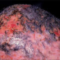
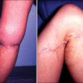
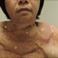
Superficial burns have intact epidermis and routinely heal independently with minimal complications within 7 to 10 days. Partial-thickness burns extend through the epidermis and into the dermis. A superficial partial-thickness burn will be moist, shiny, and blanches easily upon compression, with or without blistering. Deep partial-thickness injuries will form an eschar and extend further into the dermis, not the subcutaneous tissue. A superficial partial-thickness burn will heal spontaneously without surgical intervention, generally within 10 to 14 days. A deep partial-thickness burn should ideally heal in 2 to 3 weeks. However, these burns may often be skin-grafted because of the healing time and likelihood of scarring if left open. Full-thickness burns extend through the dermis and into the subcutaneous tissue. They will be dry, with minimal or no sensation secondary to nerve destruction. The burn may be white, dark brown, or charred in appearance and often resembles leather. Full-thickness burns may also appear cherry red with petechiae as a result of thrombosed capillaries. Full-thickness burns typically require autografting for definitive closure ( Fig. 49.1 ).
Acute burn progression, prereconstruction.
Scarring is one of the most well-known late effects of burn injury. Scar disfigurement and threat to functional impairment are known sequelae after any major burn. Access to acute burn care with essential long-term follow-up with a dedicated team is a necessity, including surgical reconstruction options.
Transition to reconstruction
Burn reconstruction is an essential component of the overall recovery of adult and pediatric burn patients. Burn care should focus on survival and quality of life with optimized restoration of function. The burn reconstruction and recovery process can take up to several years after the initial injury, depending on the depth and severity of the damage. Access to reconstructive care with a dedicated multidisciplinary team is valuable as surgical methods evolve and advance. Some of the reconstruction needs of the burn patient may include, but are not limited to, scar contracture release, unstable scarring, nonhealing wounds, dyspigmentation, pruritus management, anatomic/functional deformities, alopecia, syndactyly, heterotopic ossification (HO) or osteomyelitis, ectropion, complex tendon repair, and laser procedures.
The acutely burned patient undergoes multiple transitions in the recovery timeline, each of which may have reconstruction considerations.
The outpatient setting allows for more control over reconstruction’s nuances and multidisciplinary aspects, facilitating positive outcomes and a patient-centered approach. However, under some circumstances, reconstruction and acute burn closure coincide. Patient education begins as early as in the emergency or prehospital setting to help the patient and family understand the trajectory of acute burn care and that burns may become a lifelong diagnosis. They can result in chronic problems requiring ongoing specialized care. In the inpatient setting, the closure of functional and cosmetic areas should be approached with the prevention of poor outcomes in mind.
Reconstruction principles should be incorporated into every patient’s journey to reduce future reconstruction needs. The transition from acute inpatient care to outpatient care is an important step in the patient’s transition to independence, and they may become more empowered to participate in their care with aggressive therapy and start the rehabilitation process from burn-related critical illness. The transition from the acute care setting to inpatient rehabilitation and/or discharge is one of the most critical because this is a time when many of the final closure efforts are maturing. Losing continuity of care during this time can have devastating consequences for outcomes, especially regarding therapy goals, wound care, and specialist follow-up needs. The APPs can assist during this transition to ensure continuity of care and follow-up and provide oversight. Particular focus should be placed on educating patients and their family members about the standards of care and what to expect, as well as empowering them to participate in the care to promote good outcomes. Nursing and other support staff education and instructions should be provided in detail, especially when transitioning to home health, inpatient rehabilitation, skilled nursing facilities, correctional facilities, or any other entity that is not a specialized burn or reconstruction center.
Many patients find that the frequency of surgery during acute burn management is overwhelming and takes a significant emotional and physical toll over time. For this reason, the first outpatient appointments should be focused on achieving initial burn closure, education on scar management, and preventative measures for developing healthy, mature burn scars. Reconstruction surgery should not be pursued at this time unless there are unstable scars, autoreleased contractures, acute infection concerns, exposure of the cornea, or neurovascular bundles or if the patient is insistent upon and clinically ready to begin reconstruction efforts. Often, a burned patient’s critical and acute care is primarily physician directed from a skin closure and medical perspective. The transition to reconstruction allows patients to regain control over their lives, especially regarding surgery scheduling and priorities.
Upon referral to reconstructive care, the reconstruction patient will likely have symptomatic, functional, and cosmetic concerns. Surgical staging is usually necessary for patients with multiple areas of concern and major reconstruction needs. Some patients may tolerate multiple interventions simultaneously, but mobility and self-care considerations should be weighed with urgency to continue reconstruction efforts. In many cases, laser therapy can be effectively incorporated intraoperatively with other procedures to reduce the frequency of surgery. Generally, functional deficits will be prioritized, especially with hand reconstruction and major joint contractures. Oral commissure contractures and periorbital and oral aperture corrections are also prioritized for functional outcomes. Unstable or autoreleased areas should be given special priority and may drive more urgent surgical intervention to promote achieving and maintaining wound closure. Ultimately, prioritizing surgery is a joint effort between the surgical team and the patient, with the assistance of the multidisciplinary team. Physical and occupational therapy provides critical insight into the patient’s functional progress. ,
Reconstruction may take several years and even necessitate intermittent interventions throughout the patient’s life. Managing expectations for cosmetic and functional results should be measured with quality of life and frequency of surgery. Thorough patient education, especially at the onset of reconstruction efforts, will help increase patient compliance and improve functional and cosmetic outcomes.
Multidisciplinary approach
The reconstruction patient can have a complicated hospital stay, and a reconstruction center should have access to a robust multidisciplinary team. The patient-centered approach allows patients to take an active role in their care and decision-making, and the surgeon, with each patient, will set the tone for priorities and reconstruction timelines. APPs work with the surgeon to evaluate and treat patients, including assisting in surgery and performing routine bedside procedures in outpatient, inpatient, and intraoperative settings. Especially in nonacademic settings, APPs are pivotal in extending physicians’ capacity to provide their surgical expertise to more patients without sacrificing quality of care. APPs also facilitate communication among the multidisciplinary team, following up on consults, referrals, and continuity of treatment plans. A multidisciplinary approach to burn care has benefitted patients, families, and systems in achieving outcomes.
Provider support positions in the reconstruction outpatient setting can also be beneficial for extending care. A medical scribe is an adjunct to the team, allowing providers to spend more time facilitating care and providing patient support and education. Medical scribes also promote consistency, standardization, and increased recordkeeping accuracy, ultimately promoting efficiency among the care team. Medical photography is also essential to the reconstruction process, providing a benchmark for patients’ progression toward their care goals, documentation of complications, and giving insight for research. Any care team member can be designated to perform photography, but a dedicated professional medical photographer is ideal for the best quality documentation. According to individual institutional policies and available technology platforms, many applications are available for privacy-compliant photo repositories.
Rehabilitation and therapy services, including physical, occupational, and speech therapy, are nonnegotiable as reconstruction multidisciplinary team members. Therapy interacts with the patient at each visit to provide additional treatment modalities and recommendations for scar management, range of motion (ROM), and mobility. They will evaluate the patient postoperatively for splinting and compression needs and sometimes perform splinting intraoperatively for pediatric and complex splints. They are critical for appropriate splinting, stretching, and compression for complex scar areas. Patient education by therapy reinforces the importance of compliance, and they can identify and closely monitor functional efficacy and outcomes during reconstruction. Having in-house therapy resources does not negate the need for outpatient or in-home rehabilitation and therapy, and most reconstruction patients will benefit from continued referrals to these services several times a week when undergoing or recovering from reconstruction surgeries. Certified hand therapy especially should be maintained during hand reconstruction, per evidence-based guidelines, and at the surgeon’s discretion. ,
Nursing staff facilitate the execution of care in all patient settings and are the first line of defense in identifying new and unresolved issues. According to the Burn Nursing Scope of Practice and Competency, the burn nurse facilitates coordination of care to facilitate outcomes. Nursing staff spend much time at the bedside with the patient between routine nursing assessments and time-consuming wound care in both inpatient and outpatient settings. Nurses are the gatekeepers for the patient flow in the perioperative and outpatient settings and facilitate many administrative support functions associated with referrals and follow-up care.
Certified child life specialists (CCLS) are an essential adjunct to the reconstruction team for pediatric patients. Their goal is to minimize the trauma to the patient and provide age-appropriate interventions and behavioral tools to promote coping mechanisms and positive outcomes. The pediatric burn patient can have complex psychological and behavioral responses to health care settings that require a specialized approach to care. The CCLS is trained to help manage fear and anxiety for the patient and family members, creating a less stressful healing environment in inpatient, outpatient, and perioperative settings.
Social workers and advocacy resources are necessary because of the multidisciplinary nature of reconstruction treatment. Social support, such as assistance with lodging, transportation, prescriptions, and primary care resources, is sometimes needed for patients who may travel long distances for treatment, are underinsured, or have complicated social situations. Additional support functions may be required to facilitate completion of work, school, or leave-related documents. Disability and workman’s compensation forms, as well as other related record requests may be needed. The reconstruction provider will provide referrals to other specialists in the absence of or in conjunction with the primary care provider, and many patients require assistance with identification and scheduling with specialists familiar with the complexities of burn reconstruction care. Social workers and nurse navigators are essential advocates for patient follow-up needs and for addressing social issues that may hinder the progress of reconstruction efforts.
Multidisciplinary consultants may be needed to facilitate the reconstruction process, especially if the patient’s acute burn course is complicated. The patient’s primary care provider should ensure adequate specialist follow-up after discharge. Still, frequently, it falls to the reconstruction team to make these referrals to optimize the patient for future surgeries medically. Other specialties may be needed if the patient had a prolonged hospital stay or sustained other trauma, such as general surgery, otolaryngology, and orthopedic surgery. Genital reconstruction may be a joint effort with urology or gynecology for especially complicated cases. If amputations were performed acutely or revised as part of the reconstruction plan, a prosthetist referral would be needed once wound healing and scar maturation are achieved. Each patient’s unique needs should be considered thoroughly for specialist referral.
Initial evaluation
All new patients begin with an initial evaluation in the clinic by a multidisciplinary reconstructive team, including a surgeon, APP, occupational and/or physical therapist, and nursing. The patient may be self-referred to the burn reconstructive clinic or referred by an acute burn provider based on established referral criteria, which include overall TBSA of 20% or greater in adults or 5% in pediatrics and required autografts for closure or autografts over specific anatomy (hands, feet, face, neck, breast, perineum, or any contracture-prone joints), burn scars that are hypertrophic or symptomatic, and alopecia. The referral should be made once the patient has achieved closure of most acute burn wounds.
During this initial evaluation in our reconstructive center, the patient is first welcomed to an exam room by nursing staff, who obtain vital signs and an initial assessment. Next, the patient completes self-reporting questionnaires utilizing validated screening tools on overall health, life satisfaction, mobility, functionality, pruritus, depression, and posttraumatic stress disorder. The physical and occupational therapy team then evaluates the patient’s functional and mobility status. Modified scar goniometric measurements are obtained of affected joints. Education and treatment are provided on scar management (sun protection, moisturization, scar massage, scar gel products, interim and custom compression), desensitization, custom orthotic fabrication, serial casting, task modification with adaptive equipment, hydrotherapy, and home exercise program. ,
The provider then sits with the patient for a complete history and physical examination. The details and treatment of the acute burn injury and any previous reconstructive treatment are discussed. The patient’s personal goals, including cosmetic and functional, are also discussed during this time. After obtaining informed consent, medical photography of all burn scars and joint mobility is obtained at the initial visit and future follow-up. Plain film data persist for studies to identify HO, although microcomputed tomography may be considered if a physical exam warrants.
The surgeon subsequently evaluates the patient with the multidisciplinary team. A focused physical exam is performed, and the patient’s goals and limitations are reiterated. Potential options for surgical intervention are discussed. Patient priorities are considered with stages of operative planning and which individual procedures are best to pair together to limit surgeries without compromising patient rehabilitative success and satisfaction. The plan is staged working proximal to distal (e.g., axilla before hands). Most often, functional surgical goals are scheduled before cosmetic goals; however, the patient’s mental health and view of self are always considered.
During the discussion with the patient, several needs are systematically approached. An emphasis is placed on the importance of the social support system, which is the framework for assistance with activities of daily living (ADLs), wound care, and transportation, among others. Their mental health is also evaluated through evaluation during history, overall demeanor, and as self-reported by patients on questionnaires. If a patient’s acute injury was work related, special consideration is needed, including additional communication with the case manager and further orders for specific treatment options, equipment, or others as indicated.
At the conclusion of the visit, the provider reaffirms the plan with the patient, including expectations, and ample time is given to review the surgical and recovery processes and to answer the patient’s questions and those of their accompanying support person. After stages of the surgical reconstructive plan are created and the patient is agreeable, surgeries are submitted to insurance for the precertification process. If a patient is unfunded, self-pay options may be provided based on institutional guidelines. With written patient consent, external records are requested if the patient was treated acutely or had prior reconstructive treatment at an outside facility; if the patient has any additional comorbidities, especially those increasing the operative risk, records from those treating specialists are requested. In these circumstances, preoperative clearance is often required. Referrals are made to appropriate outside providers, including outpatient therapy (physical, occupational, certified hand, speech), neurology (nerve conduction velocity studies), orthotist, orthopedic surgery, cardiology, pulmonology, home health, prosthetist, among others. Lastly, nursing staff may apply wound care according to orders, providing the patient with a written discharge plan.
The amount and frequency of follow-up are very patient dependent. The patient may return to the clinic for necessary reevaluation (after progressing in a home exercise program or outpatient therapy, completing outside nerve or functional studies, or evaluating other providers as needed) before scheduling a surgical intervention. If outside medical records were requested, these are reviewed once received. Recommended surgery is scheduled by stage once the patient is clinically ready and agreeable to the plan. Patients quickly become familiar with their care team for their burn reconstructive journey by using a dedicated multidisciplinary approach in the clinic. As this is often a lengthy process with multiple surgeries and many clinic visits, having a good rapport with the team benefits the patient’s continuity of care.
Perioperative considerations
The surgical plan for the burn reconstructive patient is an initial guide to the operative stages. The plan is fluid, and the order of operative stages may be altered during the surgical course per the patient and surgeon. Regardless of stage, there are several considerations for all patients at each aspect of the course, including preoperative, intraoperative, and postoperative. The next sections have some guidelines and considerations to bear in mind by stage.
Preoperative planning
In the preoperative planning stage, the patient’s safety is the highest priority. If prior history or comorbidities necessitated obtaining preoperative medical clearance after the patient’s initial evaluation in the burn reconstructive clinic, that clearance must be obtained and reviewed. Patients considered at increased risk for operative intervention are discussed with the surgeon, patient, and anesthesia provider.
Various anesthetic and airway methods can be considered to determine the safest option. Individualized positioning needs and current scar contractures that may limit operative positioning, such as neck contractures, should be reviewed among the team preoperatively for safety. Discussion with the patient over perioperative expectations is also reiterated preoperatively, including dressings, splinting, ROM, the necessity for multiple follow-up visits, and possible outpatient therapy requiring reliable and regular transportation. The need for specific custom-fabricated orthotics should be communicated preoperatively with the therapy team. Additionally, with procedures that require a specialty device or bed, orders are placed early to allow time for arrival.
As burn reconstructive patients are often colonized with skin microbes, it is important to take steps to prevent the complication of postoperative infection with a twofold approach. First, the reconstruction patient is encouraged to shower the night before following an institution-specific preoperative bathing protocol. Prevention of surgical site infection (SSI) and the use of prevention strategies should be considered in all phases of the reconstruction period, including before, during, and after surgery. To reduce microbe burden, a cleansing protocol used before surgery as part of the prevention bundle for reduction of SSI has been endorsed by national organizations, including the Association of Perioperative Registered Nurses and other infection control associations. Some burn centers that utilize cultured epithelial autografts more frequently will often limit or restrict the use of certain cleansing agents, such as chlorhexidine gluconate, for as long as 6 months before the procedure secondary to its known cytotoxicity. Secondly, if available, previous acute cultures are reviewed to consider a targeted postoperative antimicrobial course. The need to obtain new wound cultures is assessed if the patient has a chronic wound or unstable scar. As with all operative interventions, postoperative pain control must be considered. Using prescribed low-dose opioids in a short course provides acute pain management for the immediate postoperative phase, to be weaned to over-the-counter antiinflammatory medications when able.
The previously mentioned considerations can take place weeks before surgery. The patient and a support person meet in the preoperative area on the morning of the procedure. At that time, education on the operation’s expectations, wound, dressing care, splinting, and postoperative follow-up is provided again, and time is allowed for a question-and-answer session. The surgeon carries out surgical markings to outline the surgical site correctly. The risks and benefits of the procedure are discussed, and informed consent of the patient is obtained and witnessed. As lasers require multiple applications for effectiveness, they are often easily paired with other acute procedures, whether initial release, reapplication of skin substitute, or ultimate autografting.
Intraoperative planning
The operative team consists of the surgeon, an APP or other first assist, the scrub technician, the circulating nurse, and the anesthesia provider. Operative goals are communicated with the team to ensure the proper equipment, suture, and dressing material are available and to guarantee the correct patient position. Medical photography is obtained preoperatively after the sterile prepping and draping is complete, intraoperatively during the release, and after the procedure, before the application of dressings.
Appropriate dressing selection and application are essential. The goal of the dressing is twofold. The first is to provide pressure to the skin substitute product to promote granulation for a neodermis or the skin graft for neovascularization and ultimate graft take. For this, a bolster-style pressure dressing holds the skin product or skin in place and prevents shear or loss. Second, the dressing serves as a barrier to protect the skin product or skin and prevent infection; therefore an antibiotic product and a layer of padding are often used. Sometimes, a silver sponge dressing with the use of a negative-pressure wound therapy device is applied for both pressure to oppose the wound bed and to provide topical antibacterial therapy. The dressings should also be applied to the area while the body part is in the maximum ROM allowed by the release so the splint may be applied at the appropriate position to optimize the release immediately postoperatively. Care should also be taken when applying the dressings; an effective splint cannot be fabricated if they are too bulky. Often, the initial postoperative dressing is left clean, dry, and intact by the patient, and all ROM is on hold until the first clinic follow-up appointment, which is on postoperative days 3 to 5, depending on surgeon recommendations. Custom fabrication of splinting is performed immediately by therapy. The preferred method is in the postanesthesia area with the patient awake; however, in children who cannot facilitate proper splinting, in those cases of painful or difficult placement or with casting, this is often performed intraoperatively.
Postoperative planning
As the postoperative timeline progresses, the patient’s home exercise program also progresses, allowing more range of the area and, conversely, less splinting until eventual night splinting only, and ultimately no splinting at all. Dressing orders also progress postoperatively. They are usually left intact 3 to 5 days at a time for the first 1 to 2 weeks and then the patient may often begin showers or sponge baths and transition to daily dressing changes after that period if the area is developing appropriately. Home health nursing needs are assessed and ordered for patients requiring frequent dressing changes or negative-pressure wound therapy device changes.
As mentioned in preoperative considerations, antimicrobial and analgesic therapies are necessary postoperatively. The decision for an empiric versus targeted course depends on previous cultures; if cultures within 6 weeks preoperatively are available, a targeted course is utilized with a short postoperative course of opioid management. If under pain management, their specialist manages postoperative pain control techniques.
All progress is assessed at the initial postoperative appointment with evaluation by the APP and therapy team. Additional medical photography is obtained, and a dressing change is performed. Therapy is available to modify any splint needs, perform postoperative goniometry, and begin education on the home exercise program. After this, the follow-up timeline begins to lengthen weekly for the first 2 weeks. Patients are encouraged to resume therapy once the skin graft or substitute has begun appropriate incorporation. Sponge bathing around the area of focus is allowed immediately postoperatively; however, there is no full showering or submersion with water. This is usually allowed about 2 weeks postoperatively for both scenarios. Lastly, the next stage of operative intervention is discussed and scheduled. Depending on the skin substitute placed, the subsequent surgery is usually in 2 to 4 weeks for additional skin substitute layering or autografting. Once autografting of that surgical site is completed, the next stage of the reconstructive plan can begin.
Common wounds in reconstructive patients
Unstable scars
Scars are considered unstable when they do not provide adequate functionality or are symptomatic. These scars tend to be associated with spontaneous reepithelialization over granulation tissue. This results in a more vulnerable top layer of skin that is more prone to cracking and shearing and may lead to chronic wounds that are difficult to heal despite topical treatment. Specific scarring locations, such as scars over joints, bony prominences, or higher-friction areas, are more prone to becoming unstable. Unstable scars may require surgical excision and skin substitute application with subsequent skin graft for resolution.
Pressure injuries
Pressure injuries can be expected in patients with larger TBSA burn injuries with prolonged hospitalization. The most common pressure injury sites are the sacrum, posterior scalp, and bony prominences on limbs. If pressure injuries are present, they may need surgical treatment concomitantly with reconstructive procedures. Educating the patients on aggressive pressure-reduction measures with frequent turning or repositioning and/or ordering low air loss mattresses when appropriate is important to prevent the worsening of injuries.
Splint-related pressure injuries can also be expected in patients with long-term splint use. It is important to ensure adequate padding is placed in splints or dressings to prevent wounds. Splint checks should be performed at each follow-up visit to ensure proper fit and account for new dressings, edema, and improved ROM.
Related posts:

Stay updated, free articles. Join our Telegram channel

Full access? Get Clinical Tree


