Fig. 6.1
Schematic representation of the temporary acute shortening technique. (a) Presentation of the limb on admission – open fracture with bone and soft tissue loss. (b) Debridement with acute shortening and stabilization in the external circular frame. (c) Proximal elongation tibial osteotomy is performed. (d) Limb length is restored by bone regeneration at the osteotomy site
Acute shortening with or without angulation enables good bone molding and quality while avoiding complicated reconstructive soft tissue procedures. The temporary gross deformity thus created offers ample soft tissue necessary to cover the bones which are placed in contact. This maneuver reduces the need for any grafting procedure that would otherwise be used to decrease wound size and bring the wound edges together completely without tension (Ullmann et al. 2006; Simpson et al. 2001). A multiplicity of surgical repeat debridements is avoided using this method, since the initial debridements can be as radical as necessary in the knowledge that the wound can be closed and the need for bone and soft tissue grafting is much reduced. Furthermore, primary vascular and nerve end-to-end repair is facilitated without the need for grafting (Lerner and Soudry 2011). It is also a preferable procedure for heavily contaminated wounds where, after debridement, suspicion remains regarding wound contamination and potential postoperative complications.
The peripheral pulses, color, and capillary refilling must be checked during acute malposition procedures, to make sure that the angulation does not cause any vascular compromise. The extent of the acute shortening must be within safe limits which allow a good distal blood flow. The acute shortening and angulation must be abandoned if signs of vascular compromise appear. For this reason, this procedure should be performed without draping the distal part of the injured limb during the operative procedure, to allow continuous checking of the peripheral pulse, capillary refilling, and Doppler examination.
If the distal blood flow is compromised, compression between proximal and distal bone fragments must be immediately released. When the bone defect is greater than the safe limit for acute shortening, the limb can be shortened during the operative procedure only to the safe limit. Then, the remaining gap between bone fragments can be gradually closed using an external fixation frame during the early postoperative period.
Performing an acute shortening or shortening-angulation procedure can achieve primary closure of the soft tissue defect without tension on the edges of the wound. For a transversely oriented soft tissue defect, acute shortening produces adequate area for contact of the wound edges. In contrast, limb shortening for a longitudinally oriented wound can result in divergence of the wound edges, thereby creating a dilemma with regard to closing the soft tissue defect. An S-shaped extension of the wound solves this problem. This simple maneuver permits the closure of the wound by the counter transfer of the conforming skin-fascia flaps (Lerner and Soudry 2011).
Once skin and soft tissue healing at the wound region is well established, the temporary artificial deformity is gradually corrected, and limb axis and length are gradually restored (usually after 3–4 weeks). The axis, shape, and length of the injured limb segment are reconstructed secondarily once the wound is healed, using techniques of gradual realignment and lengthening at the site of an additional metaphyseal corticotomy or osteotomy in the Ilizarov or Taylor external fixation frames. This is performed without the need for additional complex bone grafting procedures. Distraction forces applied to the bone also create tension in the surrounding soft tissues, specifically the skeletal muscles, and initiate the sequence of adaptive changes known as distraction histiogenesis (Ilizarov 1989a, b; Lindsey et al. 2002; Meffert et al. 2000). Double- or multiple-level elongation corticotomies should be used for dealing with large bone defects, significantly shortening total time of elongation and skeletal external fixation.
The distraction technique allows restoration of relatively large bone defects without the need for bone grafts and complicated flaps, avoiding morbidity of donor sites and other serious complications. Additionally, the mechanical quality of the distracted bone is superior to cancellous bone and structural bone grafts.
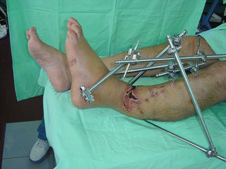
In patients who suffer from a one-side-located extensive soft tissue defect (especially wounds located on the anterior aspect of leg), the fracture site bone remains uncovered, even after performing the acute shortening procedure. In addition, further bone resection is often unacceptable. On the other hand, residual bone exposure indicates the need for soft tissue reconstruction by flaps as the only remaining alternative. However, acute shortening combined with angulation directed to the side of the main soft tissue loss to cover the exposed bone is the treatment of choice. The angulated bone fragments are fixed using a tubular external fixator or hinged Ilizarov external fixation frame (Fig. 6.2).


Fig. 6.2
Clinical image showing external fixation of the open tibial fracture after performing an acute temporary shortening and anteromedial angulation
Stay updated, free articles. Join our Telegram channel

Full access? Get Clinical Tree


