Key Words
first-degree burn, second-degree burn, third-degree burn, total body surface area (TBSA), escharotomy, tangential excision, skin graft or autograft
Synopsis
Burn care starts at the time of injury with appropriate wound care, fluid resuscitation, and nutrition. Appropriate early management of many burns can prevent infection, allow for healing without surgery, and prevent contracture formation. Knowledge of the burn wound is key to correctly classifying burn injuries and thereby determining which wounds require surgery.
Although the fundamentals of surgical burn wound management have not changed significantly in the last 30 years, we now understand that early and aggressive surgery results in higher survival rates and fewer complications. The use of topical antimicrobials has improved with many more options available, so systemic antibiotics can be reserved for true invasive infections. Early physical therapy with range-of-motion exercises and splinting can prevent many of the contractures that result in long-term disability after burn injury.
This chapter will focus on the surgical aspects of acute burn management.
Clinical Problem
Presentation
Burn injuries have a variable presentation depending on the depth and mechanism of the injury. Many burns are not of uniform depth, so there will be different degrees of injury in the same burn wound.
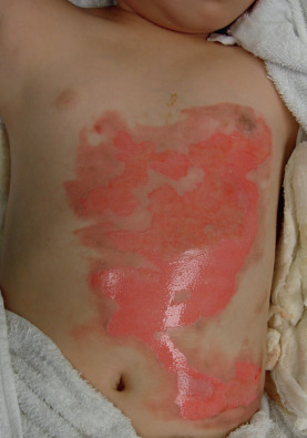
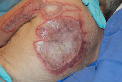
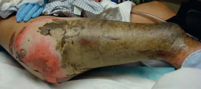
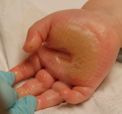
Burns that involve only the epidermis are classified as first-degree burns and do not warrant special care. Burns that involve the epidermis and a portion of the dermis are classified as second-degree or partial-thickness burns. These burns may be moist and pink or have fixed hemoglobin and appear cherry red and dry, depending on the depth of dermal involvement ( Fig. 4.1.1 ). Burns involving the epidermis and entire dermis down to the subcutaneous tissue are third-degree or full-thickness burns ( Fig. 4.1.2 ). These burns are white, yellow, or brown with a leathery texture ( Fig. 4.1.3 ). Lastly, burns extending through the entire dermis into the subcutaneous tissues, muscle, or bone are fourth-degree burns and are the most severe burns that we treat. These burns may present similarly to third-degree burns, and it is not until excision is performed that the depth is appreciated. Alternatively, they may present with black, charred eschar and exposure of the underlying structures.
Patients presenting with burn injuries should have their wounds classified using the terminology described earlier.
Etiology
Burn injuries can be described by the source of the heat: scald, flame, contact, and chemical ( Fig. 4.1.4 ). Although the mechanism of the injury may be different, the end result is the same with injury to the skin. Loss of a portion of the skin leads to lack of temperature regulation, fluid and protein loss, electrolyte imbalance, and a propensity for infection. Electrical injuries must be differentiated from other burn injuries due to their propensity for significant internal damage. The electrical current can travel through the body, emitting thermal energy internally. The cutaneous injury may be relatively small, whereas the underlying tissue necrosis may be extensive.
Associated Conditions
Burn injuries may result from a traumatic event such as a motor vehicle accident or explosion. These events put the patient at risk for concomitant traumatic injuries. The acute burn victim must be assessed for other injuries if the mechanism of the accident suggests that other injuries may be present.
Burn victims who are trapped in a house fire or other enclosed space have a risk for inhalation injury. Mechanical ventilation, bronchoscopy, and aggressive pulmonary care may be needed in addition to the care of the cutaneous burn injury.
Burn victims with diabetes and peripheral vascular disease who sustain lower-extremity and foot burns will have a protracted course and a high risk of limb loss due to inadequate blood flow to the tissues to aid in healing.
Elderly burn victims or those with significant medical comorbidities are at risk of death even with small burns under 10% total body surface area (TBSA).
Pre-Operative Management
Physical Examination
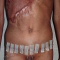
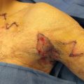
The depth of the burn injury is determined by physical examination. The color, texture, and moisture of the wound, the presence or absence of capillary refill, and the presence or absence of sensation are all findings on examination that help the physician determine the depth of the injury. Insensate, white, leathery skin that lacks capillary refill has sustained a third-degree burn. Pink, moist skin with brisk capillary refill is characteristic of a superficial second-degree or partial-thickness burn. Second-degree burns should be managed conservatively with wound care. Third- and fourth-degree burns require surgical excision and wound coverage. Deep second-degree burns can be treated conservatively initially to see whether healing will occur. If it becomes apparent over 10 to 14 days that the wound is not healing, then surgery should be performed. Full-thickness burns on the dorsum of the hands and feet, over the elbow, or over the anterior tibia should alert the physician that exposure of tendons or bones may occur with tangential excision. The surgeon should be prepared to manage these situations should they occur.
Pre-Operative Testing Needed
No specific testing is required for evaluation of the burn wound. In major burns (>20% TBSA) the hemoglobin or hematocrit should be checked before surgery and a crossmatch performed in anticipation of the need for a blood transfusion. Electrolytes should be monitored and repleted as necessary. In addition, the patient’s coagulation profile should be assessed, and fresh frozen plasma and platelets should be made available as needed.
Optimal Timing of Surgery
For full-thickness burns, surgery should occur as soon as the patient is resuscitated and is hemodynamically stable. This is usually 24 to 72 hours after injury. The wound will not improve without excision, so surgery should commence as soon as safely possible.
For deep partial-thickness burns, it may be difficult to determine initially whether the wound will heal in a timely fashion or whether it will require surgery. The surgeon’s goal is for the wound to be healed in 3 weeks. If the surgeon is unsure whether surgery is required, a period of waiting is recommended. By 10 to 14 days post-burn, the surgeon should be able to determine whether surgery is required for healing by day 21.
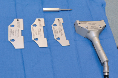
Supplies Needed
Watson knives
Weck knives
Epinephrine-soaked sponges: 5 cc 1/1000 epinephrine per 1 L normal saline
Electrocautery
Dermatome
Mesher
Burn dressings
Tumescent solution: 1 liter lactated Ringer’s or normal saline, 1 cc 1:1000 epinephrine, 30 cc 0.25% Marcaine
( Figs. 4.1.5 and 4.1.6 )