Abdominal wall reconstruction

12 Abdominal wall reconstruction


Introduction


The history of abdominal wall reconstruction is ensconced in the surgery-in-general literature. Abdominal wall hernias, which occur either congenitally (e.g., umbilical) or in an acquired fashion (e.g., postpartum, posttrauma, and postsurgery or iatrogenically), are routinely treated by general surgeons. Plastic surgery involvement in abdominal wall reconstruction came to the fore in complex cases where routine techniques exploiting local tissues and mesh would not suffice. Plastic surgery consultation became indicated for flap mobilization, regional tissue transfers, free flaps, and tissue expansion techniques. A seminal paper by Ramirez et al.1 revisited and popularized components separation as a useful technique in abdominal wall reconstruction. What has followed is an understanding of abdominal wall muscular dynamics, the role of component separation and muscle realignment as a central tenet of any reconstruction, and advances such as endoscopic or minimally invasive methods to perform component separation. Recent advances include perforator-sparing techniques, and evolution in biologic as well as synthetic meshes.


Closure of routine laparotomies itself causes many postoperative hernias. Easy to recall are the fulminant complications such as eviscerations, septic dehiscences, often despite retention sutures placed initially or at “take-back” surgery. Yet approximately 20% of laparotomies become incisional hernias in the US. Given that more than 4 million laparotomies are performed annually, this is a staggering number. Only about 90 000 are repaired annually, leaving both a huge backlog of cases that must be addressed as well as a future opportunity to deliver care to those who have limitations in activity, chronic pain, and functional issues such as chronic constipation or urinary retention from these hernias which are the sequelae of so many laparotomies.


Most surgeries are performed in the rich world – the world’s wealthiest 2 billion people get 75% of all the surgery done each year while the poorest 2 billion only get 4%.2 With increasing access to care, parts of the developing world will face similar challenges of abdominal wall reconstruction.


The ascendancy of the evidence-based medicine paradigm over the last two decades has focused attention on both the short-term failure rates of techniques as well as the long-term data. A landmark paper by Luijendijk et al.3 showed that, if followed for 10 years, the failure rates with primary suture techniques are 2 in 3, and with synthetic meshes 1 in 3. This is unacceptably high. Consequently, there has been an escalation in interest and an introduction of novel techniques in addressing these problems, as long-term data highlight the inadequacy of previous methods. The influence of evidence-based medicine has driven a resurgence of investigations into abdominal wall reconstruction techniques, as well as multispecialty collaboration between general surgeons, colorectal surgeons, trauma surgeons, minimally invasive surgeons, and plastic surgeons.


Patients and surgeons are seeking a decrease in herniation rates in the long term, not just a delay until reherniation. Quality measures, cost containment, and cost-effectiveness are paramount in the minds of institutions, payors, and credentialing committees, as we operate in a changing healthcare paradigm.


Historical perspective


Synthetic mesh-based reconstruction – a subject of fevered debate


Many abdominal hernias can, should be, and are repaired using synthetic mesh. The development of mesh was a major accomplishment and part of the long march forward in the repair of recalcitrant hernias.


The long-term failure of mesh can be ascribed to many causes, including cyclical fatigue and biofilm contamination. The repair breakdown is most often at the junction of the native tissues and the mesh, rather than through a fracture of the mesh itself. This occurs independently of suturing technique (interrupted or running) or suture material (absorbable or nonabsorbable).


The ultimate cause is posited as a failure of true biologic tissue ingrowth into the mesh. Tissue grows around the mesh (in the case of a porous mesh, the growth is around the interstices) and relies on scar strength to mitigate risk of recurrence. Biofilm contamination can lead to infection, which causes separation of the mesh from surrounding tissue, and thus failure of the repair and the need for removal of the device. Encapsulation, the body’s normal response to a foreign body by multinucleated giant cell reaction, can also lead to contraction of the mesh leading to failure at the margins where it is mechanically pulled away from native fascial edges. Encapsulation can then lead to extrusion and failure of the repair.


Currently, there is a shift towards biologic meshes and away from synthetic meshes, particularly in infected or contaminated patients, although each method of repair retains its own niche.


The following are broadly accepted contraindications to implantation of synthetic mesh: removal of previous infected mesh, enterotomies during adhesiolysis, peritonitis, repair of peristomal hernia, open abdominal wall wound, enterocutaneous or enteroatmospheric fistula, and colostomy reversal. A relative contraindication is a previous history of infection. Those with prior history of infection are at higher risk for repeat infection from indolent biofilm reactivation, hence the concern for bacterial implantation of a prosthetic device.


Discussion of biofilm pathophysiology is beyond the purview of this chapter. In brief, free-floating planktonic bacteria may become sessile, enabling adherence to various surfaces in the body, including implanted prosthetics. Numerous bacterial adaptive mechanisms have been revealed, including quorum sensing, reduced growth rate in the presence of antibiotics to decrease bactericidal vulnerability, and plasmid exchange to increase resistance to antibiotics. Approximately 95% of the biomass of a biofilm is bacteria – the remainder is a network of scaffolding that aggregates these bacteria. Scientific inquiry has accelerated on this subject but it remains a poorly elucidated topic at present, and its role in failure of abdominal wall reconstruction is only slowly being understood. Whether it is flagrant infection or insidious biofilm formation in the setting of fistulae, enterotomies, peritonitis, colostomy reversal, or open abdominal wall wounds, the odds of failure with a prosthetic in these scenarios are increased over autologous or biologic techniques which avoid synthetic mesh.


The components separation method


The technique most credited for the advancement in approach to abdominal wall reconstruction using autologous methods is the components separation method, as popularized in the modern plastic surgery literature by Ramirez et al. in 1990.1 Demonstrating clinical vignettes and elegant cadaver dissections, this manuscript advanced the technique of using bipedicled, neurotized, musculofascial advancement of the rectus abdominis muscles and internal oblique with transversus abdominis muscles. Historically, however, this technique had been mooted as early as 1916 by Gibson,8 then again in 1929 by Dixon,9 and finally in 1961 by Donald Young.4 Young described separating the transversus/internal oblique/rectus unit from the external oblique, similar in principle to components separation.


Components separation is a more nuanced way of performing the classic trauma teaching of lateral relaxing incisions. By laying bare the anatomy of the abdominal wall, Ramirez et al. showed that releasing the rectus abdominis muscle at the linea semilunaris from the external oblique muscles creates an avascular plane between the external oblique and the rectus–internal oblique–transversus complex.1 Since the segmental intercostal innervation to the rectus abdominis muscle arises deep to the internal oblique musculature, this technique preserves not only the vascularity but also the innervation to the muscle, which is principal in a stable reconstruction. Blood supply to the rectus abdominis muscle arises from segmental intercostals, as well as from the superior epigastric artery as it becomes the terminus of the internal mammary artery. The dominant blood supply is from the deep inferior epigastric artery (DIEA) as it arises from the external iliac artery.


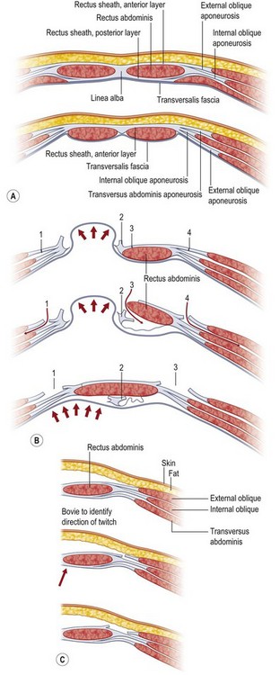
In 2000, Shestak et al.5 further described the components separation method and detailed its limitations. The surgical goal is to recreate the midline tendon (linea alba) where the paired rectus muscles are held to length, and the relentless unopposed lateral traction of the external oblique, internal oblique, and transversus complex is paired to the contralateral complex. The authors describe incising the fascia 1 cm lateral to the linea semilunaris, identifying the plane between the external oblique and internal oblique fascia (Fig. 12.1). They underscore the importance of dissecting superior to the internal oblique fascia, thus protecting the underlying nerves to the rectus abdominis muscle, and avoiding injury to the spigelian fascia (reducing the likelihood of a spigelian hernia). Extent of lateral dissection is determined by the amount of laxity present, with maximal advancement of each ipsilateral complex toward the midline of 4 cm in the upper abdomen, 8 cm at the waist, and 3 cm in the lower abdomen. An additional 2 cm can be advanced with elevation of the rectus muscle off the posterior rectus sheath (see Fig. 12.9, below).


As previously mentioned, the dominant perfusion of the soft tissues and skin of the anterior abdomen is the territory of the DIEA as well as contributions from the superficial inferior epigastric artery and from the intercostals. A notorious complication of components separation is marginal or incisional necrosis of the soft-tissue pannus, a result of devascularization of the anterior abdominal soft tissues by dividing the perforators that course through and perforate the rectus abdominis muscles. Several techniques have been advanced to minimize the risk of ischemic complications.10


Rise of the biologics – seeking the holy grail of ideal repair material


The ideal repair material should be biocompatible or inert, nonallergenic, nonresorbable/permanent, cost-effective, resilient (since no material can be resistant) to infection or contamination, kinematic and having mechanical characteristics with high tensile strength, radiolucent, accessible for repeat operative access for intra-abdominal process.


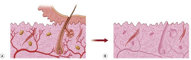
Those characteristics are potentially obtainable in many biologic meshes, which are sourced from acellular dermal matrices (human, porcine, bovine), or other large sheeted xenogeneic sources such as acellular bovine pericardium. Processing involves making these materials acellular to decrease the risk of rejection, as well as enzymatically cleaving the xenogeneic epitopes that could cause a cross-species reaction in biologic meshes derived from nonhuman sources (Fig. 12.2). Discussion of the available types of mesh is beyond the scope of this chapter.


Processing can further involve cross-linking to increase strength, although this may decrease the ability of macrophages and monocytic lineage cells to migrate into the biologic sheet. Cross-linking can make biologic tissues inaccessible to migration by cells that use metalloproteases to grow into tissues and facilitate inosculation of blood vessels into existing channels.


Biologics may be better able to tolerate exposure – and may granulate through if kept moist and not allowed to desiccate. Case studies and prospective trials have demonstrated that exposed biologic mesh may be managed by wet dressings, silver sulfadiazine dressings, or vacuum-assisted closure (VAC) utilizing negative-pressure wound therapy (NPWT).


Histological studies in both animal models as well as incidental biopsies of biologics from human surgery have demonstrated revascularization and recanalization of blood vessels and the presence of macrophages, elastin, and organized collagen in the explanted biologic biopsy.


In the setting of contamination, colonization, or infection, prosthetic repairs are at higher risk for failure and thus autologous reconstruction via flaps or biologic meshes is preferred.


Primary closure versus synthetic mesh repair


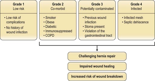
Factors such as body mass index (BMI), smoking status, nutritional status, and the presence of comorbidities (e.g., diabetes mellitus, chronic obstructive pulmonary disease (COPD)) have been investigated to see which predispose to postoperative herniation.14 The patients at highest risk of recurrence have an infected mesh or dehiscence from a septic cause. Potentially contaminated wounds (stoma, prior infection, enterotomy) are inherently higher-risk (Fig. 12.3). The problem remains multifactorial. Whether running or interrupted sutures are used, whether permanent or slow-resorbing sutures are employed, whether figure-of-eight knots or simple knots are put down, none has been conclusively shown to have a significant outcome difference.


When hernias are repaired, the long-term success of these techniques is surprisingly low. The authoritative papers by Luijendijk et al. in 20003 and by Burger et al. in 200415 indicate that, while mesh is superior to primary suture technique, even mesh repair over a 10-year follow-up period has a 32% failure rate. Numerous causes are thought to contribute to these findings – cyclical fatigue of the synthetic mesh device by constant respiratory excursion of the abdomen, lack of true bioincorporation into the mesh, or biofilm seeding leading to capsular contraction, encapsulation, and eventual extrusion.


Furthermore, each successive attempt at repair of a recurrent hernia is likely to be more complex and complicated than the previous, and, all other factors remaining constant, is less likely to be successful, since scarring is increased and local flap options may already have been exhausted.


Evidence-based medicine is now de rigueur when addressing almost any clinical concern. No longer satisfied with anecdotes, the savvy clinician demands the data – and it is this critical scrutiny that has highlighted the deficiencies of time-honored techniques and opened the surgical community to challenge the status quo. Collaboration and cross-pollination of techniques between the general surgery and plastic surgery disciplines have led to greater insights than each could have individually. The type of material used – acellular cadaveric dermis (from human or animal source) or different types of mesh (polypropylene, soft polypropylene) – remains a topic of debate in the literature, with studies revealing favorable results with either material.16,17


Basic science/disease process


Hernias and abdominal wall defects may be asymptomatic or symptomatic, and range from the minor cosmetic inconvenience to major destructive processes of the abdominal wall. Congenital umbilical hernias are usually repaired in infancy or childhood. Inguinal herniorrhaphy is similarly undertaken in childhood or in adulthood when they become symptomatic. Major defects such as gastroschisis or omphalocele are not addressed in this chapter – they require multiteam approaches and staged operations.


Acquired defects resulting from straining during heavy labor, load-bearing exercises, and childbirth are defects in the fascia that tend to enlarge, if unsupported by a truss or unrepaired. Narrow-neck hernias are at greater risk for incarceration and strangulation of bowel, whereas large-neck hernias, although more dramatic, are less likely to cause bowel trauma. Success rates with smaller defects in healthy patients are high. Some recur, and may require multiple surgeries.


Analysis of the National Safety and Quality Improvement Program (NSQIP database) highlights the surgeon’s intuition: comorbidities of smoking, diabetes mellitus, COPD, coronary artery disease, poor nutritional status/low serum albumin, immunosuppression, chronic corticosteroid use, obesity, and advanced age increase the risk for postoperative infection and failure. Infection, in turn, creates a higher risk of repair failure. Once the cascade is established, serial failures can turn a small, reparable defect into a challenge.


Other hernias begin as multiple small “Swiss cheese” defects, and when one defect is repaired, the other unrepaired defect(s) can enlarge. These defects may be dormant and arise as a consequence of old retention suture puncture points, prior stoma sites, or drain sites. Thus, some recurrences may not be true failures of hernia repair so much as failure of diagnosis of multiple defects. Ideally, there should be a preoperative computed tomography (CT) scan confirming the location and number of fascial defects, and at the time of repair there should be a wider dissection to identify occult hernias in proximity to the index hernia. Laparoscopic evaluation can provide a broader view of the abdominal wall and potentially identify previously occult defects as well.


Rarely, other failures may arise as a consequence of collagen vascular disorders. The patient with multiple failed attempts and signs consistent with Ehlers–Danlos syndrome, including hypermobility of skin and hyperextensibility of joints, should be referred to a rheumatologist for workup of collagen vascular disorders. This is relatively uncommon and incidence may be approximately 1 in 5000 live births worldwide.


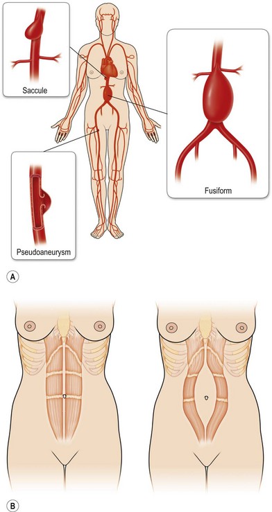
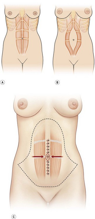
Rectus diastases are not true abdominal wall fascial defects but are pathological stretching to the linea alba either congenitally or, most frequently, postpartum. Functionally, a diastasis is analogous to an aneurysm – wherein the adventitia (fascia) and intima (peritoneum) are intact but the muscular layer is absent (Fig. 12.4). In techniques for abdominal wall reconstruction that do not centralize muscle, or cases in which the abdominal muscles have retracted beyond the possibility to be reapproximated, a functional diastasis remains, in lieu of the tendinous fusion of the paired rectus abdominis muscles. This diastasis can enlarge over time from intra-abdominal pressure, even to the point of requiring repair. Repair is achieved without intraperitoneal entry, by plicating or imbricating the defect so that the rectus abdominis muscles are returned to the midline (Fig. 12.5).


Hernias are challenging problems, and a significant fraction of hernia repairs fail, occasionally further complicated by enterocutaneous fistula formation, periprosthetic mesh infection, ulceration, and tenuous skin coverage. In particular, hernias that are re-recurrent will have the highest relapse rate and also the greatest predilection for complications.


A review of the long-term data reveals that Luijendijk et al. reported 43% versus 24% for initial recurrences and 58% versus 20% for secondary recurrences at 3 years.3 At 10-year follow-up, Burger et al. reported 63% versus 32% recurrence if primary suture techniques were used versus prosthetic mesh.15 Despite the reduction in recurrence rates, the rates remain unacceptably high.


The development and adoption of prosthetic materials have made a tremendous impact on the treatment of hernias. Synthetic mesh was introduced in the 1950s and has become a common component of the repair process. Data from the largest prospective, randomized, multicenter study show a significant decrease in recurrence rates in relatively healthy patients with small defects (<6 cm length or width) who received synthetic mesh versus those repaired primarily with suture alone.3


Diagnosis/patient presentation


Hernias should be treated as a chronic disease process – a notion that is gaining ground in surgical circles – a conglomeration of collagen disorders, excess mechanical loads, comorbidities, and outdated surgical techniques, and other poorly understood factors. Success is predicated on a systematic approach from understanding the etiology of prior failure, risk factors, metabolic status, the biology and biomechanics of repair materials, employment of an appropriate surgical technique (i.e., open or laparoscopic), to postoperative vigilance.


In the elective setting, the diagnosis of an abdominal ventral defect is made on physical exam when the patient presents with a symptomatic, intermittently painful bulge. In the emergency setting, strangulated bowel is diagnosed in the presence of a firm, painful bulge in a systemically ill patient, with fever, leukocytosis, and possible bowel obstruction. Diagnosis is made on physical exam and confirmed by CT scan. An appropriate workup includes plain films of the abdomen, complete blood count, basic metabolic panel, and lactate level. After fluid resuscitation and electrolyte correction, an urgent exploratory laparotomy must be performed. Definitive repair may or may not be performed at that time based on patient factors, comorbidities, and the contamination and condition of the wound.


Loss of domain occurs when muscle, fascia, and/or skin have necrosed or retracted and have become contracted over time and abdominal viscera extrude into the hernia sac. It is said that the bowels have “lost their domain” in the peritoneal cavity. Recreating this lost domain is the challenge and goal of the hernia surgeon.


Nonhernia abdominal defects may present from surgical oncology referral for anticipated plastic surgery closure at the time of resection of a segment of the abdominal wall. Preoperative imaging can delineate the involved structures and thus clarify the missing anatomic components in need of reconstruction, such as skin, soft tissue, muscle, fascia, and peritoneum.


Goals of repair include both form and function. Aesthetic correction of the bulge or skin-grafted deficient contour is important, but the main impact of reconstruction is on restoration of function. Re-establishment of fascial integrity restores the function of the remaining muscular elements. Once the muscles are reconnected as part of the same aponeurosis, they have paired and gain dynamic tension and countertension. In the presence of a fascial defect, the unopposed pull of the oblique and transversus abdominis muscles on either side of the defect leads to progressive lateralization of the muscles and enlargement of the defect. In a functional repair, the muscles are restored in continuity and are balanced. This improves the ability to ventilate more effectively and efficiently, as the abdominal muscles are accessory muscles of respiration. This is particularly germane in patients with concomitant pulmonary disease. Realignment of the muscle allows patients to Valsalva, generating pressure against the rectum, assisting defecation. Similarly, patients’ urinary retention improves since pressure is transmitted to the bladder to facilitate more complete emptying. Posture and back pain improve since the viscera are returned to their proper domain, and the load of the abdominal contents is replaced inside the abdomen. The moment arm of a lever is distance times force; when abdominal viscera are not contained inside the cavity, the weight of the visceral bulge causes strain on the back and postural muscles since the distance from the load axis (the spine) to the weight (viscera) is increased. With restoration of domain, the weight is returned closer to the load axis, thus decreasing the moment arm, and decreasing the strain on the supporting muscles (this is akin to why it is easier to hold a gallon of milk against the chest then to hold it at arm’s length). Studies and surveys have shown that activities of daily living improve, as do patients’ self-image and subjective well-being. Another functional improvement is regaining the ability to exercise, although in some recalcitrant cases it may be prudent never to allow that patient to have severe load-bearing activities again. In that setting, manual laborers may have to be trained in a different vocation.


In summary, repair and restoration of the abdominal wall provide cosmesis, postural and truncal stability, improvement in micturition, improvement in defecation, ability to perform activities of daily living, and also provide a satiety signal with restoration of physiologic intra-abdominal pressure, decreasing appetite.


Patient selection


Patient selection is less about declining the operation for certain individuals and more about eventually optimizing them for a bona fide chance at a durable and reliable repair. Patients who are not medically fit from a cardiopulmonary standpoint may be deferred or declined in conjunction with soliciting their wishes, risk aversion, and motivations. The only absolute contraindication is if the patient is medically unsuitable for surgical clearance.


Patients with ascites are extremely likely to have a poor outcome and should be referred to a hepatologist for management of cirrhosis or hepatic failure prior to attempt at a repair. Metastatic disease (hepatic, abdominal, or distant) is a relative contraindication, but hernia repair, as emphasized earlier in the chapter, is also about enhancing quality of life and diminishing pain. With current oncologic management, patients with metastases of certain diseases can be expected to live for years or decades, and thus cancer remains only a relative contraindication. Patients likely to need other intra-abdominal surgery may be postponed until their intra-abdominal issues are resolved, though this too remains a relative contraindication.


The largest category of denied patients is the noncompliant patient, whether this is noncompliance for weight reduction and nutritional repletion, noncompliance in nicotine avoidance, or noncompliance in seeking better glycemic control. Behavioral management, psychosocial support, and counseling can be enlisted to facilitate the goals of compliance. Those patients who lack adequate familial and social support to recover properly from surgery may also be deferred from surgery.


With weight loss in particular, patients report finding themselves in a Catch-22 situation – they cannot get their hernia fixed because they are overweight, and they remain overweight since they cannot exercise until their hernia is fixed. No easy solutions exist to this conundrum; caloric restriction, low-impact aerobic exercise, and water aerobics have been shown to be helpful. Socioeconomic barriers may prevent patients from getting these additional support modalities. For BMI > 40, a bariatric surgery referral should be considered. Patients whose BMI exceeds 40 are potential candidates for surgery if they clearly and realistically understand how their lives may change after surgery In certain circumstances, less severely obese patients (with BMIs between 35 and 40) also may be considered, since obesity-induced or exacerbated physical problems such as irreparable hernia are interfering with lifestyle or precluding or severely interfering with employment, family function, and ambulation.


Weight loss must not emperil adequate nutrition – this can be confirmed by obtaining assays of vitamin levels, albumin, prealbumin, and transferrin levels, and reticulocyte index. Even thin individuals may have diabetes or glucose intolerance. HgbA1c levels can be predictive of a diabetic diathesis and serum glucose levels greater than 110 mg/dL can suggest glucose intolerance. An endocrine consult may be helpful if either of these is found to be elevated, and an additional glucose tolerance test can be administered. Better glycemic control at the time of surgery with HgbA1c levels less than 7% is desirable to decrease the risk of perioperative complications.


Important vitamins and minerals include zinc for cross-linking collagen, vitamin C for collagen synthesis, vitamin A for counteracting the deleterious effects of corticosteroids in wound healing, folate, and thiamine. Other indicators of nutritional adequacy include albumin and prealbumin, transferrin, and reticulocyte index. The presence of elevated fasting blood glucose (above 110 mg/dL), recent smoking (less than 4 weeks’ nicotine compliance), use of bronchodilators (presence of COPD), and absence of weight reduction attempts are all potential indicators of a patient at high risk of recurrence. At-risk subpopulations should be given thiamine, folate, and multivitamins intravenously. Massive weight loss patients from bariatric surgery should undergo a preoperative planning session with their endocrinologist, primary care physician, or surgeon. This would involve checking and restoring iron levels and fat-soluble vitamins, including vitamin A, D, and, most importantly, K. Although vitamin B12 is water-soluble, the altered gastric physiology of most bariatric and gastric bypass procedures can predispose to vitamin B12 deficiency and its level specifically will need to be assessed and restored if indicated.


Smoking has multiple deleterious mechanisms of action on healing and recovery of the abdominal wall reconstruction patient. Combustion products from tobacco include hydrogen cyanide that causes oxidative decoupling and halting of cellular respiration by inhibiting the mitochondrial enzyme cytochrome C oxidase. Another combustion product, carbon monoxide, has a 200-times greater affinity for hemoglobin than oxygen, and thus decreases oxygen-carrying capacity by left and downward shifting of the oxygen dissociation curve, thus hindering release of oxygen to cells in the healing wound.


Nicotine (whether absorbed via smoking or smoking cessation medications such as nicotine gum, lozenge, transdermal patch, inhaler, or nasal spray) is a potent vasoconstrictor, enhances platelet aggregation, and directly damages the endothelial lining of blood vessels. Thus smoking and/or nicotine directly impairs microcirculation and decreases tissue oxygenation in healing wounds.

Stay updated, free articles. Join our Telegram channel

Feb 21, 2016 | Posted by in General Surgery | Comments Off on Abdominal wall reconstruction

Full access? Get Clinical Tree

Get Clinical Tree app for offline access