History of light-based therapies in medicine
The first records on using light for medical purposes date back to ancient Egypt (4000 BC), describing topical natural photosensitizer paired with sunlight for repigmenting skin of patients suffering from vitiligo.
By the end of the 18th century, ultraviolet (UV) light was introduced for sterilization after discovering that short-wavelength UV light influences bacteria. Shortly after that, in 1903, the first report on the effect of UV light on a biologic system was published when the light was used to treat lupus. , Throughout the 19th century, sunlight was used to treat European cutaneous tuberculosis.
Aside from these early reports on using light for medical purposes, Albert Einstein first described the theoretic underpinnings of lasers with the essential formulas and theoretic concepts of laser light in his 1917 paper, “The Quantum Theory of Radiation.” He explained spontaneous absorption and emission of energy by the interaction of atoms and molecules with electromagnetic energy and concluded that stimulated energy emission is possible. Townes, Gennadiyevich, and Mikhailovich further elaborated on Einstein’s fundamental work in the 1950s, and they were collectively awarded the Nobel Prize in Physics in 1964 for their contribution to quantum physics.
Simultaneously, based on Einstein’s concept, Townes and Schawlow developed the device called microwave amplification through stimulated emission of radiation (or MASER). But it was not until the 1960s that Theodore Maiman developed and successfully operated the first true laser using a ruby crystal. Leon Goldman, also known as the father of laser medicine and surgery, initiated the first clinical studies with the ruby laser in 1964. Over the following decades, the potential of lasers as a highly targeted, therapeutic treatment modality was recognized, and several additional lasers for medical purposes were developed. ,
Since Anderson and Parrish first described selective photothermolysis in 1983, the concept has evolved dramatically, and lasers of various specific wavelengths were subsequently introduced. , In 1983 Castro et al. and in 1984 Apfelberg et al. stated that lasers might effectively treat hypertrophic and keloid scars. After that, reports were published about the use of argon lasers (488 nm), neodymium-doped yttrium aluminum garnet (Nd:YAG) lasers (1064 nm), and common CO 2 lasers (10,600 nm) for the treatment of hypertrophic scarring, but with very high recurrence rates, most likely related to ablation that was too dense. , In 1994 the effectiveness of the vascular-specific flashlamp-pump pulsed dye laser (PDL) to improve erythematous and hypertrophic scars was demonstrated with lower recurrence rates. Over subsequent years the PDL (585 nm or 595 nm) became one of the most studied lasers for treating hypertrophic scars with significant long-term improvements and reduced need for surgical scar excision. ,
However, it was Manstein et al. in 2004 who introduced the concept of fractional photothermolysis, which turned out to be a significant breakthrough in the clinical use of lasers.
Laser principals
General laser basics
Lasers are devices generating energy in the form of a light beam of a specific wavelength, which is absorbed and transformed into heat by a particular tissue component with a subsequent modulating effect ( Fig. 53.1 ). , The word laser is an acronym for light amplification by stimulated emission of radiation.
The concept of a laser device. External energy supplies the laser cavity containing the laser medium where the resting atoms get excited. When more atoms are in an unstable high-energy configuration, population inversion is created, and photons are released for light amplification within the optical cavity/resonator. When sufficient intensity has been developed for complete amplification to occur, the photons are then allowed to escape through a partially reflective mirror, and the emerging beam of light gets delivered to the appropriate target.
(From Issler-Fisher AC. Ablative fractional resurfacing for burn scars & the impact on reconstructive burn surgery: Exploring the effects of a novel treatment paradigm [thesis paper]. University of Sydney; 2021.)
The same four components can be found in all lasers , , :
-
■
Active laser medium. The laser medium is a material of controlled purity, size, concentration, and shape, which determines the wavelength and name of the laser. It can be solid, liquid, or gas, comprising the atoms that release the photons when stimulated by an external energy source.
-
■
Optical cavity/resonator. The laser medium is enclosed by a resonate chamber in which the amplification process of the photons ensues by reflecting back and forth between two reflective mirrors. More and more atoms can become excited and return to their resting state, amplifying the generated energy. One of the two mirrors is partially reflective and partially transparent, allowing the amplified photons to exit in the form of a laser beam.
-
■
Energy source. The external power supply can be a light source, an electrical field, or a chemical. It is needed to excite the resting electrons in the laser medium’s atoms and create the population inversion. This means that more than one atom is excited than a lower unexcited energy state. Once the electrons fall back into their resting orbitals, energy in the form of photons is emitted and amplified in the optical cavity. The released photons are all identical and considered monochromatic (of the same wavelength).
-
■
Delivery system. The emerging beam of laser light is delivered to the target via a delivery system, usually in the form of a fiber optic or articulating arm with mirrored joints.
The distinct feature of the emerging laser light beam, compared to a standard flashlight, lamp, or conventional light, is that it is monochromatic, coherent, and collimated. , , , Monochromatic means that the photons of the laser light are of the same single wavelength (in contrast to, e.g., intense pulsed light [IPL], which consists of different wavelengths), approaching unity. Laser light is coherent, as the photons are in phase, and the waves of light travel spatially and temporally synchronized and reinforce themselves. By comparison, photons in conventional light move randomly. Lastly, laser light is collimated, which means the transmission of light waves is parallel without significant divergence of the emerging beam, which has a very high energy density. Depending on the wavelength, light energy can be visible or invisible with a spectrum of electromagnetic radiation ranging from long radio waves (wavelength >10 cm) to very short γ-rays (<10 -11 m).
Laser-tissue interactions
The laser light interacts with living tissue depending on the particular wavelength of the emitted photons. Clashing laser light with a tissue can produce desirable and undesirable effects. The laser energy can be reflected, scattered, transmitted, or absorbed. A fraction of the energy is bounced off the surface and redirected in different directions. This is called reflection, which accounts for approximately 5% of the emitted laser light. The amount of laser energy expanding spatially, passing further through tissue, and leading to irradiation over a larger tissue area is called scatter. Wavelengths between 400 and 1200 nm scatter the most. To achieve a specific biologic effect with minimal collateral damage, the light beam must be absorbed by a particular component in the skin. This exact absorption is usually a function of the wavelength and pulse duration of the laser. When the photons hit a particular molecular target in the tissue, called chromophores, the energy of the light is transformed into heat. , The laser-tissue interaction can be chemical (photochemical), mechanical (photoacoustic or photo disruptive), or thermal (photothermal). The latter is the most frequent effect exploited for plastic and reconstructive surgical purposes.
The so-called selective photothermolysis describes the mechanism whereby a particular laser targets specific chromophores. Reflection from the surface or imprecise absorption of light by chromophores leads to either no or an unwanted effect on the skin. Melanin, hemoglobin, and water (intracellular or extracellular) are the three main selectively targeted chromophores by laser devices with specific wavelengths. , Thus the wavelength must be selective for the targeted chromophore without damaging surrounding tissues for a particular therapeutic laser-tissue interaction. Increasing the external power supply can also enhance the laser beam’s penetration depth. Another important factor of selective photothermolysis is the thermal relaxation time of the treated tissue, which includes the time it takes a chromophore to dissipate two-thirds of the heat to the surrounding tissue during the laser pulse ( Fig. 53.2 ). To achieve an optimal outcome, aside from the wavelength, further factors should be considered when selecting a laser device for a specific indication, such as fluence/energy, power density, pulse width/duration, mode of delivery, and spot size (increasing spot size increases penetration).
Chromophore absorption chart.
(From Issler-Fisher AC, Waibel JS, Donelan MB. Laser modulation of hypertrophic scars. Clin Plast Surg . 2017;44[4]:757-766.)
Overview of types of laser modulation for burn scars
Laser scar modulation, particularly ablative fractional laser (AFL) devices, provides an enormously promising treatment modality to complete reconstructive burn surgery. It enables the surgeon to address various scar aspects not optimally attended to by classic reconstructive approaches. Therefore it is essential to consider the complexity of burn scars and necessary to acknowledge and individually identify the various burn scar characteristics (altered erythema and pigmentation, structural changes, tension, contour abnormality) as well as symptoms (pruritus, neuropathic pain, heat intolerance, inability to sweat, limited range of motion).
As each patient experiences scar characteristics and symptoms differently, assessing all these aspects and their relevance to the patient is essential to tailor a suitable treatment plan for each patient individually and holistically rather than just anticipating creating a more aesthetically pleasing scar.
General considerations
Overall, laser treatment is well-tolerated by patients; however, because burn survivors frequently have a different pain perception than other patient cohorts, , establishing a solid anesthetic plan with the patient is important. Pain management can include applying ice to the treatment area shortly before the treatment, topical anesthetic creams, local or regional anesthetics, sedation, and general anesthetic. The choice of anesthesia depends on various factors, including age (children usually require sedation or even a general anesthetic), comorbidities, size of scar treated, and the mode of laser treatment (e.g., ice and local anesthetic can change blood vessels, which might interfere with the targeted chromophore; ablative lasers are usually more painful than nonablative lasers).
Patients undergoing AFL resurfacing (AFR) are routinely prepped with chlorhexidine. Some burn centers recommend antiviral prophylaxis to prevent herpes simplex if the face is lasered. However, there has yet to be an international consensus on the necessity of prophylactic antibiotics, and practices vary significantly between countries. Antibiotic prophylaxis should be considered if a hypertrophic scar in a hair-bearing area is treated, especially if recurrent acute or chronic folliculitis occurs. Nonablative laser treatment does not require antimicrobial prophylaxis.
Generally, laser treatment is kept under 30% total body surface area (TBSA) to avoid a systemic inflammatory response syndrome–like response and management of postprocedural comfort, as well as time restrictions in operating theaters.
Postprocedural discomfort is often eased with ice packs or cold towels. Recommendations for postoperative wound care include antiseptic wash and emollient for several days or a dry nonadhesive dressing or petroleum gauze for 24 to 72 hours followed by emollient. Patients are advised not to be exposed to a dirty/dusty environment or extreme temperatures for 2 to 4 weeks, and adequate photoprotection (sunblock with sun protection factor >30 as well as protective clothing) is recommended for at least 12 months to minimize the risk for postinflammatory hyperpigmentation. Compression garments can generally be worn again after 3 days, and most patients return to school/work after 1 to 3 days.
Vascular lasers
Vascular lasers can effectively address erythema and associated symptoms, such as pruritus and neuropathic pain. The vascular-specific, flash pumped 585- and 595-nm PDL has probably been the most extensively studied vascular laser in the literature for burn scars. The selective photothermolysis of the blood vessels of the PDL leads to targeted vascular destruction resulting in tissue hypoxia, subsequently preventing excessive collagen deposition. Combined with collagen fiber heating and catabolism, the process results in collagen realignment and tissue remodeling. , , , Despite the broad use of this modality and the theoretic mode of action, fractional CO 2 laser treatments are more successful in decreasing hypertrophy and smoothing scars.
Lasers targeting pigment
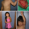
Lasers targeting pigment are used broadly for tattoo removal and undergo rapid and constant technical development. In burn scar treatment, the most common complaint is hyperpigmentation, especially in Fitzpatrick skin types 4, 5, and 6 individuals ( Fig. 53.3 ). Strict sun UV protection to prevent this hyperpigmentation is mandatory for about 12 months after burn healing. Unfortunately, the hyperpigmentation is challenging to treat because the target is not technically a distinct pigment granule such as in tattoos, where picosecond lasers have made great strides. In aesthetically prominent areas such as the face and neck, a lightening permanent makeup tattoo may lead to more satisfying results for the patient than laser treatments. IPL treatments with melasma filter and settings and Nd:YAG lasers can be attempted but require multiple treatments and maintenance throughout the years. The Nd:YAG laser has a wavelength of 1064 nm and can reach deeper layers of skin tissue than other lasers. The Nd:YAG laser can be used to remove brown age spots (solar lentigines), freckles, nevus of Ota, nevus of Ito, lumbosacral melanocytosis, Hori nevus, and café-au-lait-macules, as well as dark hair and certain tattoos.
Fitzpatrick classification of skin types I through VI.
Fractional lasers
Conventional ablative and nonablative laser resurfacing devices induce a two-dimensional thermal effect, whereas fractional lasers’ microscopic thermal tissue destruction is three dimensional. The laser beam is split into a pixelated pattern of microbeams leading to microscopic treatment zones (MTZs) and sparing surrounding tissue from where a rapid wound-healing response is initiated. These unaffected viable dermal islands act as a reservoir to promote neocollagenesis and tissue remodeling. Because of the limited collateral thermal damage, fractional photothermolysis is much safer than conventional laser resurfacing techniques.
Fractional lasers can be nonablative (introduced in 2004, heating the dermis to 50–70°C leading to irreversible tissue coagulation) or ablative (introduced in 2007, heating the dermis to >100°C inducing tissue vaporization with a narrow thermal coagulation zone around the ablated MTZs).
Nonablative fractional laser treatment for burn scars: Erbium:Glass and ResurFX
For fragile scars early after epithelialization, the nonablative fractional erbium:glass (1550 nm) and ResurFX (1565 nm) lasers have modulated the early stages of hypertrophic scar formation. Like the ablative fractional CO 2 laser (AFL-CO 2 ), small damage columns are inflicted upon the tissue without damaging the epidermis. The authors’ observation with both modalities, which are used routinely in their burn center in the early treatment stages, is that scar maturation can be hastened. The extent of hypertrophic scarring can be limited, but few studies have collaborated on these findings because of the minimal use of those entities in burn centers , ( Fig. 53.4 ).
Before and after treatment with nonablative erbrium:glass laser. Hypertrophic scar chin and neck before (left) and after (right) three nonablative erbuim:glass laser treatments, 1 month and 7 months postepithelialization.
Ablative fractional resurfacing for burn scars
AFR has led to a change in burn scar management, allowing the surgeon to approach burn scars holistically. The crucial feature of AFL is that it induces a release of tension in the spot over a larger body surface area (in contrast to a simple Z-plasty). A mechanical initial release is achieved by creating hundreds of microholes in the scar, which patients often describe as a feeling of immediate release after treatment of larger contracted areas. Once these small lesions contract again within 48 to 72 hours posttreatment, rapid wound-healing reactions induce a molecular cascade involving heat shock proteins and metalloproteinases from the islands of unaffected tissue surrounding the MTZs. The subsequent tissue remodeling initiates a decrease of scar tension via a change of dermal architecture and composition, ultimately resulting in increased pliability, flattened scar, and healing of chronic wounds in unstable scars. ,
Erbium:YAG laser.
Erbium-doped yttrium-aluminum-garnet (Er:YAG) laser skin resurfacing effectively manages several cutaneous conditions and lesions. Its main indications include treatment of photoaging, rhytids, and benign and malignant cutaneous lesions. The application in burn scar treatment has not yet been thoroughly studied.
Ablative fractional CO 2 laser.
With a wavelength of 10,600 nm CO 2 , water absorbs energy several millimeters below the skin’s surface. Various AFL-CO 2 devices featuring distinct technical characteristics amplifying safety and efficacy are available on the market.
The AFL-CO 2 UltraPulse laser (by Lumenis), including ActiveFX and DeepFX handpieces and the SCAAR FX mode, is the most used and studied device among burn centers internationally. A distinct pattern of fractional laser damage accomplished with high energy combined with a short pulse duration enables a precise and effective treatment. Different functions and handpieces of the device can address various aspects of a hypertrophic scar. More superficial ablation can correct surface irregularities and achieve a smoother scar appearance (e.g., flattening of the classic cobblestone appearance of a meshed skin graft). Dermal remodeling and associated release of tension are achieved by deeper fractional ablation. The selection of the AFL-CO 2 modality and required treatment settings allows the surgeon to modify and tailor the treatment to each patient.
Various studies have reported a significant improvement of cosmesis, functionality, and symptoms, such as neuropathic pain, pruritus, and heat intolerance, as well as improved quality of life of patients suffering from hypertrophic burn scars after treatment with AFL-CO 2 . ,
Further, because of its potential for accelerated scar rehabilitation and improved maturation, it has been anecdotally reported that treatment with AFL-CO 2 (if used carefully) can be initiated as early as the burn wounds are epithelialized. Early treatment of the panfacial area, in particular, may be beneficial for preventing and improving early eyelid ectropium and microstomia.
Laser-facilitated drug delivery.
For topical medication to be effective, the stratum corneum of the epidermis must be traversed. As such, AFL is an excellent tool to facilitate the delivery of drugs past the epidermal layer and significantly enhances the bioavailability of topically applied medications. The precise and uniform ablative channels resulting from the induced tissue vaporization provide an even distribution within the dermal layer. ,
Intralesional injections of antimitotic drugs, such as corticosteroids and 5-fluorouracil, are widely accepted as first-line therapeutic choices for small nodular pathologic burn scars. Corticosteroids are ideally injected into the papillary dermis, where collagenase production occurs. However, these drugs are usually injected with a 25- to 27-gauge needle into very dense scar tissue, often leading to local accumulation, frequently in a too-deep dermal layer, which can lead to undesired adverse effects. Therefore laser-assisted drug delivery (LADD) is an effective and safe technique to distribute drugs evenly over larger body surface areas.
It is common practice in some burn centers to use AFL to combine AFL with LADD of antimitotic medications. However, while the enhanced bioavailability of drugs administered via laser has been reported in various animal studies, it is still not entirely clear if the positive effect on hypertrophic scars is achieved because of the appropriately delivered and evenly distributed antimitotic drug, the laser-induced remodeling itself, or a combination of both mechanisms.
Although LADD for burn scars is commonly related to corticosteroids and 5-fluorouracil, there is a vast potential for delivering other medications. It is an underresearched treatment adjunct with up-and-coming prospects for scar rehabilitation.
Suggested burn scar treatment approach
General considerations
Without conclusive high-level evidence, it is generally considered that laser treatment, particularly AFR, is very efficient in immature and very mature scars and positively influences the entire rehabilitative process.
Because of the enormous heterogeneity of burn survivors, patient-specific factors must be considered, including Fitzpatrick skin type, ethnicity, burn mechanism, scar location, scar maturity, individual and personal circumstances, among others. Therefore each patient will require an individually tailored treatment approach, including one or several laser modalities combined with or without surgical interventions. The ideal time point for the start of laser interventions is undetermined; however, as mentioned earlier, treatment with AFL can be initiated as soon as the wound is epithelialized (as soon as 6 weeks after healing), potentially reducing contracture formation, increasing range of motion, and accelerating scar rehabilitation. However, particularly in a very immature scar, the treatment choice and settings must be altered to skin type, ethnicity, scar location, and scar maturation. To achieve an effective outcome, all laser treatments must be repeated multiple times; however, the number of required laser treatments varies significantly between patients. This also applies to the required interval between each laser session. It is recommended to wait at least 6 to 8 weeks between laser treatments; intervals can range between 6 weeks and more than 6 months. For example, patients with a light Fitzpatrick skin type with an immature scar may require longer treatment intervals because of persisting postprocedural erythema.
Another crucial factor to consider is acknowledging the scarred areas connected by the kinetic movement chain. In almost all burn survivors, there are thicker areas or thin contracture lines when stretched, which are exposed to movements of the greatest range of motion across joints following the laws of functional kinematics. Laser scar modulation allows the treatment of all interconnected structures by reducing the overall tension with the lowest recurrence rates. This can aid in avoiding recurrent contractures, especially if combined with surgical contracture releases, which means that not just the “contracture center” should be treated but the surrounding scar area that causes the contracture area to be tethered.
Related posts:


Stay updated, free articles. Join our Telegram channel

Full access? Get Clinical Tree


