Introduction
The restoration of skin integrity is fundamental in the treatment of burn injuries. Skin is a complex, multilayered, multifunctional structure that, in normal conditions, is maintained by both the cells within the structure and systemic cell migration. It is a dynamic tissue with cell responses essential in the repair process and normal homeostasis. The skin’s regenerative capacity is rapidly overwhelmed as the damage from a burn injury extends into the dermal tissue, with the wound healing response resulting in scar repair.
The appropriate clinical use of cell-based therapies in burn injury relies on assessing the injury in terms of the depth of injury, the percentage of total body surface area (TBSA) involved, and the body site. A clear understanding of the extent of the skin injury in the context of the patient’s capacity to heal will drive the selection of the appropriate therapeutic intervention (see Chapter 9 ). The planning of the interventions needs comprehensive knowledge of the current techniques available and the capacity to undertake the intervention in the environment of operation.
Cell transfer dates to the beginning of tissue transfer for wound repair. The split-thickness skin graft (SSG), the most frequently used surgical intervention, relies on the cells within the graft remaining viable during vascularization and integration; the process is described as take. In recent decades, cell-based therapies have emerged initially in response to the need to repair skin loss in areas exceeding SSG donor site capacity (see Chapter 49 ). The improvements in knowledge of the burn pathophysiology (see Chapter 7 ), resuscitation (see Chapter 8 ), infection control (see Chapter 11 ), nutrition (see Chapter 22 ), and advanced wound care have facilitated the early survival of patients with major burn, but long-term survival and the quality of survival hinges on timely skin repair, including the exploration of cell-based therapies (see Chapter 14 ).
Cells within the skin construct originate from all three embryologic layers and relate to their function within the tissue, expressing specific characteristics across different body sites. The predominant cell in the epidermis is the keratinocyte, and in the dermis, the fibroblast; as such, much attention has been paid to these cells during the evolution of cell-based therapies for cutaneous repair. However, there is much to learn from the interactions of the cells related to the vasculature, innervation, skin adnexal structures, and the melanocytes, dendritic, and immune cell populations ( Fig. 51.1 ). Understanding the capacity for skin regeneration and the cellular activity when the tissue is stressed has been pivotal in understanding the capacity for the cells to be cultured and investigated in vitro. Cells are the building blocks for tissue regeneration; the interaction with the extracellular matrix (ECM) in everyday and healing situations has been extensively studied, with the production of the ECM by the cells being directly related to the expression of cell phenotype.
Skin is a complex multicellular structure that includes many specialized cells in addition to the keratinocyte and fibroblast.
Tissue engineering, combining engineering principles with biology, has expanded over the last few decades, aiming to provide skin repair solutions. The goal is to improve clinical outcomes by providing alternatives to SSG for skin repair. There are many issues to consider in developing a skin repair solution.
Incorporating technology into the clinical model of care (MOC) requires understanding the indication for surgery and the timing of the surgical intervention (see Chapter 9 ). The time taken to produce an autologous cell-cultured solution may be prohibitive, such that off-the-shelf allografts have been explored or noncellular temporizing solutions used until the cultured cell solution is available (see Chapter 14 ). The need for a timely approach has also led to the development of point-of-care cell harvesting for immediate application to the prepared wound bed. The sources of the cells to facilitate the repair have included allograft and autograft cultured keratinocytes and fibroblasts; more recently, stem cells such as mesenchymal stem cells (MSCs) and adipocyte-derived stem cells (ADSCs) are being investigated for skin repair. All cells, either cultured or minimally manipulated, must demonstrate wound adherence to be effective in proliferation, migration, and differentiation to develop the skin construct.
Further, the incorporation of a cellular therapy into the MOC needs attention not only to the timing but also the technique of surgical debridement and wound bed preparation such that there is a framework for the cells to express the appropriate phenotype, as seen with the tissue-guided regeneration with the use of dermal scaffolds, such as Integra Dermal Regeneration Template. There is a need to shift focus to regenerative rather than scar repair to reduce the injury’s impact and restore function.
Fundamental to using cell therapies is the capacity to cover large wound areas promptly. The increased coverage area can be achieved simply by meshing an SSG or using the Meek technique. There is a limit to the expansion; the scar quality is increasingly compromised as the expansion ratio increases. The use of laboratory-based tissue expansion allows the coverage of extensive areas from a modest donor site. The cells are prepared by dissociating the tissue and releasing them into suspension for immediate delivery to the wound, or cultured in the laboratory as sheets or in composite skin-like structures. The capacity for expansion gives flexibility regarding the body site of the donor site, and therefore the opportunity to match the donor and recipient sites. The expansion of the wound area covered also ensures the reduction in donor site morbidity, reducing the size of the donor site harvested and reducing the need for secondary donor site harvesting.
There is a complex research, development, and governance pathway in developing all techniques, with approvals required before clinical use. There are a range of products currently available, but none to date address all the issues of full-thickness skin repair. The techniques available must be well understood to integrate them into a given MOC for optimal outcomes and to provide the platform of knowledge for the next generation of products. , The techniques and their utilization will be discussed in the context of introducing cell therapies into burn wound repair.
Background to the problem
Skin grows and self-organizes to tissue with specific body site characteristics, continuously replenishing over time. The cells within the epidermis and dermis are constantly dividing with ECM turnover and epidermal integrity retention. The rate of cell proliferation changes with age in response to trauma and in association with dermatologic conditions. The capacity for regeneration is overwhelmed by all but trivial injuries. Research into cellular responses to wound healing has opened opportunities for skin cell therapies. Burn injury is common and is frequently associated with skin damage that exceeds the regenerative capacity of the wounded area, and skin integrity is restored by scar repair. The scarred skin area will not provide a full functional restoration of the tissue. The time taken to heal is the strongest predictor of scar outcome.
The priority for significant burn injury is survival, but we also need to focus on the functional and aesthetic quality of the outcome. Acute burn care knowledge has advanced with the rapid institution of supportive measures facilitating survival (see Chapter 26 ). The need to repair the skin deficit has driven innovation in advanced wound care systems, skin substitutes, and cell-based therapies. The donor site’s availability limits the capacity to heal the wound using traditional SSG. Indeed, the capacity to restore skin integrity is essential for survival, but it comes at the cost of the noninjured skin donor sites and the scar quality of a SSG repair. SSG’s do not replace the full thickness of the skin and the donor sites and often do not match the recipient sites.
The timing of burn surgery can have a significant impact on the outcome for the patient. The early initiation of surgical intervention can aid in reducing the severity of the burn wound, minimize the extent of tissue damage, reduce the risk of infection, and limit the progression of inflammation and the extent of blood loss. , This in turn can lead to improved wound healing, reduced scarring, and improved function, reinforcing the need for expedient skin restoration. To facilitate early surgery, the current widely used options for skin repair in major burns where SSG donor sites are limited include the following:
-
■
Use cadaver allograft and serial harvesting of the available SSG donor sites to replace the allograft as the patient donor site heals (see Chapter 13 ).
-
■
Use dermal substitute scaffolds, such as Integra or biodegradable temporizing matrix, which require up to 3 weeks to restore the dermal elements, and then epidermal repair with thin SSG, which can be meshed and used in conjunction with cultured epithelial autograft (CEA) sheets or epithelial cell suspension (see Chapter 49 ).
-
■
Harvest an autologous skin biopsy for cell culture using laboratory-based tissue expansion to increase the number of cells available to regraft as CEA cell sheets or a cell suspension, which can be available at the point of care. The CEA takes up to 3 weeks to be ready for clinical use when cadaver allograft or dermal substitutes can close the wound (see Chapter 14 ). The multicell-layered cultured skin substitutes may take some weeks to produce.
Although cell-based therapies have been increasingly used since the 1980s in the context of major burns, there remains a significant need to achieve the goal of a one-stage regenerative skin repair at the time of burn wound excision. Further, there is a need to improve skin repair beyond an SSG scar in nonmajor burn injuries. Current cell therapies provide the opportunity to provide an epithelial repair with preservation of the dermal wound bed or in conjunction with thin meshed SSG, improving scar outcome and reducing donor site morbidity. With a focus on regenerative repair and the understanding of the turnover of the cells and ECM in the skin construct, there is the opportunity to explore the concept of treatment of the established scar with cell therapies.
When considering all the factors involved in surgical decision-making, we now also consider what opportunities advanced wound care, skin substitutes, and cell therapies provide. Cells are the building blocks for life and can change phenotype, as seen when the skin heals by scarring. The aim is skin regeneration, so we need to understand what is needed for regenerative skin repair:
-
■
Source of cells capable of differentiation
-
■
A framework for cell migration
-
■
Three-dimensional (3D) spatial information of the wound area
-
■
A feedback mechanism to guide self-organization
-
■
Delivery techniques of the cells and matrix
-
■
Fibrosis management for tissue integration
Cell-based therapies provide the source of cellular material, and research and development over the last decades have provided some insight and solutions to the needs listed. By manipulating the surface with various biologic and innovative dressing systems, the time to heal can be reduced. For the opportunities of cell-based therapies to be realized, there is a need to understand the role within the continuum of care and integrate them with advanced wound technologies.
Applied anatomy
The complex structure of the skin is continuously responding to internal and external changes. Despite the multiple cell types derived from all three embryologic layers in the skin construct (see Fig. 51.1 ), most focus has been on the keratinocyte to restore epidermal integrity and the fibroblast for dermal repair. Restoring an epidermal layer is a priority, limiting fluid losses and reducing the risk of infection. In a deep wound, epidermal repair alone will result in scarring, so attention has been paid to the introduction of the fibroblasts as a suspension, in a cultured construct, or endogenously as a dermal scaffold or SSG meshed widely, thus providing an environment for fibroblasts to express the appropriate phenotype.
The cell therapies in clinical use have addressed some aspects of skin repair, specifically dermal structure for durability and epidermal repair for fluid and infection control ( Fig. 51.2 ). Cells cannot be considered in isolation from the ECM, providing the framework for cell adhesion and expression. The ECM is dynamic, with the turnover rate varying with age and pathology. Regarding the adnexal structures, , cells related to immune functioning, vascularizations, and innervation, despite extensive research, the clinical progress to date has been limited. These are all potential areas for future development as the understanding of the role of exogenous versus endogenous cell populations increases along with the role of manipulating the architecture and chemistry of the frameworks for cell proliferation migration and differentiation.
The choice of cell therapy and skin substitute needs to be done with the knowledge of the extent of the injury and the capacity to heal. Superficial wounds will heal with conservative treatment. As the wound extends deeper into the dermis, salvage of the dermis can be facilitated by using cells to provide the epidermal integrity. As the dermis needs to be introduced into the wound, meshed split-thickness skin graft (SSG) and cell therapy combinations can allow a larger area of cover for a given donor site and fade the mesh pattern. Dermal restoration can be achieved with a dermal scaffold, which will guide the cells migrating from the wound bed to form a new dermis, in situ tissue-guided regeneration, with a combination of traditional and cell-based therapies for the epidermal repair.
Classification of cell source and delivery
Cell therapy is defined as the use of living cells to restore tissue function and has been used for many decades; the most common example is blood transfusion. The first reports of the use of skin cell therapy date to the 1950s, with the current clinical use of CEA initially reported in the early 1980s. There is increasing interest in systemic manipulation of the inflammatory response to facilitate endogenous cell migration into the wound for repair, enhancing the intrinsic processes that have become overwhelmed. However, there are currently no approved systemic therapies in clinical practice, and discussion will be limited to introducing cells into the wound site (see Chapters 14 and 49 ).
Cell therapy can be classified according to the source of the cells used, both allograft and autologous, concerning the following:
-
■
Capacity for differentiation
-
■
Extent of laboratory manipulation
-
■
Final phenotype
The aim is for the cell delivered to the wound to have biologic activity to facilitate regenerative healing. The capacity for the cells to remain in situ and become integrated into the developing tissue relates to their origin, phenotype, and capacity to regenerate.
Stem cells are defined as undifferentiated cells capable of asymmetric replication with the retention of the replication capacity while facilitating a line of differentiated cells into mature tissue-specific cells. The capacity of the local stem cells within the skin is frequently overwhelmed in burn injury. The systematic response results in many cell types migrating to the wound area, and the phenotype change results in a scar repair. The aim of cell therapy is the introduction of a cell population into the wound that will restore the surface of the skin and reduce the ongoing inflammatory response, modulating the environment to facilitate regeneration.
Stem cells are classified based on their regenerative potential. Embryonic stem cells are pluripotential cells that can differentiate into any cell in the body. Adult stem cells are resident in most tissues but in small numbers such that in trauma and disease, their capacity to regenerate is overwhelmed. The largest populations derived from the bone marrow, MSCs, and the adipose tissue, ADSCs, are classified as multipotential and capable of differentiation into some tissues, including skin. All cell types can be harvested from the patient for processing and reapplication to the prepared wound site or from a suitable donor. The time taken for processing has led to the exploration of the use of allogeneic cells, such as MSCs, considered immune-privileged with a reduced risk of rejection. The MSCs can be banked and potentially available during the acute burn phase. The ADSC has been harvested for immediate use and postlaboratory processing for delayed application. There is the potential for such stem cell populations to differentiate into the skin adnexal structures, which would improve the quality of skin repair in the future.
As indicated, the most frequently used cell therapy in skin repair has been the differentiated cells, specifically keratinocytes and fibroblasts harvested from the individual or used as an allograft. In 1975 Rheinwald and Green demonstrated that disaggregated epidermal cells could be isolated and serially subcultured in vitro. The viable epithelial sheets suitable for grafting were produced. It was reported that epidermal cells grown in vitro did not consistently express the HLA-DR transplantation antigens, hence the exploration of the clinical use of cultured allografts in wound healing. The use of allograft cells had significant appeal as the concept lends itself to the off-the-shelf approach. Banked allograft cells can be rapidly expanded to meet the needs of the clinical timing. The drawback of allograft use is the lack of permanency, with the cells being replaced over time. The temporizing nature can be used specifically for the biologic activity related to the cells assisting in tissue salvage and epithelization. The allogeneic skin substitutes include those from natural sources, such as cadaver allograft and human amniotic membrane, processed and frozen or freeze-dried stored ready for use; these will express the immunologic crucial HLA-DR antigen by the Langerhans cells and therefore will be rejected over time.
The stored cells require a delivery system or technique; allograft can be delivered as an intact sheet of cells or as a cellular suspension, and a suitable dressing system must protect the wound. Alternatively, the cells may be grown or mounted on a dressing system, such as TransCyte, made of neonatal foreskin fibroblasts in a silicone sheet, frozen, and stored for use in partial-thickness burn injuries. The need to provide skin coverage in inadequate donor sites can be addressed by a temporizing technique involving allograft or by laboratory-based tissue expansion by culturing elements of the uninjured skin.
Since the initial reports in the 1980s, for example, the survival of patients with significant burns treated with cadaver skin allografts and subsequently autologous keratinocyte cultures and children with full-thickness burns to the thigh demonstrated a superior aesthetic outcome with cultured keratinocyte sheets expanded compared with the areas simultaneously treated with meshed SSGs. , The clinical experience has led to an understanding of the performance of cell therapies in wounds of different depths. In a full-thickness wound, epithelial cells alone will result in a fragile scar repair; therefore adding the dermis is essential. In the dermis, the fibroblast is key to establishing and maintaining the ECM, influencing the phenotype. There is a role in the use of epithelial cells in partial-thickness wounds, including donor sites, and in combination with traditional meshed SSG as well as in combination with dermal scaffolds.
The integration of epithelial cells into clinical practice has been varied, with issues relating to the availability, cost, and complications. Understanding cell biology guides understanding of the potential functionality in the debrided burn wound bed. Take of the cell therapy is via cell-cell and cell-extracellular matrix adhesion, not vascularization as with traditional SSG. Therefore the wound debridement must be meticulous, as contaminating bacteria can disrupt the adhesion, leading to poor take. The clinical use of cell therapy requires consideration of the dressing systems used, as the surface is fragile and requires protection until maturity is achieved. Timing is also a consideration; the laboratory culture process of 3 weeks is a significant concern when aiming to attain expedient wound healing to reduce long-term scarring.
The clinical observation was that the more immature CEA sheets performed better on the burn wound with less blistering and secondary breakdown. The option to harvest the cells from the feeder layer before achieving confluence allowed the epithelial cells to be used clinically from day 5 in the laboratory as a cell suspension. The cells need to be delivered evenly across the wound surface, which was achieved using an aerosol spray system. The clinical observation was then made that the more extensive wounds treated with autologous cell suspension had a superior scar outcome in some cases than those treated with traditional SSG. The use of laboratory-based cell therapies is complex, costly, and therefore difficult to justify in more minor burn wounds. The next step in the evolution of the technology was the development of a point-of-care cell harvesting device. They were harvesting autologous cells from an SSG’s dermoepidermal junction (DEJ) by enzymatic and mechanical processing undertaken in a medical device, such as the ReCell kit. Using cells harvested at the point of care allows cell expansion in a similar ratio as achieved in the culture process with the wound bed replicating the laboratory feeder layer. The technique requires a small donor site; therefore the donor site can be more closely matched with the recipient site to facilitate matched skin characteristics. The mixed cell population, including melanocytes, has the potential to reestablish the pigment load in the skin, restoring standard color.
When using cell therapies of all types of surgical debridement techniques, it needs consideration that the wound becomes the tissue culture environment. Surgery was undertaken with meticulous attention to detail to salvage the dermal framework where possible while ensuring all necrotic and contaminated tissue was excised. Equivalent healing with potentially improved scar compared with meshed SSG with a smaller donor site has been demonstrated. The timing of cell availability for clinical use is critical in surgical planning based on the patient’s assessment, the team’s experience, and the availability of cell-based materials.
The clinical use of cell-based therapies requires a delivery system that is robust and reliable. Initially, the cells were delivered in sheet form, mimicking skin grafts. Carrier dressings allow transport from the laboratory to the operating room. The knowledge of the cell behavior in a range of conditions (e.g., differentiation of the keratinocytes at an air-liquid interface) allowed the development of more substantial sheets of cells. Cells were introduced into various carriers and developed into laboratory-based composite skin substitute sheets for clinical use. The structured sheets are familiar, but as with a traditional skin graft, they require the surgeon to tailor the sheet to the underlying wound bed. The take by revascularization needs accurate application and fixation of the graft to the wound.
As indicated earlier, the cell therapy may be applied to the wound as a suspension in some systems and as an aerosol spray, facilitating the spread of the cells across the wound. There are systems, including the codelivery of cells with peptides, that assist with cell adhesion to the wound. The development of 3D printing technology, such that cells can retain viability, has led to research in differential placement of cell populations. 3D technology has been used in the laboratory to develop composite skin substitutes that combine 3D printed matrices with the accurate placement of cells. Understanding the chemistry that can support cells and facilitate phenotypic development to produce tissue has expanded rapidly. The concept of a point-of-care 3D printed solution for skin repair is now being explored using various printing techniques, such as coaxial, ink drop, and electro spun devices.
Recent progress has been made concerning the skin adnexal structures with an understanding of the architecture, chemistry, and placement of cells needed to facilitate the phenotype development. Growing hair has been a long-term goal with limited clinical use but an area of great promise based on extensive basic science knowledge. Further, the knowledge of the interaction of the developing skin construct and the immune system and healing processes will influence the integration of the repair.
Principles of management
Burn wound care is complex and requires a dynamic approach to decision-making as the patient and wound progress over time, and the use of cell therapies needs to be taken in the context of the following:
-
■
Would the wound heal without intervention?
-
■
What are the risks involved in surgery?
-
■
When is the best time to have the surgical procedure?
-
■
What technique will result in the best outcome related to the injury?
In the complex decision-making matrix, priority must be given to the factors that influence the clinical outcome:
-
■
%TBSA
-
■
Depth of injury
-
■
Anatomic location of injury
-
■
Age of the patient (see Chapter 29 )
-
■
Comorbidities and associated injuries
In addition, the timing of the surgery will influence which techniques are most appropriate for repair along the clinical journey.
Knowledge of all the techniques available for skin repair with a complete understanding of the indications is essential in integrating cell therapies into the MOC. The clinical needs of skin restoration have driven wound regeneration research and development. The clinical use of the available cell-based therapies with detailed analysis reported is essential in continuing the innovation to achieve regenerative skin repair.
Based on the diagnosis of depth and extent of injury and considering the patient’s condition, a decision to proceed with surgery is made. Preparation for surgery involves optimizing the patient’s condition based on understanding the role of resuscitation and the factors that will influence healing, such as infection control and nutrition (see Chapter 45 ).
The wound preparation involving infection control and potential tissue salvage can begin before surgery with attention to edema control and techniques, such as negative pressure. The diagnosis of the extent of the injury will also influence the timing of the planned intervention, bringing together the patient’s needs, the experience and knowledge of the clinicians (see Chapter 2 ), and the operative environment with available techniques.
Meticulous wound bed preparation is key in burn surgery, especially when using cell-based therapies. The burn-injured tissue can be debrided using a combination of techniques: tangential excision, sharp dissection, diathermy, ultrasonic scalpel, and dermabrasion. The wounds are irrigated with copious quantities of warm sterile saline, and homeostasis is achieved using a combination of cautery and topical adrenaline and tourniquets where appropriate. If tumescence is used, then attention must be paid to understanding what tissues are salvageable. The chemical and enzymatic techniques have been developed specifically to debride only necrotic tissue, salvaging all viable elements in the wound bed. Facilitating tissue salvage with dermal salvage allows the use of cells in isolation to increase the speed of healing with the associated reduction in scar risk.
The principle of dermal salvage is linked to the capacity to restore epidermal integrity rapidly, reducing the ongoing inflammation and secondary tissue loss. Timing is essential when preserving the dermis as the inflammatory response builds within the tissue, which is also associated with an increased risk of bleeding during surgery. The development of the point-of-care system for delivery of cell suspension harvested from the DEJ can be used off the shelf at any time. Early surgical intervention will reduce the toxic load of the burn injury, reducing the ongoing drivers of inflammation and infection risk, and has the potential to reduce the time to healing and therefore scarring. Surgical planning involves all areas of the injury, the debridement, the repair, and the timing. Effective surgical planning and communication in surgery are of the utmost importance and are linked with surgical time. A surgical planning tool is useful in ensuring all team members are fully informed and assists in data tracking ( Fig. 51.3 ).
Surgical planning sheet to facilitate surgical preparation for the team, ensuring the correct equipment is available and tracking the site of the skin repair.
Wound bed preparation needs to be thoughtful and undertaken with a clear understanding of the opportunities and concerns of the chosen repair technique. In addition, consideration needs to be given to the source of the cells and, precisely, the donor site requirements. The donor site is an iatrogenic injury, and every effort should be made to limit morbidity. The opportunity for cell expansion will allow a larger area to be covered, reducing the donor site size. For example, when harvesting skin in the ReCell kit or for CEA, the depth must be just beyond the DEJ. Using the dermatome on the setting of 0.003 to 0.005 in (0.08–0.13 mm) will yield a sufficient cell load and rapid healing with a capacity to reduce the donor site scar risk and allow for reharvesting if needed. The donor site will heal more rapidly if some harvested cells are delivered to the surface, reducing scar risk.
The delivery system dictates the application of the cell therapy to the prepared wound bed. Still, the principle of intimate and stable fixation to the wound bed is critical, as with any skin grafting, to ensure take. The mechanism of take of the cell therapy will vary; initially, the adhesion of the cells to the wound surface is essential in anchoring the construct or cells to the surface. Cell adhesion may be enhanced by the introduction of fibrin into the delivery system. The use of polymers to augment cell adhesion needs to be undertaken with the understanding that concentration is critical to avoid the prevention of migration of the cells. Once delivered to the wound, the cell therapies used in isolation or combination with other techniques require a dressing system that will protect the surface and facilitate the migration, proliferation, and integration of the developing skin construct. The cells introduced into the wound as a suspension are dissociated, and the surface integrin expression is modulated to facilitate adhesion. Similar to the healing process by secondary intention, the cells at the margin of the wound change character to reduce the intercellular adhesion to allow migration and proliferation across the wound surface to restore epithelial integrity. The cells introduced will have a range of phenotypes, and some will be terminally differentiated but retain a biologic capacity to influence wound healing with the potential to enhance the migration of systemic cells into the area.
The requirement of establishing vascularization is achieved by the ingrowth of the cells from the wound bed and the development of new vascular structures. The dressing system needs to afford protection by providing a barrier to infection and assisting in controlling hemostasis and edema. The layered dressing system allows for shear between the layers rather than on the wound surface, allowing modest mobilization of the patient postsurgery.
In summary, all the principles of burn surgical planning, preoperative optimization, meticulous debridement, hemostasis, infection control, accurate placement, and fixation of the skin repair product need to be considered. Every effort should be made to match as closely as possible the repair with the anatomy of the wound in terms of depth, surface area, and body site. The dressing system needs to be tailored to the requirements of the specific techniques, considering the basic principles of physical protection, infection control, and comfort ( Fig. 51.4 ). Early surgical intervention is associated with improved patient outcomes and with associated improved mortality. In planning surgical intervention, it is essential to understand the potential risks and how the risk profile relates to the time since the injury.
The model of care has developed with the aim of tissue salvage when possible, excision of burn tissue between day 2 and 5 postinjury, focused on minimizing the time to healing. %TBSA , Percentage of total body surface area.
Cases
The case studies will demonstrate the use of autologous cells harvested during surgery in line with the MOC highlighted in Fig. 51.4 . The MOC has developed with cell therapies in over 5000 cases, initially as CEA in the early 1990s, evolving to using point-of-care harvested autologous cells in over 5000 cases.
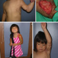
Pediatric scald injury is the most common burn injury in children, with the potential for devastating lifelong consequences (see Chapter 3 ). The temperature profile of the injury is frequently less than 62°C below the temperature at which the collagen is denatured and the dermal framework is retained. First aid with cool, clean water within the first 3 hours of injury will reduce the impact of the injury. Infection control is essential with topical antimicrobial dressings until clinical assessment. The superficial injuries are treated conservatively, and if healed within 14 days, the risk of scarring is modest (see Chapter 47 ). The capacity to rapidly repair the epithelial layer will reduce the time to healing. Wound bed preparation aims to salvage the dermis using dermabrasion or ultrasonic scalpel, achieving a clean, dry wound bed. Cells are harvested from a thin SSG donor site matched to the recipient site when possible. The x1.5 cm 2 SSG is processed in the ReCell point-of-care device by enzymatic and mechanical debridement, producing a mixed suspension of cells from the DEJ. The 4 mL of cells are delivered to the wound as an aerosol using a nozzle attached to a standard 5-mL syringe containing the cells. A layered dressing system is firmly applied and remains intact for 7 days if the patient remains well. The outer dressings will shell off, often leaving the primary protective layer intact. This may be soaked off in a bath or soaked with oil for removal the subsequent days.
The healed surface is cleaned, moisturized, and protected by a light compression vest. By 6 weeks, the skin construct matures, and the child has returned to life as normal. At 6 months, the area had no visible scarring.
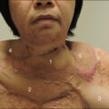
In adults, flame burns are the most common, frequently resulting in deep dermal or full-thickness injury. The use of laboratory-based cell therapies for nonmajor injuries is prohibitively expensive and involves a time delay. Although traditional SSG should result in a healed wound, it will always be associated with a scar, and the donor site area must be adequate for wound cover. The use of cell-based therapies has the potential to reduce the scar and donor site area. Clinical assessment at 48 hours is focused on answering the question, “Will the burn wound heal within 14 days?” If not, then surgery is planned. The donor site will depend on the depth of the burn wound and the potential for dermal salvage. In areas of deep dermal injury, a thin meshed SSG will be used to introduce a papillary dermal element along with the cells harvested from the DEJ. In middermal burns, the cells can be used in isolation. The donor site area needed will be measured to ensure it is kept to a minimum, the posterior lateral thigh is frequently used, the skin is thicker, and it is out of the line of sight of the patient if a scar persists. Burn wound debridement is achieved using scarp dissection, dermabrasion, and hemostasis. A thin meshed graft (1.5:1, 2:1, 3:1) is widely spread onto the deep areas of the wound. The cells harvested from 4 cm 2 of the SSG in a volume of 10 mL are sprayed onto all the scars, including the donor site. The dressings are prepared and placed under the wound area before spraying the cell suspension. Any cells that run off are caught in the dressings and applied to the wound as it is wrapped. The bulky dressing is held firmly with an elastic tubular sock to facilitate edema control and allow modest mobilization. The outer dressings are debulked at 48 hours to enable the inspection of the intact inner layers and replacement of outer layers with a lower profile, allowing progressive mobilization. As the surface integrity is restored between 7 and 10 days, massage commences, and the area is protected with a light compression sock. The restoration of the pigment and fading of the mesh pattern is seen in 6 weeks, with the scar continuing to fade in 6 months. VSS is 0.