Differential Diagnosis.
Pustular eruptions in the neonate include a large range of infectious and non-infectious causes (Box 9.2). Neonatal herpes is most feared because of the serious sequelae. Gram preparation and Tzanck smears or direct immunofluorescence with monoclonal antibodies are helpful. Final diagnosis is obtained after culturing for S. aureus. The same symptoms are caused rarely by group B streptococci, which are of a more invasive nature [4].
Therapy and Complications.
Local therapy is sufficiently effective for limited lesions. Possible topical preparations are 5% sulphur in zinc paste, mupirocin ointment or fusidic acid ointment or gel. If accompanied by lymphadenopathy or fever, oral therapy with flucloxacillin is instituted. However, methicillin resistance has been observed in 60–70% of neonatal staphylococcal infection [5]. In these cases oral treatment with clindamycin or another antimicrobial agent is advised based on local antibiotic resistance patterns.
Systemic complications such as lung abscesses, pneumonia and osteomyelitis are rare and suggest underlying immunodeficiency. In such cases intravenous antibiotic therapy is indicated.
Neonatal Impetigo
Aetiology.
Some strains of S. aureus produce an exfoliative exotoxin. This toxin is able to split the superficial epidermis at the level of the granular layer. These exotoxins cleave desmoglein 1, a desmosomal protein necessary for keratinocyte cell–cell adhesion [6]. When the exotoxins enter the blood circulation, involvement of the entire skin is possible and can lead to the staphylococcal scalded skin syndrome (SSSS) [7]. Staphylococcal scalded skin syndrome was first described in neonates by Von Rittershain [8]. At present, three exfoliative exotoxins have been described [9]. Only two exotoxins, exfoliatin A and B, are associated with neonatal impetigo and SSSS [10]. The higher potential for developing disseminated and even systemic disease in neonates is brought about by less efficient metabolism and excretion of the toxin (see Chapter 54) [11].
Pathology and Clinical Symptoms.
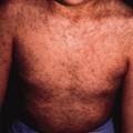
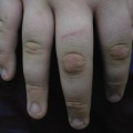
Cytological smears and histology are identical to those of pustular infection [11]. Neonatal impetigo may occur on the second or third day of life. This starts with pustules, which develop into flaccid bullae filled with yellow fluid. The bullae burst easily, leaving a circular erosion with a peripheral collarette of scale. The infection is limited to the epidermis but may extend very easily and may result in SSSS. Both impetigo neonatorum and SSSS result from the secretion of exfoliatin A or B by S. aureus.
Differential Diagnosis.
Pustular eruptions must be distinguished from impetigo (see Pustular staphylococcal infection, above). Development of SSSS may occur quickly. Eruptions such as mastocytosis, bullous herpes simplex, pressure bullae, Brocq ichthyosis and others may mimic SSSS [12].
Therapy and Complications.
Impetigo neonatorum is treated in the same way as pustular staphylococcal infection, but systemic treatment is required more often to prevent the development of SSSS. However, the infection is mainly limited to the epidermis and generally does not result in systemic disease. Localized infections are treated as detailed above. Widespread infection requires systemic treatment with penicillinase-resistant penicillin or cephalosporins.
Staphylococcal scalded skin syndrome is always treated systemically via intravenous therapy. It is sometimes lethal, especially in the neonatal period.
Other Bacterial Infections
Other specific neonatal infections are omphalitis, neonatal cellulites and abscesses and circumcision infections. If induced by S. aureus, such infections may progress into impetigo and SSSS. Another important bacterial pathogen in the neonatal period is Listeria monocytogenes.
Omphalitis
Omphalitis neonatorum has a low incidence in developed countries (about 0.7%) but is more frequently encountered in the developing world (about 6%) [13]. Improper severing of the umbilical cord, application of oily substances to the umbilical stump and other unhygienic practices during the neonatal period are important predisposing factors [14]. Late separation of the umbilical cord may lead to infection and inflammation: omphalitis. In most cases, Gram-negative organisms are responsible for omphalitis, particularly Escherichia coli and Klebsiella pneumoniae. Infections with group A streptococci and staphylococci have also been observed. Polymicrobial infection is not uncommon. Gram staining is a reliable and easy method for initial identification of the micro-organism [14]. A persistent omphalitis may indicate immunodeficiency [15]. Cellulitis occurs when inflammation spreads beyond the umbilicus. When the inflammation extends to the subcutaneous tissue and underlying fascia, necrotizing fasciitis develops [16]. Necrotizing fasciitis is associated with a mortality rate of 50%. Thus, early diagnosis is strongly indicated, and should be followed by surgical débridement.
Neonatal Cellulitis and Abscesses
Neonates are susceptible for cellulitis and abscesses following skin injury. Scalp abscesses are associated with the use of scalp electrodes. Also, puncture wounds from blood sampling or intravenous catheters are sites where cellulitites or abscesses are found. The micro-organisms involved are those that colonize the neonate during passage through the birth canal. Besides S. aureus, E. coli, group A and B streptococci can be isolated. Gram stain and culture of the abscess content will reveal the diagnosis. In cases of Gram-positive cocci in clusters, S. aureus is the most probable micro-organism and treatment can be started with a penicillinase-resistant penicillin (oxacillin or flucloxacillin). Gram-positive cocci in chains most probably reflect group A or B streptococci and can be treated with small-spectrum penicillin. Gram-negative rods should be treated with a third-generation cephalosporin. Surgery should be considered when fluctuation or abscesses are diagnosed.
Breast abscess is a special entity that appears in full-term neonates within the first 8 weeks. Usually, unilateral swelling of the breast in combination with redness and warmth appear about 1 week after birth. Under influence of transplacentally acquired maternal hormones the glandular breast tissue, especially of the full-term neonate, is stimulated and therefore more susceptible to infections. The disease is seen more often in girls than in boys. Diagnostic procedures and treatment are the same as described above [17].
Circumcision and Infections
Circumcision has proven to have preventive effects on the development of penile cancer [18] and also leads to a lower risk of urinary tract infections. Pathological phimosis and paraphimosis induce inflammation of the glans penis and the prepuce (balanitis, posthitis, balanoposthitis). These infections are extremely painful and primarily affect uncircumcised male babies. Chronic balanoposthitis may result in scarring and secondary phimosis [19].
The circumcision of a neonate is a low-risk procedure [20]. The most common complications are local infections and bleeding. Any infection should be treated directly.
Listeriosis
Listeriosis is a septicaemic disease caused by Listeria monocytogenes, which is a small aerobic non-spore-forming Gram-positive rod. The micro-organism is isolated from animals and soil. Transmission is via contaminated food, especially products made from unpasteurized milk. Early-onset infection is acquired in utero; late-onset infection may be acquired after birth. Late-onset infection usually affects full-term healthy infants. The infection develops soon after birth to weeks thereafter [21]. The infection is accompanied by grey–white maculopapules with vesicles or pustules [1].
The organism can be recovered in routine cultures, but special techniques may be necessary to isolate and identify Listeria. The treatment of choice is the combination of amoxicillin in conjunction with an aminoglycoside. Listeria is not sensitive to cephalosporins.
References
1 Praag van MCG, Rooij van RWG, Folkers E et al. Diagnosis and treatment of pustular disorders in the neonate. Pediatr Dermatol 1997;14:131–43.
2 Oranje AP, Folkers EF. The Tzanck smear: old, but still of inestimable value. Pediatr Dermatol 1988;5:127–9.
Related posts:


Stay updated, free articles. Join our Telegram channel

Full access? Get Clinical Tree


