In psoriasis, there are several clearly defined subtypes: guttate psoriasis, plaque psoriasis, flexural psoriasis, pustular psoriasis, erythroderma and psoriasis with arthritis.
A series of over 1200 patients with psoriasis, aged 1 month to 15 years, was described by Morris et al. [12]. They found an equal gender distribution and a positive family history in 71%. Plaque psoriasis was the most common type overall, affecting 34%. A psoriatic napkin rash with dissemination, affecting 45%, was the most common type of psoriasis in the under-2 years age group. Population studies indicate that both genetic and environmental factors interact to precipitate the development of psoriasis. The most frequently observed variant of psoriasis is the plaque type, followed by guttate psoriasis and juvenile psoriatic arthritis [11].
In a retrospective epidemiological study in India, data from 419 children (less than 14 years of age) with psoriasis were studied. A positive family history was present in only 19 (4.5%) patients. The extensor surfaces of the legs were the most common initial site affected [105 (25%) cases], followed by the scalp [87 (20.7%)]. Classic plaque psoriasis was the most frequent clinical presentation [254 (60.6%) patients], followed by plantar psoriasis [54 (12.8%)]. Nail involvement was observed in 130 (31%) cases. All types of nail changes described in psoriasis were seen in these patients. Pitting was the most common nail change, followed by ridging and discoloration. Five children (1.1%) (three girls and two boys) had psoriatic arthropathy. Precipitating factors that brought about the onset of the disease or were associated with exacerbation could be recalled in only 28 (6.6%) patients. Koebnerization was observed in 27.9% of patients. Pruritus was the most frequent symptom, reported by 365 (87.1%) children [7].
In 1262 patients (aged 1 month to 15 years) presenting to a tertiary referral pediatric dermatology department in Australia, a high rate of positive family history at 71% was reported. Twenty-six per cent of children had a history of a psoriatic napkin rash and facial involvement occurred in 38% of children. Plaque psoriasis was the most common type overall, affecting 430 patients (34%). Three hundred and forty-five children were less than 2 years of age. An entity defined by the authors as psoriatic napkin rash with dissemination was the most common type of psoriasis in the under-2 year age group, affecting 155 (45%) patients [6].
An analysis of phenotypic variation in psoriasis as a function of age at onset and family history showed that there is an increased severity of skin and nail disease in early onset psoriasis when psoriasis is familial [13].
References
1 Verbov J. Psoriasis in childhood. Arch Dis Child 1992;67:75–6.
2 Benoit S, Hamm H. Psoriasis im Kinders- und Jugendalter. Klinik und Therapie [Psoriasis in childhood and adolescence. Clinical features and therapy]. Hautarzt 2009;60:100–8.
3 Lehman JS, Rahil AK. Congenital psoriasis: case report and literature review. Pediatr Dermatol 2008;25:332–8.
4 Lever WF, Schaumburg-Lever G. Histopathology of the Skin. Philadelphia: J.B. Lippincott, 1983.
5 Nanda A, Kaur S, Kaur I et al. Childhood psoriasis: an epidemiologic survey of 112 patients. Pediatr Dermatol 1990;7:19–21.
6 Morris A, Rogers M, Fischer G, Williams K. Childhood psoriasis: a clinical review of 1262 cases. Pediatr Dermatol 2001;18:188–98.
7 Kumar B, Jain R, Sandhu K et al. Epidemiology of childhood psoriasis: a study of 419 patients from northern India. Int J Dermatol 2004:43:654–8.
8 Farber EM, Nall ML. The natural history of psoriasis in 5600 patients. Dermatologica 1974;148:1–18.
9 Lerner MR, Lerner AB. Congenital psoriasis: report of three cases. Arch Dermatol 1972;105:598–601.
10 Telfer NR, Chalmers RCJ, Whale K et al. The role of streptococcal infection in the initiation of guttate psoriasis. Arch Dermatol 1992;128:39–42.
11 Farber EM, Nall L. Childhood psoriasis. Cutis 1999;64:309–14.
12 Morris A, Rogers M, Fischer G et al. Childhood psoriasis: a clinical review of 1262 cases. Pediatr Dermatol 2001;18:188–98.
13 Stuart P, Malick F, Nair RP et al. Analysis of phenotypic variation in psoriasis as a function of age at onset and family history. Arch Dermatol Res 2002;294:207–13.
Guttate psoriasis
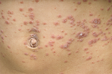
Guttate psoriasis (Fig. 80.3) occurs most often in children and young adults. The term is derived from the Latin gutta (a drop) and refers to the drop-like appearance of cutaneous lesions in this form of psoriasis. Guttate psoriasis appears abruptly, often a week after a streptococcal pharyngitis [1]. An association with perianal streptococcal dermatitis has been described [2–4].
The lesions of guttate psoriasis vary from 2–3 mm to 1 cm in diameter, and are round and slightly oval. The small papular lesions have characteristic overlying silvery scales. The lesions are scattered more or less symmetrically over the body, and are particularly common on the trunk and proximal parts of the extremities. Lesions occur frequently on the face, ears and scalp, and are unusual on the palms and soles. Guttate psoriasis usually persists for 3–4 months and resolves spontaneously. It can continue for more than 1 year. After an attack of guttate psoriasis most patients experience a recurrence within the next 3–5 years [5].
References
1 Farber EM, Nall L. Childhood psoriasis. Cutis 1999;64:309–14.
2 Honig PJ. Guttate psoriasis associated with perianal streptococcal disease. J Pediatr 1988;113:1037–9.
3 Patrizi A, Costa AM, Fiorillo L et al. Perianal streptococcal dermatitis associated with guttate psoriasis and/or balanoposthitis: a study of five cases. Pediatr Dermatol 1994;11:168–71.
4 Herbst RA, Hoch O, Kapp A et al. Guttate psoriasis triggered by perianal streptococcal dermatitis in a four-year-old boy. J Am Acad Dermatol 2000;42:885–7.
5 Nanda A, Kaur S, Kaur I et al. Childhood psoriasis: an epidemiologic survey of 112 patients. Pediatr Dermatol 1990;7:19–21.
Plaque psoriasis
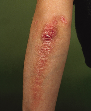
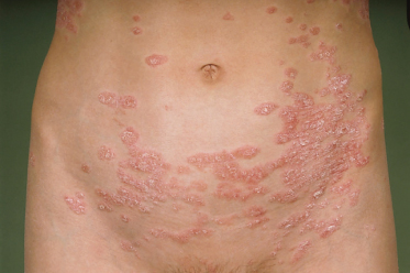
The erythematous plaques are well defined with sharp borders (Figs 80.4–80.6). The silvery grey scale on the surface of the lesion is easily removed. Psoriasis may occur exclusively on the scalp for many years. Sharply demarcated lesions can present on the extensor surfaces of the knees and elbows and on the trunk. Lesions are often symmetrically distributed. The size of the lesions is highly variable, and psoriatic plaques may spread peripherally without central healing. In children, plaques are often smaller and the scales finer and softer than in adults. In dark-skinned children the scale may be so subtle that the condition presents as hypopigmentation and the scale becomes obvious only when the lesions are scratched.


