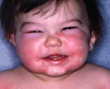
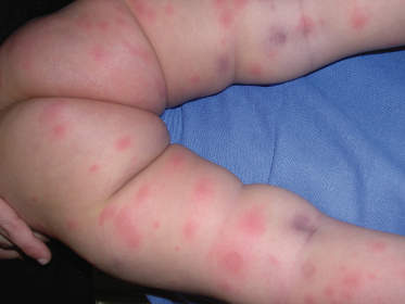
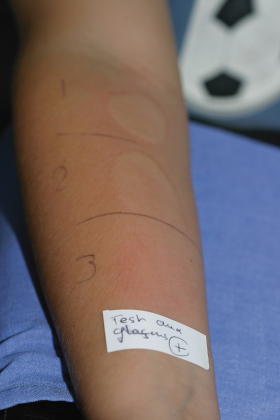
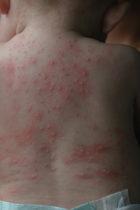
Angio-oedema (Fig. 74.2) describes swelling of the subcutaneous or submucosal tissues. Angio-oedema associated with urticaria is due to histamine release and should be differentiated from hereditary angio-oedema (angioneurotic oedema or Quincke’s oedema) where the subcutaneous swelling is usually isolated and due to extravasation of fluid from an abnormal kinin-generating system (see Pathogenesis).
Serum sickness-like reactions are also frequently classified in the spectrum of urticaria. These are characterized by an urticarial skin eruption associated with swelling of the extremities, arthritis and fever.
History [10,11].
Many different names have been given to this condition. Hippocrates recognized the association of urticaria with nettles, and this word was incorporated into the name used for urticaria in several languages. In the middle of the 18th century, Zedler called the disease urticario (Latin urere, to burn), and in 1792 Franck used the now commonly accepted name urticaria.
In 1882, Quincke reported on the acute oedema which now bears his name and Osler, in 1888, was the first to note that angio-oedema can occur in families. Tremendous progress has been made during the last century, including the synthesis of histamine, and the discovery of mediators of inflammation, immunoglobulin E (IgE) and mast cells. The first antihistamine was synthesized in 1937 by Bovet and Staub.
Pathogenesis.
Urticaria is due to a local increase of permeability of capillaries and small venules. A large number of mediators play a role but histamine, derived from mast cells in skin, appears to be the major mediator in acute urticaria [8,12]. Other agents, including prostaglandins, leukotrienes, eosinophil and neutrophil chemotatic factors, platelet-activating factor and interleukin 1, may also play a role in producing the lesions of urticaria [8,12,13]. Mediator release can be initiated by immunological or non-immunological stimuli, and urticaria is sometimes classified into allergic and non-allergic. However, in many situations, and especially in chronic urticaria and reactions to medications, it may not be possible to determine whether the effect is due to a pseudo-allergic reaction, an IgE-mediated reaction or an immune complex reaction.
Immunological Mechanisms
The majority of acute urticarias are mediated by IgE antibody to ingested, injected or topically applied allergens [7,8,12]; such reactions occur more frequently in atopic patients [7]. On the other hand, chronic urticaria is mostly due to non-specific histamine release, is not associated with atopy [3], and serum IgE levels are usually normal. However, in some cases, an autoantibody has been identified in the serum of some patients with chronic urticaria; this IgG autoantibody cross-links α-subunits of high-affinity IgE receptors of basophils and mast cells in skin [13,15].
Complement Abnormalities.
The anaphylatoxins C3a and C5a may induce histamine release, either directly or in association with immune complexes [8,12]. Sera, drugs, infections and systemic disorders may cause immune complex-mediated reactions, which correspond to the clinical entities serum sickness and urticarial vasculitis. Hereditary complement defects may cause urticaria, especially C2 and C4 deficiencies [15,16].
In hereditary angio-oedema/C1 inhibitor deficiency, the absence or dysfunction of C1 inhibitor results in repeated episodes of subcutaneous swelling due to the release of bradykinin and C2-kinin mediators [17].
Non-Immunological Mechanisms [3,8]
Causes of mediator release include chemical stimuli such as food (eggs, shellfish) or drugs (penicillin, aspirin, curare, morphine and radiocontrast materials) and/or physical stimuli (cold, heat, pressure or sun).
Aetiology.
Both acute and chronic forms of urticaria may be caused by a wide variety of factors [2–5,18–25]. A presumptive cause is identified or suspected in 40–92% of cases of acute urticaria [2–5,18–22] and in 16–65% of cases of chronic urticaria [23,25]. The main causes of acute and chronic urticaria in children are summarized in Box 74.1. The aetiological features are somewhat different from those of adults. In infants less than 6 months of age, the main cause is food (cow’s milk) allergy (Fig. 74.3). In older children, infections and drugs are the most frequent causes of acute urticaria, and physical factors are usually the aetiology of chronic urticaria [2–5,18–25].
Box 74.1 Main Causes of Childhood Urticaria
Infections
- Viral: hepatitis B, infectious mononucleosis, influenza, adeno- and enterovirus infection
- Parasitic (in endemic areas only)
Drugs
- Penicillin and cephalosporins
- Non-steroidal inflammatory drugs (e.g. aspirin, ibuprofen)
- Histamine liberator drugs: codeine, radiocontrast products
Food
- Cow’s milk proteins (especially in children <6 months of age)
- Eggs (especially uncooked egg white)
- Nuts and peanuts
- Fish and seafood
- Exotic fruits (cross-reactivity with latex proteins)
- Food additives (tartrazine, sodium metabisulphite)
Insect Bites
- Hymenopteras
Physical Agents (Chronic Urticaria)
- Cholinergic urticaria, cold urticaria and dermatographism
- Idiopathic
Infections
Various infections are associated with both acute and chronic urticaria [2–5,18–26]. Respiratory infections are frequently associated with acute urticaria in children. The role of viruses and parasites is clearly established [2–5,18–29]. However, the relationship between fungal or bacterial infections and urticaria is less well documented. Pathophysiology is not well understood; protein substances of the infective agent may act as antigens and cause a type I allergic reaction or, less commonly, a type III response.
Viruses
Viruses are the most common infectious cause of urticaria in childhood. Urticaria occurs in 30% of cases of serum hepatitis during the preicteric phase [27–29]. The exact mechanism is unknown. In the prodromal and early phases of hepatitis B infection, viral antigen levels may exceed that of antibodies, resulting in a serum sickness-like illness. Hepatitis viruses A and C have also been reported to be causative organisms [27–29]. Urticaria may be associated with a large number of other viral disorders [2–5,18–22]: herpes, varicella zoster, rubella, measles, mumps, Epstein–Barr virus, cytomegalovirus, influenza, adenovirus and enterovirus infections. The rash of parvovirus B19 infection (fifth disease) may present with pruritic urticarial lesions.
Bacteria
Various bacterial infections have been reported to cause urticaria: streptococcal pharyngitis, dental abscesses, sinusitis and urinary tract infections. and recently Mycoplasma pneumoniae [30]. However, the cure rate of urticaria after treatment of these conditions is highly variable. In addition, medication given for infection, especially aspirin (a well-known histamine-releasing agent) and antibiotics, should not be ignored as a possible cause of urticaria.
Parasites
Parasitic infestations should be considered in individuals from endemic areas, especially if blood eosinophilia and elevated IgE are present. When abdominal complaints are noted, the faeces should be examined for cysts and ova. Helminths and, less commonly, Toxocara canis infestation, trichinosis and distomatosis can induce urticaria [31]. In industrial countries, the role of parasites in urticaria is often overestimated, and usually parasite eradication does not improve urticaria [19,21,22].
Fungi
Much has been written about the potential role of Candida as an inducer of urticaria. However, fungi and yeasts, even in the presence of positive immediate-type skin tests, do not generally play a causative role. The cure rate of urticaria after treatment with antifungal drugs and proven elimination of the yeast organism is not encouraging [19].
Drugs
Drug intolerance is also a frequent cause of acute urticaria [2–5,18–22,32–35] but in most cases, the eruption results from a pseudo-allergic urticaria mimicking allergic urticaria [35]. Many drugs have been incriminated, but β-lactams and aspirin are the most common. Usually the reaction is an ordinary acute urticaria, which develops from a few minutes to a few days after the medication has been administered. Less frequently, anaphylactic shock or a serum sickness-like reaction may occur [9,36,37]. Identification of drug-induced urticaria is of practical importance, so that the agent may be avoided unless its use is absolutely necessary. The causative drug should be determined based on careful questioning about chronological data; the diagnostic and predictive value of in vitro and skin tests is low [38]. Drug additives (preservatives and dyes) may be the true inducers of urticaria.
Penicillin and other antibiotics can induce acute urticaria or anaphylactic shock; reactions are much more severe when the drug is given by injection. Penicillin allergy is associated with a family history of penicillin allergy and polymorphism in the IL4 gene and genes involved in penicillin metabolism [7]. Penicillin is a cause of both IgE-mediated reactions and pseudo-allergic urticaria; β-lactams and cephalosporins may also cause severe serum sickness [9,36,37]. Among antibiotics, sulphonamides and tetracyclines (minocycline) are also leading causes of urticaria.
Aspirin.
Aspirin and other non-steroidal anti-inflammatory agents (indometacin, ibuprofen), all of which affect prostaglandin synthesis, are major causes of urticaria [34]. Symptoms occur from minutes to hours after ingestion of the drug, and may persist for weeks after a single exposure. Aspirin can also have an exacerbating effect in many patients with chronic urticaria or urticaria of other aetiologies such as infection.
Other Drugs.
Local anaesthetics, such as lidocaine and amethocaine, can induce severe hypersensitivity reactions. Substances such as insulin or corticosteroids have also been incriminated. Morphine, codeine, quinine and curare act as direct histamine liberators and cause urticaria in a variable proportion of normal individuals. A similar mechanism may account for the 1–2% of individuals who develop urticaria or angio-oedema during radiocontrast studies.
Food Allergy
Food allergy is a classic cause of acute and recurrent urticaria [7,39–44]. Food proteins can induce a specific allergy (nuts, fish, eggs) or a non-specific histamine release (shellfish). The true frequency is unknown, as many individuals recognize this association and do not pursue a further evaluation. Symptoms can start immediately after food ingestion (fish, berries, nuts, eggs) or may be delayed for several hours (cereals, milk, chocolate, vegetables). While the food is in the mouth, the patient experiences immediate itching, burning and swelling of tongue and buccal mucosa. Urticaria, often starting in the perioral area, and angio-oedema are often associated. Nausea, vomiting, diarrhoea and, in explosive reactions, anaphylaxis may follow [7,43]. The list of food ingredients implicated as causes of urticaria is long, but in France five foods are noted to be responsible for 80% of all food allergies in children under 15 years of age: eggs, peanuts, cow’s milk, fish and mustard [44].
Specific Allergy.
In some cases, very small quantities of a food provoke serious attacks, including anaphylaxis, and an IgE-mediated reaction is suspected. In these cases prick tests and specific IgE antibodies to the food allergen are of value. Diagnostic information correlating reaction rates and specific IgE concentrations has been reported for peanuts, milk, sesame and several tree nuts. In addition, IgE epitope mapping of milk and peanut proteins has recently improved the diagnosis and determination of allergy severity [7].
Cross-reactivity exists with some food antigens, as well as reactions to Umbelliferae and other vegetables (carrot, potato, tomato). Cross-reactions may also be observed with fruits and pollens (apple/birch pollen), and with fruits and contact allergens (kiwi/latex proteins). In several publications, food allergy appears more frequent in atopic children [22,41,44]. The natural history of food sensitivity has not been well documented, but studies suggest that cow’s milk and egg sensitivity is usually lost in early childhood, whereas peanut, nut and seafood sensitization tends to be long-lasting [19,42].
Non-Specific Histamine Release.
Crustacea (shellfish), egg white, thiamine (cheese) and strawberries have a non-specific histamine-releasing effect.
Food Additives [45].
Food dyes, especially the yellow dye tartrazine, cochineal red and additives (sodium benzoate, sodium metabisulphite), are implicated in both acute and chronic urticaria. Many patients with chronic urticaria react to one or another of these substances in blinded challenge studies. However, it is highly probable that reactions to these chemical agents are due to a pharmacological rather than an allergic mechanism. Results of dietary avoidance have been variable, suggesting that dye/additive intolerance is only partially responsible for symptoms.
Inhalants and Contact Allergens
Pollens, mould spores, animal dander and other allergenic airborne substances are more likely to induce rhinitis and asthma. However, they do occasionally induce acute or chronic urticaria. The causative allergen is usually ragweed, but grass and tree pollen may play a role [19]. Mite allergens may cause urticaria either through direct penetration of the skin or through inhalation. Contact with some animals, particularly cats, dogs and horses, may produce urticaria in exposed sites; this condition is most commonly seen in atopic subjects. Conversely, atopy is not an associated condition in cases of urticaria or angio-oedema following Hymenoptera stings (bee, wasp, hornet) [46]. Many different species of caterpillars cause intensely pruritic dermatitis, including urticaria [47,48]. Recently, latex protein contact allergy has been the subject of much interest. Indeed, this allergy is seen in children, especially those who have been operated on in early infancy for spina bifida or congenital malformations. The sensitization may be due to repeated contact with latex (e.g. surgeon’s gloves) [49]. The diagnosis can be suspected because of previous urticarial reactions or angio-oedema after contact with balloons or exotic fruits (cross-reaction) [22,49]. Prick tests and specific IgE antibodies are of interest, but direct contact tests under careful medical supervision may be necessary to confirm the diagnosis.
Vaccination and Serum Reactions
Vaccines can cause anaphylactic reactions because of allergenic constituents such as foreign serum and egg proteins on which the viruses are grown. However, studies show that children allergic to eggs can be immunized safely with viruses grown on eggs [50]. Cases of urticaria have been reported with tetanus vaccine, measles vaccination, hepatitis B [51] and haemophilus B vaccination [52]. Serum sickness was frequent in cases of administration of antiserum of horse or rabbit origin, as well as rabies vaccination. Reactions may also occur with antilymphocyte serum and snake antivenom treatment [53].
Physical Agents
An interesting group of urticarias are those caused by physical agents, since this aetiology is the most frequent in cases of chronic childhood urticaria [19,23–25]. In addition, in some patients with ordinary acute or chronic urticaria of unknown cause, dermographism and lesions at the site of pressure are commonly seen.
Dermatographism.
The weal and flare reaction results from stroking of the skin. Dermatographism is frequent in normal populations (1.5–4.5%) but can also be seen in association with chronic or acute urticaria of unknown origin [19,54]. A special condition is the association with diffuse mastocytosis (see below).
Temperature
Both heat and cold may induce immediate as well as delayed urticaria. Cold urticaria is an important cause of recurrent symptoms in childhood (Fig. 74.4), being reported in as many as 8% of cases [19]. Both inherited and acquired forms exist [19,54–56]. Symptoms, in addition to urticaria and angio-oedema, may include wheezing, hypotension or syncope. Manifestations generally resolve only after return to a warm environment. Cold urticaria may be associated with viral infections including mononucleosis, hepatitis and measles [56].
Cholinergic Urticaria
The lesions of cholinergic urticaria characteristically consist of small, 2–3 mm weals (Fig. 74.5). These are precipitated by exercise, emotional stress and warm temperature, and are among the most common forms of urticaria in children and adolescents. A spontaneous remission is possible.
Solar Urticaria
This is a rare form of urticaria caused by light exposure, with lesions occurring on exposed skin area. Erythropoietic protoporphyria and systemic lupus erythematosus should be considered in children (see Differential diagnosis). Visible light (400–760 nm), as well as ultraviolet A (UVA) (320–400 nm) and UVB (280–320 nm), can cause urticaria. Phototesting may help in the diagnosis, and the wavelength to which the patient is sensitive can be determined with a monochromator.
Aquagenic Urticaria [57]
This condition is rare in children; urticaria appears after contact with water, regardless of the temperature.
Pressure and Vibration
In vibratory urticaria, weals develop immediately. Lesions in pressure-induced urticaria may be delayed for as long as 4–12 h.
Urticaria Associated with Exercise [58]
Urticaria or anaphylaxis can be precipitated by vigorous exercise; most cases are idiopathic, but some occur in association with food sensitivity, especially to cereals. A prominent symptom in these cases is generalized pruritus followed by urticaria and angio-oedema.
References
1 Champion RH, Roberts SOB, Carpentier RG et al. Urticaria and angioedema: a review of 554 patients. Br J Dermatol 1969;81:588–97.
2 Hannuksela M. Urticaria in children. Semin Dermatol 1987;6:321–5.
3 Bailey E, Shaker M. An update on childhood urticaria and angioedema. Curr Opin Pediatr 2008;20:425–30.
4 Legrain V, Taïeb A, Sage T et al. Urticaria in infants: a study of 40 patients. Pediatr Dermatol 1990;7:101–7.
5 Mortureux P, Léauté-Labrèze C, Legrain-Lifermann V et al. Acute urticaria in infancy and early childhood. Arch Dermatol 1998;134:319–23.
6 Bourrier T. Anaphylactic shock in the infant. Arch Pediatr 2000;7:1347–52.
7 Sicherer SH, Leung DY. Advances in allergic skin disease, anaphylaxis, and hypersensitivity reactions to foods, drugs, and insects in 2008. J Allergy Clin Immunol 2009;123(2):319–27.
8 Czarnetzki BM. Urticaria. Berlin: Springer-Verlag, 1986.
9 Hebert AA, Sigman ES, Levy ML. Serum sickness-like reactions from cefaclor in children. J Am Acad Dermatol 1991;25:805–8.
10 Czarnetzki BM. The history of urticaria. Int J Dermatol 1989;28:52–7.
11 Champion RH. Urticaria: then and now. Br J Dermatol 1988;119:427–36.
12 Powell J, Powell S. Mechanisms underlying urticaria. Hosp Med 2000;61:470–4.
13 Greaves MW. Chronic urticaria. N Engl J Med 1995;332:1767–72.
14 Du Toit G, Prescott R, Lawrence P et al. Autoantibodies to the high-affinity IgE receptor in children with chronic urticaria. Ann Allergy Asthma Immunol 2006;96(2):341–4.
15 Gelfand JA. The role of complement in urticaria and angioedema. Clin Immunol Rev 1981;1:257–309.
Stay updated, free articles. Join our Telegram channel

Full access? Get Clinical Tree


