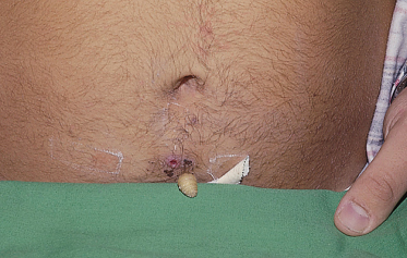
Fig. 69.2 Cordylobia anthropophaga. Larva penetrating the skin of the abdomen.
Courtesy of Dr Fausto Alonso.
Diagnosis.
After removing any scab, the posterior segment of the larva is seen as a 1- to 3-mm dark line in the centre of the ‘boil’. After occlusion with Vaseline or oil, air bubbles appear and the larva tries to reach the surface [6].
Treatment.
Removal can be performed with forceps or by manual expression after application of paraffin oil. Others have reported application of bacon slices to the larval apertures in order to attract the larvae; the larvae can then be removed with tweezers [3,4]. In later stages of the infestation, however, this approach is often unsuccessful [4]. Dermatobia hominis larvae are best removed with a cruciate incision after subcutaneous injection of 1% lidocaine for local anaesthesia [6]. Sealing of the larval aperture, partial removal or rupture of the larvae should be avoided as it can lead to formation of a foreign body granuloma, secondary infection or calcification [1–4,7,8].
Prevention of African Myiasis.
Clothes should be dried inside, and ironed on both sides to destroy eggs [3].
Dermal Myiasis (‘Creeping Eruption’)
Gasterophilus species (horse bot fly, warble fly) and Hypoderma species (cattle bot fly) are common parasites of horse and cattle, respectively, with a worldwide distribution. They can occasionally infest humans and lay their eggs on the hair of the host, where the larvae hatch and rapidly penetrate the skin. Gasterophilus larvae (length about 1 mm) cause a superficial erythematous serpentine duct that is migratory (about 1–2 cm/day, ‘creeping eruption’). The lesions are pruritic and mainly located on the arms. Hypoderma species cause deeper migratory cysts resembling a boil or cellulitis. The maggots (length up to 2.5 cm) migrate slowly for considerable distances. Hypoderma ovis has been reported to invade the central nervous system. Other causes of creeping eruptions include cutaneous larva migrans (Ancylostoma species), Strongyloides, Gnathostoma and Fasciola hepatica.
Diagnosis.
This is made as outlined above. Dermal myiasis can induce intense hypereosinophilia [9].
Treatment.
Therapy consists of the removal of the superficial Gasterophilus larvae with a needle or, in the case of Hypoderma, with a cruciform incision [1–3,7,8].
Wound Myiasis
Wound myiasis, also referred to as ‘traumatic cutaneous myiasis’, originates from the deposition of eggs into wounds. It can be caused by over 50 different species attracted by the odours of exudates and decaying wounds. Facultative myiasis producers are of worldwide distribution (Calliphora, Lucilia, Phormia, Musca, Fannia species). Among the obligatory myiasis producers are Wohlfahrtia magnifica
Related posts:


Stay updated, free articles. Join our Telegram channel

Full access? Get Clinical Tree


