Miscellaneous Epidermal Disorders 
Acanthosis Nigricans (AN) ICD-9: 701.2  ICD-10: L 83
 Asymmetric velvety thickening and hyperpigmentation of the skin, chiefly on the neck, axilla, groins, and other body folds.
 May be hyperkeratotic and associated with skin tags.
 A cutaneous marker related to heredity, obesity, endocrine disorders (particularly diabetes), drug administration, and malignancy.
 Insidious onset; in malignancy, rapid.
Classification
Type 1: Hereditary Benign AN. No associated endocrine disorder.
Type 2: Benign AN. Endocrine disorders associated with insulin resistance: insulin-resistant type II diabetes mellitus, hyper-androgenic states, acromegaly/gigantism, Cushing disease, hypogonadal syndromes with insulin resistance, Addison disease, and hypothyroidism.
Type 3: Pseudo-AN. Associated with obesity; more common in patients with darker pigmentation. Common in metabolic syndrome. Obesity produces insulin resistance.
Type 4: Drug-Induced AN. Nicotinic acid in high dosage, stilbestrol in young males, glucocorticoid therapy, diethylstilbestrol/oral contraceptive, and growth hormone therapy.
Type 5: Malignant AN. Paraneoplastic, usually adenocarcinoma of gastrointestinal or genitourinary tract; less commonly, bronchial carcinoma and lymphoma.
Epidemiology
Age of Onset. Type 1: during childhood or puberty; other types dependent on associated conditions.
Etiology and Pathogenesis
Dependent on associated disorder. In a subset of women with hyperandrogenism and insulin intolerance and AN, loss-of-function mutation in the insulin receptor or anti-insulin receptor antibodies can be found (types A and B). It is postulated that excess growth factor stimulation in the skin leads to proliferation of keratinocytes and fibroblasts. In hyperinsulinemia AN, excess insulin binding to insulin-like growth factor 1 receptor and fibroblast growth factor receptor has also been implicated. In malignancy-associated AN, transforming growth factor ß released from tumor cells may stimulate keratinocyte proliferation via epidermal growth factor receptors.
Clinical Manifestation
Insidious onset; in type 5 rapid. First visible change is darkening of pigmentation.
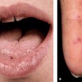
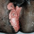
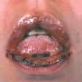
Skin Lesions. All types of AN: Darkening of pigmentation, skin appears dirty (Fig. 5-1). As skin thickens, it appears velvety; skin lines accentuated; surface becomes rugose, mammillated. Type 3: velvety patch on inner, upper thigh at site of chafing; often has many skin tags in body folds and neck. Type 5: hyperkeratosis and hyperpigmentation more pronounced (Fig. 5-2A). Involvement of oral mucosa and vermilion border of lips (Fig. 5-2B). Hyperkeratosis of palms/soles, with accentuation of papillary markings: “Tripe hands” (Fig. 5-2C).
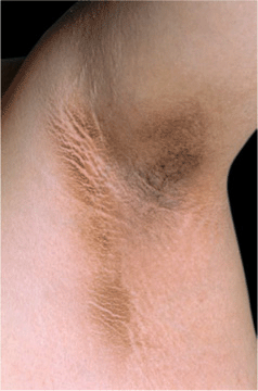
Figure 5-1. Acanthosis nigricans Velvety, dark-brown to gray thickening of the skin of the armpit with prominent skin folds and feathered edges in a 30-year-old obese woman from the Middle East. There were similar changes on the neck, the antecubital fossae, and on the knuckles.
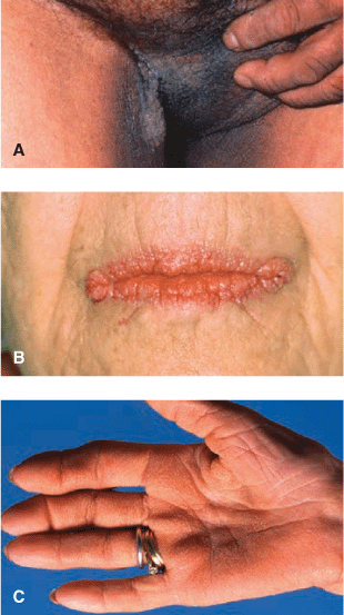
Figure 5-2. Acanthosis nigricans, type 5 (malignant) (A) Verrucous, papillomatous grayish-brown plaques in groins, medial aspects of thigh, and scrotum. Similar lesions were found on neck and all other body folds. The patient had weight loss and wasting and gastric adenocarcinoma was found. (B) Verrucous and papillomatous growths on the vermillion border of lips. Oral mucosa was velvety with deep furrows of the tongue. (C) Tripe palms. Palmar ridges show maximal accentuation resembling the mucosa of the stomach of a ruminant.
Distribution. Most commonly, axillae; (Fig. 5-1), neck (back, sides), groins (Fig. 5-2A), anogenitalia, antecubital fossae, knuckles, submammary, umbilicus. In type 5, also periocular, peroral, mammilary, and palms (tripe palms) (Fig. 5-2C).
Mucous Membranes. Oral mucosa: velvety texture with delicate furrows. Type 5: Mucous membranes and mucocutaneous junctions commonly involved; warty papillomatous thickenings periorally (Fig. 5-2B).
General Examination
Examine for underlying endocrine disorders in overweight to morbidly obese persons; in type 5 wasting, search for malignancy.
Diagnosis and Differential Diagnosis
Clinical Findings. Dark thickened flexural skin: Confluent and reticulated papillomatosis (Gougerot-Carteaud syndrome), pityriasis versicolor, X-linked ichthyosis, retention hyperkeratosis, and nicotinic acid ingestion.
Laboratory Examinations
Chemistry. Rule out diabetes mellitus; metabolic syndrome
Dermatopathology. Papillomatosis, hyperkeratosis; epidermis thrown into irregular folds, showing various degrees of acanthosis.
Imaging and Endoscopy. Rule out associated malignancy.
Course and Prognosis
Type 1: Accentuated at puberty and, at times, regresses when older. Type 2: Depends on underlying disturbance. Type 3: May regress after significant weight loss. Type 4: Resolves when causative drug is discontinued. Type 5: AN may precede other symptoms of malignancy by 5 years; removal of malignancy may be followed by regression of AN.
Management
Symptomatic. Treat associated disorder. Topical keratolytic and/or topical or systemic retinoids may improve AN but all in all not very effective.