Skin Signs of Vascular Insufficiency 
Atherosclerosis, Arterial Insufficiency, and
Atheroembolization ICD-9: 440  ICD-10: I70
 Atherosclerosis obliterans (ASO), especially of the lower extremities, is associated with spectrum of cutaneous findings of slowly progressive ischemic changes.
 Symptoms range from intermittent claudication with exertional muscle pain and fatigue to limb ischemia with rest pain and tissue damage and acute ischemia.
 Cutaneous findings range from dry skin, hair loss, onychodystrophy, gangrene, and ulceration.
 Atheroembolism is the phenomenon of dislodgment of atheromatous debris from a proximal affected artery or aneurysm with centrifugal microembolization and resultant acute ischemic and infarctive cutaneous lesions.
 More common with advanced age and invasive procedures.
 Manifestations are blue or discolored toes (“blue toe”), livedo reticularis, and gangrene
Epidemiology
Age of Onset. Middle aged to elderly. Males > females.
Incidence. Atherosclerosis is the cause of 90% of arterial disease in developed countries, affecting 5% of men >50 years; 10% (20% of diabetics) of all men with atherosclerosis develop critical limb ischemia.
Risk Factors for Atherosclerosis. Cigarette smoking, hyperlipidemia, low high-density lipoprotein, high low-density lipoprotein (LDL), high cholesterol, hypertension, diabetes mellitus, hyperinsulinemia, abdominal obesity, family history of premature ischemic heart disease, and personal history of cerebrovascular disease or occlusive peripheral vascular disease.
Pathogenesis
Atherosclerosis is the most common cause of arterial insufficiency and may be generalized or localized to the coronary arteries, aortic arch vessels to the head and neck, or those supplying the lower extremities, i.e., femoral, popliteal, anterior, and posterior tibial arteries. Atheromatous material in the abdominal or iliac arteries can also diminish blood flow to the lower extremities as well as break off and embolize downstream to the lower extremities (atheroembolization). In addition to large-vessel arterial obstruction, individuals with diabetes mellitus often have microvasculopathy (see Section 15, p. 384).
Atheroembolism. Multiple small deposits of fibrin, platelet, and cholesterol debris embolize from proximal atherosclerotic lesions or aneurysmal sites. Occurs spontaneously or after intravascular surgery or procedures such as arteriography, fibrinolysis, or anticoagulation.
Clinical Manifestation
Atherosclerosis/Arterial Insufficiency of Lower Extremity Arteries
Symptoms. Pain on exercise, i.e., intermittent claudication. With progressive arterial insufficiency, pain and/or paresthesias at rest occur in leg and/or foot, especially at night.
Pallor, cyanosis, livedoid vascular pattern (Fig. 17-1), loss of hair on affected limb. Earliest infarctive changes include well-demarcated maplike areas of epidermal necrosis. Later, dry black gangrene may occur over the infarcted skin (purple cyanosis → white pallor → black gangrene) (Fig. 17-2). Shedding of slough leads to well-demarcated ulcers in which underlying structures such as tendons can be seen.
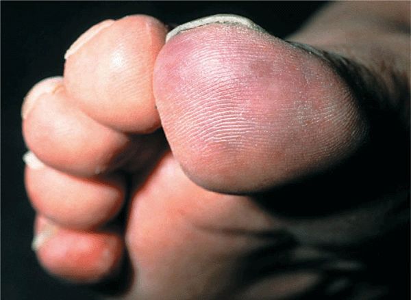
Figure 17-1. Atherosclerosis obliterans, early The great toe shows pallor and there is mottled, livedoid erythema on the tip of the toe. In this 68-year-old diabetic man, the iliac artery was occluded.
Figure 17-2. Atherosclerosis obliterans (A) There is pallor of the forefoot and mottled erythema distally with incipient gangrene on the great toe and the second digit. This is a female diabetic with partial occlusion of the femoral artery. The patient was a smoker. (B) More advanced gangrene of the second to the fifth toe, the great toe is ebony white and will also turn black.
General Examination. Pulses. Pulse of large vessels usually diminished or absent. In diabetic patients with mainly microangiopathy, gangrene may occur in the setting of adequate pulses. Temperature of foot: cool to cold.
Bürger Sign. With significant reduction in arterial blood flow, limb elevation causes pallor (best noted on plantar foot); dependency causes delayed and exaggerated hyperemia. Auscultation over stenotic arteries reveals bruits.
Pain. Ischemic ulcers are painful; in diabetic patients with neuropathy and ischemic ulcers, pain may be minimal or absent.
Distribution. Ischemic ulcers may first appear between toes at sites of pressure and beginning on fissures on plantar heel. Dry gangrene of feet, starting at the toes or at pressure sites (Fig. 17-2B).
Atheroembolization
Symptoms. Acute pain and tenderness at site of embolization.
Skin Lesions. Violaceous livedo reticularis on legs, feet, but also as high up as buttocks. Ischemic changes with poor return of color after compression of skin. “Blue toe” (Fig. 17-3): indurated, painful plaques often following livedo reticularis on calves and thighs that may undergo necrosis (Fig. 17-4), become black and crusted, and ulcerate. Cyanosis and gangrene of digits.
Figure 17-3. Atheroembolism after angiography A mottled (“blue toe”), violaceous, vascular pattern on the fore-foot and great toe. The findings were noted after intravascular catheterization and angiography in an individual with ASO.
Figure 17-4. Atheroembolism with cutaneous infarction Violaceous discoloration and cutaneous infarctions with a linear arrangement on the medial thigh of a 73-year-old woman with atherosclerosis, heart failure, and diabetes.
General Examination. Pulses. Distal pulses may remain intact.
Laboratory Examinations
Hematology. Rule out anemia, polycythemia.
Lipid Studies. Hypercholesterolemia (>240 mg/dL), often associated with rise in LDL. Hyper-triglyceridemia (250 mg/dL), often associated with rise in very low-density lipoproteins and remnants of their catabolism (mainly intermediate-density lipoprotein).
Dermatopathology of Atheroembolism. Deep skin and muscle biopsy specimen shows arterioles occluded by fibrosis with multinucleated giant cells surrounding biconvex, needle-shaped clefts corresponding to cholesterol crystal microemboli.
Doppler Studies. Show reduced or interrupted blood flow.
Digital Plethysmography. With exercise can unmask significant atherosclerotic involvement of lower extremity arteries.
X-Ray. Calcification can be demonstrated intramurally.
Arteriography. Atherosclerosis is best visualized by angiography. Ulceration of atheromatous plaques seen in abdominal aorta or more distally.
Diagnosis and Differential Diagnosis
Clinical suspicion confirmed by arteriography and deep skin biopsy (atheroembolism).
Differential Diagnosis. Intermittent Claudication. Pseudoxanthoma elasticum, Bürger disease (thromboangiitis obliterans), arthritis, gout.
Painful Foot. Gout, interdigital neuroma, flat feet, calcanean bursitis, plantar fasciitis, rupture of plantar muscle.
Ischemic and Infarctive Lesions of Leg/Foot. Vasculitis, Raynaud phenomenon (vasospasm), disseminated intravascular coagulation, cryoglobulinemia, hyperviscosity syndrome (macroglobulinemia), septic embolization (infective endocarditis), nonseptic embolization, drug-induced necrosis (warfarin, heparin), ergot poisoning, intra-arterial injection, livedo reticularis syndromes, external compression (popliteal entrapment).
Course and Prognosis
Arterial insufficiency is a slowly progressive disease, punctuated by episodes of complete occlusion or embolism. Atherosclerosis of coronary and carotid arteries usually determines survival of patient, but involvement of lower extremity arteries causes significant morbidity. Balloon angioplasty, endarterectomy, and bypass procedure have improved prognosis of patients with atherosclerosis. Amputation rates have been lowered from 80% to <40% by aggressive vascular surgery. Atheroembolism may be a single episode if atheroembolization follows intra-arterial procedure. May be recurrent if spontaneous and associated with significant tissue necrosis.
Management
Prevention. Goal of management is prevention of atherosclerosis.
Medical Management of primary hyperlipidemia: by statins, diet, and exercise. Reduce elevated blood pressure. Discontinue cigarette smoking. Encourage walking to create new collateral vessels. Position ischemic foot as low as possible without edema. Heparin and warfarin. IV prostacyclins. Analgesics.
Surgical Management. Endarterectomy or bypass for iliac occlusions. Debridement of necrotic tissue locally. Amputation of leg/foot: indicated when medical and surgical management has failed.
Thromboangiitis Obliterans (TO) ICD-9: 443.1  ICD-10: I73.1
 A rare inflammatory occlusive disease of medium sized and small arteries and veins.
 Predominantly in males, 20–40 years of age.
 Very strong association with smoking.
 An angiitis clinically indistinguishable from TO occurs in persons consuming cannabis.
 Clinical manifestations are cold sensitivity; ischemia: claudication of leg, foot, arm, or hand.
 Peripheral cyanosis, ischemic ulcers, gangrene (Fig. 17-5), and superficial thrombophlebitis.
 Therapy: smoking cessation, analgesics, wound care; antiplatelet agents, prostacyclins, pentoxifylline, angioplasty, sympathectomy, amputation.
 Synonym: Bürger disease.
Figure 17-5. Thromboangiitis obliterans Infarctive necrosis on the great toe of a 28-year-old man. The lesion is exquisitely painful. (The yellowish-brownish color is from iodine disinfection).
Thrombophlebitis and Deep Venous Thrombosis
ICD-9: 671.2  ICD-10: I 80 ICD-9: 433.40
  ICD-10: I 80.2
 Superficial phlebitis (SP) is an inflammatory thrombosis of a superficial normal vein, usually due to infection or trauma from needles and catheters.
 Inflammatory thrombosis of varicose vein usually in the context of the chronic venous insufficiency (CVI) syndrome.
 Deep venous thrombosis (DVT) is due to thrombotic obstruction of a vein with or without an inflammatory response.
 Occurs due to slow blood flow, hypercoagulability, or changes in the venous walls.
 The most common predisposing factors and causes are listed below.
Predisposing Factors and causes of Deep Venous Thrombosis
Common Factors
Major surgery
Fractures
Congestive heart failure
Acute myocardial infarction
Stroke
Pregnancy and postpartum
Spinal cord injuries
Shock
Less Common Factors
Sickle cell anemia
Homocystinuria
Protein C or S deficiency
Oral contraceptives
Malignancies
Venous varicosities
Previous history of venous thrombosis
Leiden factor V mutation
Severe pulmonary insufficiency
Prolonged immobilization
Antithrombin III deficiency
Antiphospholipid antibodies
Ulcerative colitis
Etiology and Pathogenesis
The thrombus originates in an area of low venous flow. An occlusion of a vein by thrombus imposes a block to venous return, which leads to increased venous pressure and edema in the distal limb. An inflammatory response to the thrombus causes pain and tenderness. If the venous pressure is too high, arterial limb flow may rarely be compromised and ischemia of the distal limb may occur. The thrombus in the vein often has a free-floating tail, which may break off to produce a pulmonary embolus. Organization of the thrombus in the vein destroys the venous walls, and this leads to the postthrombotic syndrome.
Clinical Manifestation
Patients complain of pain or aching in the involved limb or notice limb swelling. Some patients may have no symptoms. Pulmonary embolus may be the first indication of DVT.
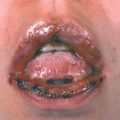
Superficial thrombophlebitis is diagnosed by the characteristic induration of a superficial vein with redness, tenderness, and increased heat (Fig. 17-6A). DVT presents with a swollen, warm, tender limb (Fig. 17-6B) with prominent distended collateral veins. Pitting edema may occur but is not always present, and a tender cord may be felt where the vein is thrombosed. With iliofemoral thrombophlebitis, the limb is swollen from the foot to the inguinal region and tenderness is not present in the limb, but collateral veins may form from the thigh to the abdominal wall. Two types are recognized: the limb may be very pale and painful (phlegmasia alba dolens) (Fig. 17-6B) or may be cyanotic and painful with cold digits if the arterial inflow is also compromised (phlegmasia coerulea dolens). In thrombosis of calf veins, the calf and foot are swollen and warm, and there is deep tenderness of the calf, often without a palpable cord.
Figure 17-6. Superficial phlebitis and deep venous thrombosis (A) A linear painful erythematous cord extending from the popliteal fossa to the mid-calf in a 35-year-old man who had moderate varicosities. Phlebitis occurred after a 15-h flight. (B) The leg is swollen, pale, with a blotchy cyanotic discoloration, and is painful. The episode occurred after abdominal surgery (the circular marks are from a compression bandage).
Migratory phlebitis describes an inflammatory induration of superficial veins that migrates within a defined region of the body; may be associated with thromboangiitis obliterans and malignancies. Mondor disease (sclerosing phlebitis) is an indurated, subcutaneous vein from the breast to the axillary region that during healing leads to a shortening of the venous cord, which puckers the skin.
Laboratory Examinations
Venous imaging by color-coded duplex ultrasound and Doppler examination reveals an absence of flow or of the normal respiratory venous flow variations in proximal venous occlusions. For thrombophlebitis of the calf veins, intravenous [125I] fibrinogen or a venogram gives a definite diagnosis.
Differential Diagnosis
Lymphedema, cellulitis, erysipelas, superficial phlebitis, and lymphangitis. An uncommon differential diagnosis is rupture of the plantar muscle, which produces pain, swelling, and ecchymotic areas in the dependent ankle area.
Management
The treatment of SP is compression, antiplatelet drugs, and nonsteroidal anti-inflammatory agents.
The treatment of DVT is anticoagulation. IV heparin. The partial thromboplastin time (PTT) should be 1.5–2 times normal. Low-molecular-weight heparin is also effective. Warfarin can be started orally at the same time and should overlap heparin for 5 days until the necessary factors for blood clotting are depressed. Elastic stockings and compression are mandatory and should be worn for at least 3 months; zinc paste–impregnated bandages (Unna boot) and ambulation should be started as soon as symptoms subside.