Warts were recognized as infectious diseases by the Ancient Greeks and Romans. Since the 19th century genital warts have been viewed as viral infections. After it was demonstrated in the early 1900s that the virus was transmitted from cell-free filtrates of warts, papillomaviruses were identified in a number of vertebrate species. Molecular cloning led to a scientific development which largely enhanced the study of the biological and biochemical properties of papillomaviruses. Sequencing allowed the identification of the open-reading frames (ORF) as putative viral genes and the investigators determined the function of viral genes. The recognition and increasing appreciation of the fact that a subset of HPV was closely associated with cervical cancer led to the establishment of HPV as the new model of viral tumorigenesis [2–4].
Classification of Papillomaviruses.
The International Committee on the Taxonomy of Viruses designated papillomaviruses as a separate family, the Papillomaviridae. Historically, HPV has been classified into cutaneous, mucosal and epidermodysplasia verruciformis (EV) types. Classification of HPV was initially based on DNA hybridization.
The genome consists of approximately 8000 base pairs and is divided functionally into three regions. The early region contains eight ORFs (E1–E8) and encodes genes for viral transcription, cellular transformation and DNA replication. The late region contains two ORFs and encodes for structural proteins, capsid proteins L1 and L2, and proteins expressed inside virally infected cells. The regulatory or long control region, which is located between the early and late regions, contains transcription enhancer genes and controls viral replication and gene expression.
Phylogenetic trees have been constructed based on alignment of known DNA sequences in the early (E6, E7) and late (L1) regions of the genome. Since each papillomavirus is specific to the species it infects, there are a few hundred types of papillomaviruses.
A range in homology has led to the following classifications: genus, species, type, subtype and variant. There are 12 genera, designated by the first 12 letters of the Greek alphabet (alpha, beta, gamma, delta, epsilon, zeta, theta, iota, kappa, lambda, mu, nu, xi, omikron, pi papillomavirus) [2,5,6].
Epidemiology and Prevalence.
Epidemiological studies have revealed that worldwide, verrucae vulgaris are induced predominantly by four major HPV types: HPV-1, -2, -27 and -57. It is clear that other HPV types, e.g. HPV-4 and -65, also appear in small proportions in common warts. The prevalence of verrucae vulgaris in meat-handlers and fish-handlers is mostly due to HPV-7 and HPV-4 infection and HPV-66 is a novel HPV type to be associated with verrucae vulgaris. Mixed infections of HPVs in one lesion of cutaneous warts have been identified. The prevalence frequencies of double infection involving two different types of HPVs within cutaneous warts are repeatedly observed. With multiplex PCR, these infections are easily and accurately identified.
Warts follow acne and atopic dermatitis in frequency as cutaneous conditions seen in paediatric dermatology clinics. The occurrence rate has more than doubled in the last 30–40 years. Skin warts are estimated to occur in up to 20% of school-aged children. They are rare before the age of 5 years, with the greatest incidence between the ages of 10 and 16 years [7–9].
Immunity
In the majority of individuals, HPV infections are transient and asymptomatic. About 40% of childhood warts resolve within 2 years. The incubation period of HPV varies from several weeks up to more than a year. Viral warts can occasionally persist for many years in otherwise healthy individuals. Older individuals may have some immune resistance to infection since there are a low number of cases in this population [3]. It is debated whether HPV induces immunomodulation resulting in local impairment of the immune response, or whether pre-existing minor abnormalities of delayed-type hypersensitivity might be responsible for persistence of the virus [10]. The human clearance of HPV involves a protective skin barrier, and interactions between innate immunity and acquired immunity. Early exposure to the wart virus may confer a period of immunity, especially in a young child with a few asymptomatic warts that are likely to resolve spontaneously within months. The immune defence of the infant, HLA type, the presence of HPV antibodies, and differences in the host genome may protect the young child.
HPV infection plays an important role in the transmission of HPV DNA between family members, and may be transmitted via saliva during an infant’s nursing. The mother’s warts are associated with persistent oral and genital HPV carriage [11]. Deficiencies in cell-mediated immunity cause problems with persistent infection.
The rare autosomal recessive condition epidermodysplasia verruciformis (EV), which starts in early infancy or childhood, is associated with an abnormal immune response to HPV [12]. Secondary immunodeficiency states – acquired immune deficiency syndrome, malignancies or organ allograft recipients as a result of immunosuppressive therapy – are also associated with frequent and persistent HPV infection [13]. Warts may have a tendency to either disappear or to be refractory to all treatments. Resolution rates may be adversely influenced by the HPV type (as with mosaic warts), the extent and duration of the warts and suppression of the host’s cell-mediated immune response [3].
Warts can also be classified by mode of regression. Palmoplantar warts regress by humoral vascular reaction and cellular extravasations. Common warts regress following the activation of infiltrating cytotoxic T-cells, natural killer cells and macrophages. In the case of plane warts, inflammatory cells and antiviral cytokines simultaneously appear. Intermediate warts (HPV types 10, 27, 28, 29) are essentially common warts seen in patients with depressed cellular immunity, so variable inflammatory cellular infiltrate and rates of regression can be seen [4,14].
WHIM Syndrome
This is the association of warts, hypogammaglobulinaemia, recurrent bacterial infections and myelokathexis. Severe chronic neutropenia appears with hyperplasia of the mature myeloid compartment in the bone marrow [15]. WHIM syndrome is associated with a heterozygous truncating mutation of the CXCR4 gene. The CXCR4 receptor is bound by CXCL12, a chemokine that regulates cardiogenesis and haematopoiesis, among others [4].
Mode of Transmission
Subclinical and Latent Forms
Among healthy individuals, persistent subclinical infections can be demonstrated on mucosal surfaces by application of 3% acetic acid, as in the procedure of colposcopic examination of the cervix or genital tract. High-risk HPV DNA can be detected in the oral and genital mucosa of infants during the first 3 years of life and occasionally in immunocompetent and immunosuppressed individuals [11]. Latent papillomavirus represents the presence of HPV DNA in clinically and histologically normal skin and mucosa. HPV infections might be transmitted horizontally from other family members. It is important to remember this to decrease the number of false allegations of sexual abuse. Identification of possible sexual abuse is still by detailed and appropriate history taking, physical examination, and correlation with the ‘socio’ clinical context [16].
Autoinoculation
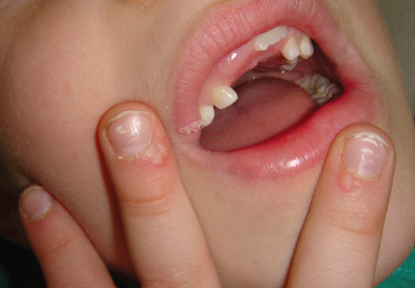
An important mode of spreading the virus is via autoinoculation (Fig. 47.2). The leading sites of HPV infection are the extremities and the face. Hand warts are transferred by contact to other cutaneous sites including the lips, nose and face [17]. Infection occurs when the basal epithelial cells of the mucosa or skin are exposed to the virus. Trauma also facilitates the spread of viruses [1].
Heteroinoculation
Viral warts are spread by direct contact through fomites or indirectly by contaminated surfaces. Papillomaviruses can remain infectious for long periods. Viral transmission is facilitated by maceration of the skin. There is an increased risk of reinfection in those who have had viral warts previously [18]. The prevalence of HPV DNA using sensitive assays is high, but does not necessarily equate with a live virus. Fomite transmission is suggested by several studies, but there are different options in the literature. There is no specific antiviral treatment for subclinical HPV infection. The most frequently reported risk factors for genital HPV infections are related to sexual behaviour, the partner’s sexual history, and age [7,19–21].
Iatrogenic Transmission
Iatrogenic transmission of the virus may occur indirectly through the use of inadequately sterilized instruments [1]. In addition, HPV DNA can be detected in the peripheral blood mononuclear cells, sera or plasma of patients with cervical cancer or HPV-associated head and neck squamous cell carcinoma. It should therefore be considered whether peripheral blood mononuclear cells might serve as a carrier of HPV during the course of HPV infection [22].
Vertical Transmission
Human papillomavirus infection of normal skin is acquired very early in infancy. An important mode of spreading the virus to the neonate is transmission of HPV either in utero or during delivery from mother to infant, which can result in the development of laryngeal papillomas or anogenital warts which may become clinically apparent only months or years later. Viraemia has not been shown to occur with HPV [17,23].
Sexual Transmission
The prevalence of childhood HPV infection varies widely in the literature. Most children who have been sexually abused will not show carriage of the virus. The period from sexual HPV exposure to development of external genital warts (EGWs) in adolescents and adults is about 3 months, but is unknown in children [24–26].
Autoinoculation or heteroinoculation from non-genital warts may be a source of EGWs in children. It has been recognized that the phylogenetically related mucosal HPV types 2, 27, and 57 can be detected in cutaneous lesions, as well as oral and genital lesions [11]. HPV-27 with HPV-3 is commonly found as a non-genital HPV in genital lesions of children. HPV subtypes in children do not show high degrees of site specificity for either mucosal or cutaneous sites, as is seen in adults [17].
Role of HPV in Non-Wart Skin Conditions.
Human papillomavirus can often be detected in common dermatoses, leading to a speculation that it may play a role in the development of conditions such as psoriasis vulgaris, lichen sclerosus and naevus sebaceus [27]. Epidermodysplasia verruciformis HPV types may play a role in the hyperproliferation of skin. Anti-HPV-5 antibodies have been demonstrated in epidermal repair processes (second-degree burn, autoimmune skin diseases, bullous disorders, psoriasis vulgaris). The mechanism by which hyperproliferative HPV types may trigger a widespread epidermal disorder is currently unknown [28].
Clinical Features
HPV Infections of the Skin
The clinical appearance of viral warts varies according to the type of the virus, the anatomical site involved and the host’s response to the infection [13].
Clinical appearance can be defined by morphology (common warts, flat warts, filiform warts, tinea versicolor-like warts, doughnut warts, epidermal cyst-type warts, punctate warts, pigmented warts, keratoacanthoma, subclinical or latent infection) or by location (common warts, palmoplantar warts, periungual warts, respiratory papillomatosis, oral papillomatosis (Heck disease), condyloma acuminatum, verrucosus carcinoma). Warts can also be categorized by the host immune response [4].
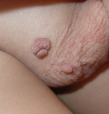
Common warts occur primarily in children and represent 70% of all skin warts (Fig. 47.3). Plantar and flat warts occur in a slightly older population. Warts can be painful when involving the soles or located near the nails, especially when inflammation develops. If they are located on visible areas, they can be socially unacceptable [8]. Common warts present a variety of clinical patterns and appear as hyperkeratotic papules with a rough, irregular surface and can occur in almost any area of the body. HPV types 2 and 4 are the most common, followed by types 1, 3, 7, 10, 26, 27, 29 and 57. Common warts may become exophytic and vary in size from tiny flesh-coloured papules to hyperkeratotic fissured masses 1–2 cm in diameter. Thrombosed capillaries are often seen after paring away the hyperkeratotic tissue [13].
Filiform/digitate warts are long, with a narrow base and verrucous tip, usually seen on the face and neck, around the lips, eyelids or nares. These variants of common warts are benign and easy to treat [4].
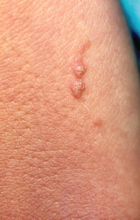
Verruca plana/plane warts/flat warts are more common among children and young adults and develop by autoinoculation. They are smooth, flesh-coloured or lightly pigmented yellow-brownish papules with hyperkeratosis, usually 2–4 mm in size. These lesions are common on photo-exposed areas, such as the face and neck and the dorsum of the hands. The Koebner phenomenon (or, more properly, pseudokoebnerization) often appears (Fig. 47.4). HPV-10 and -28 related plane warts are more hyperkeratotic and can resemble early common warts.
When flat warts are spread over a wide surface area, they may be slightly hyper- or hypopigmented, and may appear like tinea versicolor. This appearance is not usual and is more commonly limited to patients with EV, HIV infection or other immune deficiency. In EV, warts spread rapidly and may progress to bowenoid papulosis or Bowen disease (squamous cell carcinoma in situ) [3]. Regression of these lesions may occur, which is usually heralded by inflammation. Flat wart HPV types include 3, 10, 28, 41 and 49, followed by types 5, 8, 12, 14, 15, 17, 25 and 30.
Deep plantar warts/myrmecia. The name ‘myrmecia’ originates from the Greek meaning ‘ant hill’. The lesions appear as small papules and progress to deep lesions with a rough keratotic surface. A well-demarcated rim of compressed keratin surrounds a softer substance. Plantar warts often grow into the deeper layers of skin because of the pressure (callus-like warts typically involve HPV-1, -4, -7, -10). Plantar warts should be treated to lessen pain and transmission of infection [13]. The incubation period is 1–20 months. They grow deep and tend to be more painful than common warts. Treatment responsiveness is generally good. The most common type of deep palmoplantar wart is HPV-1, followed by HPV-2, -3, -4, -27, -29, -57 and -63.
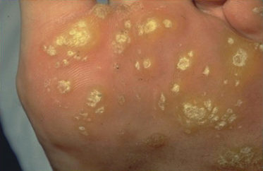
Mosaic warts are formed by smaller plantar warts, which are composing plaques. They are coalescent, usually seen on the palms and soles. Mosaic warts are important for their persistence and difficulty of treatment. Mosaic wart types are HPV-1, -4, -7 and -10 (Fig. 47.5) [4].
Punctate warts (HPV-60, -63, -65) are rare and localized endophytic hyperkeratotic papules, usually located on the palms. Occasionally warts can present a high degree of pigmentation (HPV-60), mimicking a primary melanocytic process.
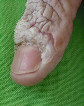
Periungual and subungual warts (benign HPV-1–4, -7, -10; premalignant HPV-16, -34) are painful; their treatment is often difficult due to the physical blockade created by the nail itself. When treating a wart in this location, the nail matrix may incur damage (Fig. 47.6).
A cystic wart appears as a nodule on the weight-bearing surface of the sole (plantar epidermoid cysts). The nodule is usually smooth but may become hyperkeratotic. If the lesion is incised, cheesy material may be expressed. Cystic wart types: HPV-60, -63, -65 [29].
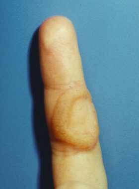
Doughnut/ring warts are seen after the use of destructive therapies. They occur around the periphery of a wart that has been treated previously (Fig. 47.7).
Stay updated, free articles. Join our Telegram channel

Full access? Get Clinical Tree


