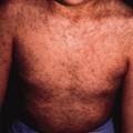
Fig. 43.2 Erythema with a glazed appearance on the plantar foot with fissuring which is accentuated at the great toe.

Interestingly, JPD has not been seen as frequently in the paediatric dermatology clinic in recent years, according to one set of authors who suggest a decreasing frequency of the condition [8]. It may also be that JPD is now more commonly recognized by pediatricians and other primary care providers and not referred as often to dermatologists.
Differential Diagnosis.
Tinea pedis may be considered in the differential diagnosis but it would be unusual without interdigital involvement. A potassium hydroxide preparation and mycological studies can help to rule out this condition if it is suspected. Allergic contact dermatitis may also be considered but it is typically characterized by more intense pruritus, along with involvement of the dorsal foot and lichenification in long-standing cases [16]. Of note, rubber components were found to be the principal allergens in one study of allergic contact dermatitis of the feet, followed by chromated leather and adhesives [17]. Since secondary sensitization is rare in JPD, patch testing is not typically necessary. Psoriasis may also affect the feet but would probably be accompanied by thick, silvery scale and often involves the non-weight bearing areas of the plantar foot.
Histology.
Related posts:


Stay updated, free articles. Join our Telegram channel

Full access? Get Clinical Tree


