32 Surgical approaches to the hand and wrist
INTRODUCTION
The intricate anatomy of the hand dictates the use of specific incisions and approaches to expose various areas or structures of the hand and wrist. Each approach is aimed to minimize manipulation or trauma to the surrounding structures and to prevent the compromise of eventual function of the hand while giving the surgeon adequate exposure to the area of interest. Approaches to common surgical problems in the hand are presented here.
GUYON’S CANAL APPROACH
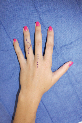
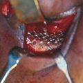
Guyon’s canal is an enclosed fibro-osseous space at the volar/ulnar base of the hand traversed by the ulnar artery and nerve. Its borders include the pisiform (ulnar wall), the hook of the hamate (radial wall), the volar carpal ligament (roof), and the transverse carpal ligament (floor). The exposure can be used to address fractures of the pisiform and the hook of the hamate, as well as pisotriquetral arthritis, or release of the ulnar nerve in ulnar tunnel syndrome.
Prior to beginning the approach, it is important to identify and mark the pisiform and the hook of the hamate. The pisiform is found immediately distal to the wrist flexion crease in line with the flexor carpi ulnaris (FCU) tendon. The hook of the hamate is located 2 cm distal and 2 cm radial to the pisiform. Starting at the hook of the hamate, a zigzag incision is extended proximally, just radial to the pisiform. It is important not to cross the flexion crease perpendicularly. Cutaneous branches of the ulnar nerve run through the subcutaneous tissues here, so careful subcutaneous dissection is imperative. Proximally, the FCU tendon is identified and mobilized. The ulnar nerve and artery are deep and just radial to the tendon, with the nerve typically superficial and ulnar to the artery. The neurovascular bundle can be followed distally, deep to the volar carpal ligament and palmaris brevis muscle, which are both divided longitudinally. Advancing deeper, the motor branch of the ulnar nerve dives below a fibrous arch at the origin of the hypothenar muscles. This branch runs adjacent to the hook of the hamate on its way to innervate the interosseous muscles. This branch must be protected during excision of the hook of the hamate as it is dissected away from surrounding soft tissue. As the neurovascular bundle is released, it can be retracted to give safe access to the pisiform and hamate.
APPROACH TO THE METACARPAL BONE
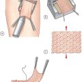
Approach to the metacarpal bone is typically achieved with a dorsal longitudinal incision either directly over the metacarpal shaft or in the intermetacarpal space if two metacarpal bones need to be accessed. The location of the incision is centered over the area of interest (e.g., in the case of shaft fracture, over the midshaft of the metacarpal bone, while for carpometacarpal [CMC] arthroplasty or arthrodesis, it is placed more proximally) (see Figure 32.1). Overlying extensor tendons must be identified and protected when necessary. An attempt must be made to also identify and protect small superficial dorsal branches of the radial and ulnar nerves. The periosteum is then incised longitudinally along the length of the metacarpal shaft, with care taken to preserve the interosseous muscles on either side. Using a periosteal elevator, the muscles along with the periosteum are mobilized to give access to the bone while avoiding circumferential stripping and keeping exposure to a minimum.
DORSAL APPROACH TO THE WRIST
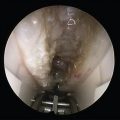
The dorsal midline approach to the wrist provides excellent exposure to the carpal bones (except the trapezium and pisiform), as well as the extensor tendons at the level of the wrist, the second through fifth CMC joints, and the triangular fibrocartilage. It is the utilitarian approach to the dorsal wrist, and its multiple uses include repair of extensor tendons, wrist fusion, radial styloidectomy, repair of the intercarpal ligament, perilunate dislocation, open reduction internal fixation (ORIF) of the distal radius and carpal fractures, and proximal row carpectomy, among others. 1 , 2
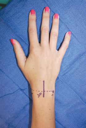
A 6- to 8-cm mid-dorsal incision is centered over the radiocarpal joint. To appropriately identify the position of the radiocarpal joint, one should mark the radial styloid and ulnar styloid, as well as the Lister tubercle on the dorsum of the wrist (Figure 32.2). The incision can be extended as needed. Care must be taken distally to identify and avoid the superficial radial and ulnar dorsal nerves at the base of the third metacarpal. Superficially, the subcutaneous fat is incised in line with the skin incision, revealing the extensor reticulum. Incise the extensor retinaculum over the third dorsal compartment and release the extensor indicis proprius (EIP) tendon. The retinaculum will be opened ulnarly by exposing the contents of the fourth dorsal compartment.
Retracting these tendons radially and ulnarly will expose the underlying dorsal radiocarpal joint capsule and the dorsal radiocarpal ligaments. The capsule can then be incised using an inverted T-shaped incision (direct longitudinal approach) or a ligament-sparing approach. 3 If the approach needs to be extended proximally, retinacular flaps will be raised on the radial and ulnar sides to expose the second through fifth dorsal compartments.
When the entire proximal row and midcarpal row need to be accessed, a ligament-sparing approach is best utilized. In this technique, a radially based dorsal capsular flap is raised by incising along the fibers of the radio-triquetral ligament and dorsal intercarpal ligaments and connecting them over the dorsal horn of triquetrum.
DORSAL ULNAR APPROACH
This approach provides access to the dorsal ulnar side of the wrist, including the ulnar carpal bones, the TFCC, distal radioulnar joint, and the ulnar head. Incision is placed over the fifth dorsal compartment. A similar mid-dorsal incision through the skin can also be used. Incision is centered over the area of interest. The extensor retinaculum is incised over the fifth dorsal compartment. The extensor digiti minimi (EDM) is mobilized and retracted. The joint can then be exposed with careful dissection with minimal dorsal capsulotomy, avoiding the TFCC and dorsal radioulnar ligament. 4 At risk in this approach is the dorsal sensory branch of the ulnar nerve during subcutaneous dissection. 5
Related posts:

Stay updated, free articles. Join our Telegram channel

Full access? Get Clinical Tree


