Abstract
This chapter provides an overview of the pertinent musculoskeletal anatomy of the hand and wrist. Radiographic landmarks and descriptions of various views are provided. Several common fracture patterns are introduced, with representative radio-graphic images.
3 Radiographic Anatomy
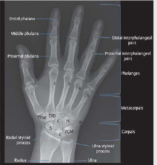
I. Bony Anatomy of the Hand
See ► Fig. 3.1.
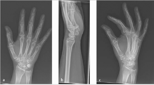
II. Radiograph Views
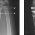
Three view of the wrist should be obtained: Posteroanterior (PA), lateral, and oblique views (► Fig. 3.2).
Posteroanterior (PA) view—obtained with wrist and elbow at shoulder height; the radius and the ulna are parallel (► Fig. 3.2a).
Able to see the extensor carpi ulnaris groove radial to the midportion of the ulnar styloid.
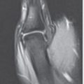
Lateral view—obtained with elbow adducted to the side; shoulder, elbow, and wrist are in plane (► Fig. 3.2b).
Perpendicular to the PA view.
Oblique view—hand is rotated externally 45 degrees from the PA position with fingers extended (► Fig. 3.2c).
Helpful in the trauma setting, including distal radius fractures, metacarpal fractures.
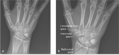
III. Joint Spaces: Parallelism and Symmetry
The joint spaces of the wrist normally have a width of 2 mm or less (► Fig. 3.3).
Radiocarpal joint is slightly wider than the rest, carpometacarpal joints are slightly narrower.
The capitolunate joint is used as reference to which other joint spaces can be compared.
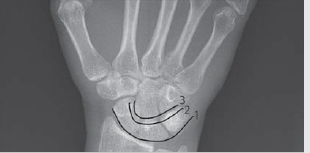
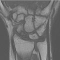
IV. Carpal Arcs
Gilula’s lines are three arcs drawn on the PA view, which are used to assess the alignment of the carpal bones (► Fig. 3.4):
First arc—smooth curve outlining the scaphoid, lunate, and triquetrum.
Second arc—the distal concave surfaces of the same bones.
Third arc—proximal curvature of the capitate and hamate.
Break in arc indicates fracture, ligamentous injury, and lunate/perilunate dislocation.
V. Shape of Carpal Bones
Proximal row
Scaphoid: Shaped like a twisted peanut.
Largest bone in the proximal carpal row.
Lunate: Crescent-shaped
Normally trapezoidal in shape, but can appear triangular if displaced.
Triquetrum: Three-sided.
Pisiform: Pea-shaped.
Distal row
Trapezium: Irregular-shaped four-sided.
Trapezoid: Four-sided
Capitate: Largest bone in the distal carpal row.
Hamate: Shaped like a hook.
Related posts:
 6 Fractures, Dislocations, and Ligament Injuries of the Hand
 8 Fractures and Dislocations of the Distal Radius and Ulna
 12 Tendinitis, Tendinosis, and Dupuytren’s Contracture
 15 Extensor Tendon Injury, Repair, and Reconstruction
 Private: 20 Vascular Pathology of the Hand and Upper Extremities, including Kienbock’s Disease
 Private: 25 Fractures of the Pediatric Hand
Stay updated, free articles. Join our Telegram channel

Full access? Get Clinical Tree


