Systemic Parasitic Infections 
Leishmaniasis ICD-9: 085.9  ICD-10: B55
 Etiology. Many species of obligate intracellular protozoa Leishmania; predominant species are:
 New World: Leishmania mexicana complex, Viannia subgenus
 Old World: L. tropica, L. major, L. aethiopica
 Vector. Sandflies. Old World: Phlebotomus. New World: Lutzomyia
 Pathogenesis. Infection of macrophages in skin, naso-oropharyngeal mucosa, and the reticuloendothelial system (viscera). Diversity of clinical syndromes due to particular parasite, vector, and host species.
Clinical Syndromes
Cutaneous leishmaniasis (CL) characterized by development of single or multiple cutaneous papules at the site of a sandfly bite, often evolving into nodules and ulcers, which heal spontaneously with a depressed scar.
• New World cutaneous leishmaniasis (NWCL)
• Old World cutaneous leishmaniasis (OWCL)
Diffuse (anergic) cutaneous leishmaniasis (DCL)
Mucosal leishmaniasis (ML)
Visceral leishmaniasis (VL); kala-azar; post–kala-azar dermal leishmaniasis (PKDL)
Synonyms: NWCL: chiclero ulcer, pian bois (bush yaws), uta. OWCL: Baghdad/Delhi boil or button, oriental/Aleppo sore/evil, bouton d’Orient. ML: Espundia. VL: Kala-azar (Hindu for black fever)
Epidemiology and Etiology
Infection in humans is caused by 20 Leishmania species (Leishmania and Viannia subgenera). Stages of parasite: Promastigote: flagellated form found in sandflies and culture; amastigote: nonflagellated tissue form (2-4 μm in diameter); replicates in macrophage phagosomes in mammalian hosts.
Transmission. Vector-borne by bite of infected female phlebotomine sandflies (2-3 mm long), which become infected by taking blood meal from infected mammalian host. About 30 species of sandflies have been identified as vectors. Sandflies are weak noiseless fliers; they rest in dark, moist places, typically most active in evening and nighttime hours. Other modes: congenital and parenteral (i.e., by blood transfusion, needle sharing, laboratory accident).
Reservoirs. Varies with geography and leishmanial species. Zoonosis involves rodents/canines.
Vectors. Transmitted by 30 species of female sandflies of genus genera Lutzomyia (New World) and Phlebotomus (Old World).
Prevalence. Estimated 12 million people infected worldwide. 1.5-2 million new cases annually; 350 million individuals are at risk of infection. 50% of new cases are in children. 75,000 individuals die annually of ML.
Geography. All inhabited continents except Australia; endemic in focal areas of 90 countries. Tropics, subtropics, southern Europe. More than 90% of cases of CL occur in Afghanistan, Algeria, Iran, Iraq, Saudi Arabia, Syria, Brazil, and Peru. Climates: Range from deserts to rain forests, rural to urban.
Host Defense Defects. Leishmania-specific anergy: patients develop DCL. Poor immune response or immunosuppression (HIV disease): VL. Hyperergic variant: Leishmaniasis recidivans caused by L. tropica.
Pathogenesis
The clinical and immunologic spectrum of leishmaniasis parallels that of leprosy. CL occurs in a host with good protective immunity. MCL occurs in those with an intense inflammatory reaction. DCL occurs with extensive and widespread proliferation of the organism in the skin but without much inflammation or tendency for visceralization. VL occurs in the host with little immune response and/or in immunosuppression. Unlike leprosy, extent and pattern are strongly influenced by the specific species of Leishmania involved. Additional factors that affect the clinical picture: number of parasites inoculated, site of inoculation, nutritional status of host, and nature of the last nonblood meal of vector. Infection and recovery are followed by lifelong immunity to reinfection by the same species of Leishmania. In some cases, interspecies immunity occurs.
Clinical Manifestation
Primary lesions occur at site of sandfly bite, usually on exposed site.
Incubation Period. Inversely proportional to size of inoculum: shorter in visitors to endemic area. OWCL: L. tropica major, 1-4 weeks; L. tropica, 2-8 months; acute CL: 2-8 weeks or more.
Symptoms. Noduloulcerative lesions usually asymptomatic. With secondary bacterial infection, may become painful.
NWCL: L. mexicana complex. Small erythematous papule develops at sandfly bite site, evolving into ulcerated nodule (Fig. 29-1). Enlarges to 3-12 cm with raised border. Nonulcerating nodules may become verrucous. Lymphangitis, regional lymphadenopathy. Isolated lesions on hand or head usually do not ulcerate. Eventually lesion heals with a depressed scar. Ear lesions may persist for years, destroying cartilage (chiclero ulcers) (Fig. 29-2).
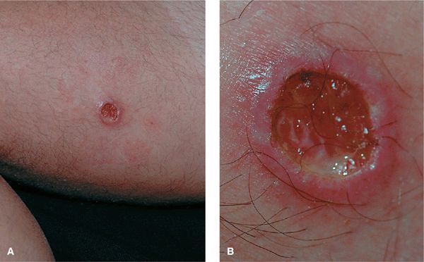
Figure 29-1. New World cutaneous leishmaniasis: ulcer on thigh A 42-year-old with HIV disease noted a painless lesion on the medial thigh 6 weeks after returning from Mexico (A) ulcer with rolled borders and base with granulation tissue (B). Leishmania were seen on lesional biopsy. L. mexicana was isolated on tissue culture of lesional biopsy.
Figure 29-2. New World cutaneous leishmaniasis: chiclero ulcer A deep ulcer on the helix at the site of a sandfly bite. This variant typically occurs in leishmaniasis acquired in Central and South America.
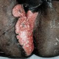
ML. Characterized by naso-oropharyngeal mucosal involvement, a metastatic complication of CL. Mucosal disease usually becomes evident several years after healing of original cutaneous lesions; cutaneous and mucosal lesions can coexist or appear decades apart. Edema and inflammatory changes lead to epistaxis and coryzal symptoms.
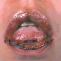
In time, nasal septum, floor of mouth, and tonsillar areas destroyed (Fig. 29-3). Results in marked disfigurement (referred to as espundia in South America). Death may occur due to superimposed bacterial infection, pharyngeal obstruction, or malnutrition

Figure 29-3. Mucocutaneous leishmaniasis: espundia Painful, mutilating ulceration with destruction of portions of the nose. (Courtesy of Eric Kraus, MD.)
OWCL. Begins as small erythematous papule, which may appear immediately after sandfly bite but usually 2-4 weeks later. Papule slowly enlarges to 2 cm over a period of several weeks and assumes a dusky violaceous hue (Figs. 29-4, 29-5). Eventually, lesion becomes crusted in center with a shallow ulcer and raised indurated border = volcano sign. In some cases, the center of the nodule becomes hyperkeratotic, forming a cutaneous horn. Small satellite papules may develop at periphery of lesion, and occasionally subcutaneous nodules along the course of proximal lymphatics. Peripheral extension usually stops after 2 months, and ulcerated nodule persists for another 3-6 months, or longer. The lesion then heals with a slightly depressed scar. In some cases, CL remains active with positive smears for 24 months (nonhealing chronic CL). The number of lesions depends on the circumstances of the exposure and extent of infection within the sandfly vector. May result in multiple lesions, up to 100 or more (Figs. 29-4, 29-5).
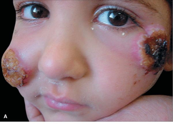
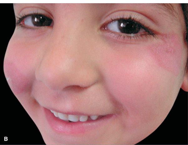
Figure 29-4. Old World cutaneous leishmaniasis: face A 7-year-old Jordanian girl with painful lesions on the cheeks for 6 weeks. (A) Large crusted nodules with surrounding edema on both cheeks. (B) Three weeks after successful therapy (sodium stibogluconate pentostam injections; 15 mg/kg per day IM injection for 21 days), lesions have healed with minimal residual erythema and no scarring. (Courtesy of Mohammad Tawara, MD.)
Figure 29-5. Old World cutaneous leishmaniasis Multiple, crusted nodules on the exposed back, arising at sites of sandfly bites. Many of the lesions resemble a volcano with a central depressed center, i.e., volcano sign.
DCL. Resembles lepromatous leprosy; large number of parasites in macrophages in dermis; no visceral involvement. In Old World, occurs in 20% of individuals with leishmaniasis in Ethiopia and Sudan. In South America, attributed to a member of L. braziliensis complex. Presents as a single nodule, which then spreads locally, often through extension from satellite lesions, and eventually by metastasis. In time, lesions become widespread with nonulcerating nodules appearing diffusely over face, trunk. Responds poorly to treatment.
Leishmaniasis Recidivans (LR). Complication of L. tropica infection. Dusky-red plaques with active, spreading borders and healing centers, giving rise to gyrate and annular lesions. Most commonly affects face; can cause tissue destruction and severe deformity.
PKDL. Sequel to VL that has resolved spontaneously or during/after adequate treatment. Lesions appear ≥1 year after course of therapy with macular, papular, nodular lesions, and hypopigmented macules/plaques on face (Fig. 29-6), trunk, and extremities. Resembles lepromatous leprosy when lesions are numerous. Develops in 20% of Indian patients treated for VL caused by L. donovani and in a small percentage of Ethiopian patients with VL caused by L. aethiopica.
Figure 29-6. Indian post-kala-azar dermal leishmaniasis. Coalescent erythematous dermal papules and nodules over the face in a picture similar to leonine facies. (Used with permission from Raj Kubba, MD.)
VL. Can remain subclinical or become symptomatic, with acute, subacute, chronic course. Inapparent VL cases outnumber clinically apparent cases. Malnutrition is risk factor for clinically apparent VL. Bone marrow, liver, spleen are involved. Term kala-azar (Hindi for “black fever,” some patients had gray color) refers to profoundly cachectic febrile patients with life-threatening disease. Patients present with fever, splenomegaly, pancytopenia, and wasting.
Differential Diagnosis
Acute CL. Insect bite reaction, impetigo, ecthyma, furuncle, Mycobacterium marinum infection, furuncular myiasis, chancre.
Diagnosis
Clinical suspicion, confirmed by demonstrating:
• Intracellular nonflagellated amastigote in biopsy of skin, mucosa, liver, lymph nodes or aspirate of spleen, bone marrow, lymph node.
• Flagellated promastigote in culture of tissues (requires up to 21 days).
Course
In general, NWCL tends to be more severe and progressive than OWCL.
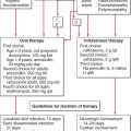
Treatment
Antimony-containing compounds meglumine antimoniate and sodium stilbogluconate (Fig. 29-4) are given systemically. Other drugs used to treat leishmaniasis: amphotericin B, ketoconazole, miltefosine, paromomycin, and pentamidine.