28 Other Orbital Fractures
Abstract
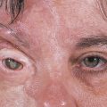
“Other Orbital Fractures” discusses fractures involving the orbit other than orbital wall blowout fractures or zygomatic complex fractures. Le Fort II mid-facial fracture lines pass through the frontal processes of the maxilla, the lacrimal bones, and the orbital floors, may be accompanied by orbital wall blowout fractures, and may involve the lacrimal drainage system. A Le Fort III fracture detaches the entire facial skeleton from the skull base, involving the medial and lateral orbital walls and orbital floor. Naso-orbital fractures, one of the most common patterns of fracture affecting the facial skeleton, are most often the result of a severe impact across the bridge of the nose. Orbital roof fractures, which may also involve the brain, the cribriform plate, and the frontal sinuses, are usually caused by severe blunt trauma or occasionally by penetrating injuries such as falling on a pointed object. Patients may present with apparently only a minor periocular laceration. Such injuries can lead to very serious neurological consequences.
28.1 Midfacial Fractures
The site and extent of midfacial fractures depends on the type of impact and its direction and degree of severity. It is useful to have an understanding of the Le Fort classification of these fractures, although pure LeFort fractures are rarely encountered in clinical practice: they are often quite asymmetrical.
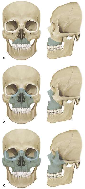
28.2 Le Fort Classification
A Le Fort I fracture is a transverse fracture through the lower part of the maxilla above the teeth. The orbit is not involved (Fig. 28‑1a).
A Le Fort II fracture has a pyramidal configuration involving the nasal, lacrimal, and maxillary bones. The fracture lines extend through the frontal processes of the maxilla, through the lacrimal bones, the floor of the orbits, and the area of the zygomaticomaxillary suture lines and involve the pterygoid plates (Fig. 28‑1b). Orbital wall blowout fractures may be present, and the lacrimal drainage system may also be involved.
A Le Fort III fracture represents a true craniofacial dysjunction in which the entire facial skeleton is detached from the skull base and suspended only by soft tissues. This fracture involves the medial and lateral orbital walls and orbital floor. The fracture lines extend through the superior aspect of the nasal bones, through the frontomaxillary suture area, through the ethmoid sinuses and medial orbital walls, below the optic canal to the inferior orbital fissures, and through the frontozygomatic suture areas (Fig. 28‑1c).
Related posts:

Stay updated, free articles. Join our Telegram channel

Full access? Get Clinical Tree


