21 Nonablative Facial Skin Rejuvenation
Introduction
Signs of aging can be considered to occur by both intrinsic and extrinsic factors. The intrinsic aging occurs as a reflection of time by which there is slow irreversible tissue degeneration. The extrinsic factor, or photoaging, is a term described as the chronic effects of skin exposure to elements, primarily ultraviolet radiation. 1 Photodamaged skin accumulates elastin-containing fibrils in the papillary dermis and mid-dermis, a process referred to as solar elastosis. 2 Collagen, which makes up 80% of the dermis, becomes disorganized and displays increased breakdown and architectural changes in the network formation. Clinically, these changes result in rhytids, skin laxity, dyschromia, and telangiectasias. Smoking can expedite the normal aging process of the skin, contributing to increased skin laxity and rhytids. Dermabrasion, chemical exfoliation, and resurfacing lasers are the gold standard ablative treatment modalities for improving overall facial appearance. These ablative treatments result in epidermal effacement in addition to stimulating collagen synthesis in the dermis. 3 Treatment of the dermis while leaving the epidermis intact reduces the healing process. Attempts to minimize risk and patient downtime have led to an increased interest in nonablative techniques for facial skin rejuvenation.
The public demand for noninvasive, nonablative methods of rejuvenation have surpassed that of ablative devices. 4 The key to nonablative rejuvenation is the prevention of epidermal injury. 5 Currently, nonablative techniques use optical, electrical, or acoustical methods to transfer energy through the epidermis in order to selectively damage dermal collagen and stimulate a subsequent synthesis of reactive collagen while largely preserving the integrity of the epidermis. 6 The principle mechanism of these nonablative devices is creation of spatially selective thermal injury. By reaching the threshold temperature for collagen denaturation of 60° to 65°C, contraction of collagen in the dermis occurs immediately along with an inflammatory response that results in delayed generation of new collagen reorganization and thickening. Uncontrolled heating may lead to temperature elevations at greater depths in the tissue that may lead to necrotic injury, irregular wound contraction, fat loss, and fat atrophy. 7 Likewise, excessive temperature elevations at the surface may create unacceptable first- or second- degree burns that negate the advantage of a nonablative therapy.
Optical technologies for nonablative resurfacing can be classified into three main groups: mid-infrared lasers that target the dermis; visible lasers such as the pulsed dye laser (targeting primarily the dermal vasculature); and intense pulsed light sources (presumably the dermal vascular as well). Intense pulsed light devices are not lasers, and rely upon incoherent broadband light sources. 4 In addition to the laser systems, radiofrequency (RF) and ultrasonic devices can be used for nonablative rejuvenation. There are many different nonablative devices available ( Table 21.1 ). It is difficult to truly compare the devices due to the various treatment regimens, settings, subjective outcome measures, and constantly changing commercial landscape. In addition, the selection of a device also depends on the targeted pathology in the skin—for example, wrinkles, texture changes, telangiectasias, dyschromias, etc. A major advantage of many nonablative devices is the ability to treat darker skin types who would not be candidates for traditional ablative resurfacing treatments due to the high risk of hypo- or hyperpigmentation. The lack of downtime and low risk profile make nonablative treatments an attractive alternative to treatments that damage the epidermis during therapy in patients seeking facial rejuvenation. 8 With most nonablative modalities, serial treatment is advocated to achieve gradual, cumulative improvement, and the final cosmetic result is generally not immediately apparent as new collagen deposition occurs over months. 9 This chapter reviews some of the common current nonablative treatment devices available and their mechanisms.
Light Systems
Intense Pulsed Light
Intense pulse light (IPL) systems are high-intensity light sources that emit incoherent polychromatic light over a broad wavelength spectrum (515 to 1,200 nm). This polychromatic light can be manipulated with filters to achieve selective photothermolysis. Selective photothermolysis refers to the ability to target a particular structure or tissue while leaving the surrounding structures unharmed. IPLs produce good results when treating pigmented and vascular lesions, but improvement in skin texture and rhytids have been less impressive. 10 IPL achieves improvement in pigmented and vascular dyschromias by targeting both melanin and hemoglobin. Indications for IPL include hair removal, facial telangiectasias, pigmentation, poikiloderma, scars, and facial rejuvenation. 5 Treatment for facial rejuvenation typically consists of a series of three to six procedures performed at 3- to 4-week intervals. There is no downtime and the patient may return to all usual activities immediately following the procedure. Patients can expect the pigmented lesions to darken 48 hours after the procedure and slough within 1 week. Common side effects seen using IPL include edema, erythema, blisters, and crusting. Less frequent complications include hypo- or hyperpigmentation and scarring. 10
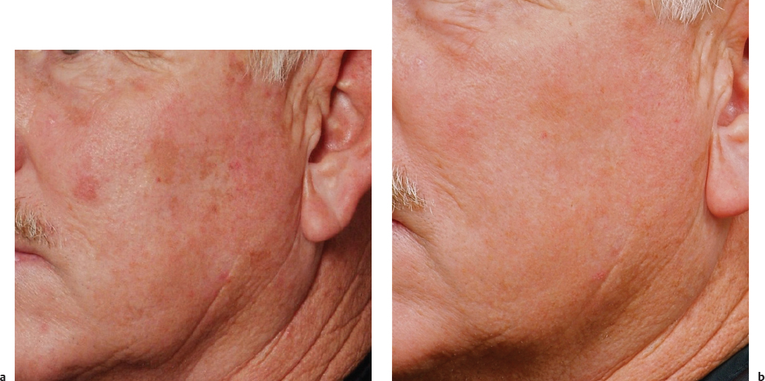
In the skin, photodynamic rejuvenation refers to the use of the combination of IPL with a photodynamic therapy (PDT) such as 5-aminolevulinic acid (ALA), though lasers can be used to activate this drug as well. 11 Administration of exogenous ALA combined with activation with a laser (595 nm) has been described to treat actinic keratosis. Using a broader wavelength such as an IPL with ALA has been shown to be clinically effective in the treatment of precancerous skin conditions and provide significant improvement in photodamage, mottled pigmentation, fine lines, tactile roughness, and skin tightness ( Fig. 21.1 ). 12 , 13
1,100–1,800 nm Infrared
Titan (Cutera) emits light at wavelengths of 1,100–1,800 nm with a long multisecond duration pulse. 14 Water is the chromophore for this device. Because a local peak for water absorption is near 1,450 nm, the light is filtered at wavelengths of 1,400–1,500 nm in order to slow the overall rate of energy absorption and allow a more prolonged, uniform temperature profile. 15 , 16 Volumetric heating of tissue (primarily the dermis) is produced to depths of 1–3 mm, but some heat is conducted to depths of 5 mm under select conditions. 15 As in other modalities, dermal heating results in new collagen production and remodeling which produces skin contraction and tightening. Ideal patients are those with mild to moderate skin laxity who seek tightening. Patients also report improvement in skin texture, reduction in pore size, and reduction in fine wrinkles. 17 Side effects include temporary erythema. 15 Treatment is safe for darker skin, types IV and V. 17
Related posts:


Stay updated, free articles. Join our Telegram channel

Full access? Get Clinical Tree


