Contact Napkin Dermatitis
Primary Irritant Contact Napkin Dermatitis
Irritant contact dermatitis is by far the most prevalent form of napkin dermatitis. In fact, this ‘chafing’ napkin rash is probably the most common skin problem of infancy. It is precipitated by the constant moisture, occlusion and frictional forces that occur under a napkin and which compromise skin integrity. With maceration of the stratum corneum, the skin barrier function is impaired, predisposing it to secondary irritants. These secondary factors include urinary ammonia, increased urine pH (due to urea-splitting bacteria), faecal proteases and lipases, Candida albicans, bacterial overgrowth and frequent washing, especially with detergent soaps [1,2]. Urinary ammonia was for many years mistakenly believed to be the primary instigator of irritant napkin dermatitis. It is now felt to increase inflammation only in already compromised skin [3]. In some cases, irritant contact dermatitis may arise from topically applied medicaments designed to protect the napkin area, as has been described with Oilatum Plus® (J. Harper, personal communication).
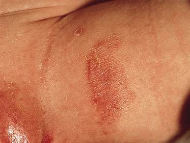
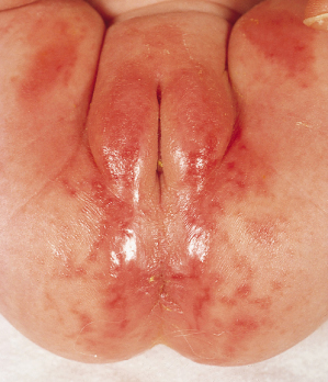
Clinically, irritant contact napkin dermatitis is characterized by a glazed, confluent erythema, sometimes resembling a burn. There may be erythematous papules, oedema and scaling of the involved skin (Fig. 20.1). When the eruption begins to resolve, a wrinkled parchment-like appearance may be noted. The rash, however, may wax and wane. This form of napkin rash primarily involves the skin where contact with the napkin is greatest, e.g. the convexities of the buttocks, medial thighs, mons pubis and scrotum or labia majora. The intertriginous areas are generally spared. An early manifestation of this type of napkin dermatitis, especially in infants less than 4 months of age, is mild perianal erythema [4].
Fig. 20.1 Irritant contact napkin dermatitis. Erythema involves primarily convexities of the napkin area.
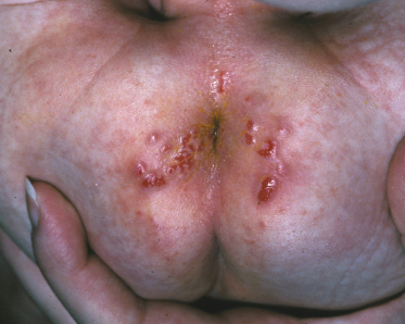
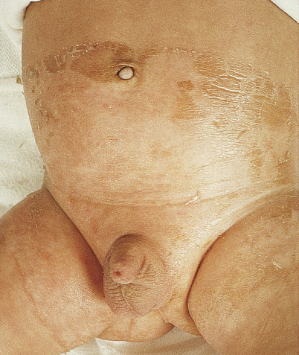
There are two less common morphological subtypes of irritant contact napkin dermatitis. One is the so-called ‘tide water’ mark dermatitis in which an erythematous macular eruption is band-like and confined to the margins of the napkin area on the thighs (Fig. 20.2) or abdomen. This characteristic rash results from the exaggerated chafing that may occur at the edges of the napkin. Skin integrity is compromised by the frequent cycles of wetting and drying [5] combined with the traumatic friction due to the plastic border of a disposable napkin. There is also a severe form of irritant contact napkin dermatitis known as Jacquet dermatitis. This presents with papuloerosive lesions that have a punched-out or crater-like appearance (Fig. 20.3). These ulcers have also been referred to as ‘ammoniacal ulcers’. This eruption, when it occurs, tends to affect older, napkin-wearing children. In male infants, ulceration involving the glans penis and urinary meatus may cause discomfort or difficulty urinating.
Allergic Contact Napkin Dermatitis
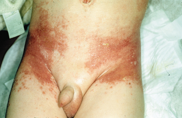
A true allergic contact dermatitis may complicate another type of napkin rash or present de novo. This is an uncommon occurrence, particularly in children under 2 years of age. Allergic contact dermatitis is considered, however, if a child does not respond appropriately to treatment. It should also be suspected if the application of a potential allergen causes the napkin rash to become more intense or to spread. Contact allergy may develop to certain medicaments applied to the skin such as those containing paraben, lanolin or neomycin. Allergic sensitivity may also occur to chemicals contained within disposable napkins or to napkin covers (as with Disperse dyes used to colour napkins) [6] or an irritant reaction may develop to laundry detergents used with cloth napkins (Fig. 20.4) [7].
Fig. 20.4 Toxic napkin dermatitis (induced by bleaching solution).
Courtesy of Professor Arnold Oranje.
Morphologically, allergic contact dermatitis begins with erythema and small vesicles that rupture, leading to an eczematous eruption. The vesicular phase may not be apparent after the first few days of the rash. Allergic contact napkin rash typically complicates a primary irritant napkin rash and therefore follows the same distribution, i.e. the convex surfaces of the skin under occlusion. There may, however, be prominent flexural involvement, particularly if the sensitivity is to a topical preparation which concentrates within the skinfolds [8]. The characteristic pattern of contact dermatitis to rubber components around the proximal thighs and the waistline associated with disposable napkins has been dubbed the ‘holster sign’ [9,10] (Fig. 20.5).
Fig. 20.5 Allergic contact dermatitis secondary to rubber components in the napkin: the ‘holster sign’.
Courtesy of Dr Neil Prose.
Simple Intertrigo
Intertrigo is an inflammatory process that occurs in areas where skin rubs against skin, as in the folds of the groin, posterior thighs and intergluteal cleft. Heat, moisture and sweat retention, coupled with friction, cause maceration, inflammation and sometimes skin erosion. Intertrigo may present with sharply demarcated moist erythema confined to the skinfolds. In contrast to a candidal napkin rash, there is little associated scale and no satellite lesions.
As intertrigo is essentially due to friction and retained moisture, it may be considered a subtype of irritant contact dermatitis. The tropical climate of the napkin area makes the napkin-wearing child susceptible to the development of simple intertrigo; however, it is most often seen in overweight infants [8].
In cases of intertrigo, there may be secondary infection with Candida albicans or bacteria. Also, clinical presentation of intertrigo may be indistinguishable from other disorders in which the role of C. albicans is controversial, such as infantile seborrhoeic dermatitis or inverse napkin psoriasis. This overlap in clinical features may make definitive diagnosis difficult.
Granuloma Gluteale Infantum
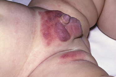
Granuloma gluteale infantum is a rare nodular eruption which may arise within an area of pre-existing irritant napkin dermatitis. It is characterized by firm, painless, reddish-brown to purple nodules varying in size from 0.5 to 4 cm (Fig. 20.6). These nodules usually appear on the buttocks and inner thighs and occasionally on the lower abdomen. Lesions have also been reported outside the napkin area involving the folds of the axilla and neck. These angioma-like swellings can be ominous in appearance, resembling lesions of Kaposi sarcoma or lymphoma. The nodules may persist for weeks to months but they eventually regress spontaneously. When the lesions resolve, atrophic scars may remain.
The aetiology of granuloma gluteale infantum is unclear. Several factors, however, have been suspected. Because this condition invariably arises in an area of pre-existing napkin dermatitis, it may represent a localized cutaneous response to long-standing inflammation. There is no correlation, however, between the severity of the napkin rash and the incidence of this nodular eruption [11]. Lesions may appear even when the napkin rash is resolving. C. albicans has been considered an aetiological factor, but Candida is frequently cultured from common irritant napkin rash and the development of granuloma gluteale infantum in these patients is rare. Finally, fluorinated topical steroids have also been implicated in the development of these nodules as in most reported cases relatively potent topical steroids were used [12]. At this time, however, there is no convincing evidence to support this.
References
1 Berg RW. Etiology and pathophysiology of diaper dermatitis. Adv Dermatol 1988;3:75–98.
2 Weston WL, Lane AT, Weston JA. Diaper dermatitis: current concepts. Pediatrics 1980;66:532–6.
3 Leyden JJ, Katz S, Stewart R et al. Urinary ammonia and ammonia producing microorganisms in infants with and without diaper dermatitis. Arch Dermatol 1977;113:1678–80.
4 Rasmussen JE. Diaper dermatitis. Pediatr Rev 1984;6:77–82.
5 Koblenzer PJ. Diaper dermatitis: an overview. Clin Pediatr 1973;12:386–92.
6 Alberta L, Sweeney SM, Wiss K. Diaper dye dermatitis. Pediatrics 2005;116(3):e450–2.
7 Jacobs AH. Eruptions in the diaper area. Pediatr Clin North Am 1978;25:209–24.
8 Schanzer MC, Wilkin JK. Diaper dermatitis. Am Fam Physician 1982;25:127–32.
9 Larralde M, Raspa ML, Silvia H et al. Diaper dermatitis: a new clinical feature. Pediatr Dermatol 2001;18:167–8.
10 Roul S, Ducombs G, Leaute-Labreze C et al. ‘Luky Luke’ contact dermatitis to the rubber components of diapers. Contact Dermatitis 1998;38:363–4.
11 Sweiden NA, Salman SM, Kibbi AG et al. Skin nodules over the diaper area: granuloma gluteale infantum. Arch Dermatol 1989;125:1706–7.
12 Uyeda K, Nakayasu K, Takaisski Y et al. Kaposi sarcoma-like granuloma on diaper dermatitis. Arch Dermatol 1973;107:605–7.
Infectious Napkin Dermatitis
Candida Napkin Dermatitis
Provided there is sufficient warmth and moisture, as occurs under an occlusive napkin, C. albicans can proliferate on the skin surface and penetrate the stratum corneum. It can then activate complement via the alternative pathway and induce inflammation [1].
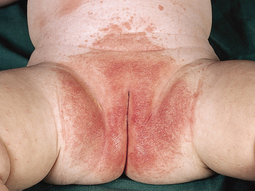
Morphologically, candidal napkin dermatitis is perhaps the most characteristic of the napkin rashes. It presents with sharply marginated, intensely red, scaly plaques with satellite papules and pustules (Fig. 20.7). These satellite lesions may have peripheral scale. In some severe cases of candidiasis, there may be widespread skin erosion. When candidiasis involves the genitalia, there is usually confluent erythema involving the entire scrotum or labia. This is unlike psoriasis, in which genital involvement is usually more localized and in which the lesions are more sharply demarcated.
The diagnosis of candidal napkin rash is primarily based on its characteristic morphology. Potassium hydroxide (KOH) scrapings may reveal pseudo-hyphae and help to confirm the diagnosis. Scrapings may be negative, however, if they are taken from inflamed areas or in long-standing cases in which the inflammatory response has led to the death of the Candida organism. Yield on KOH is greatest from scrapes from the periphery of the rash or a fresh papular or pustular lesion.
Children presenting with candidal napkin dermatitis frequently have a recent history of treatment with broad-spectrum antibiotics. Diarrhoea also makes an infant more susceptible to candidiasis. The oral cavity should be examined to rule out concomitant thrush. When the rash is chronic or recurrent, seeding from the gastrointestinal tract or from a maternal candidal vaginitis should be considered. Other potential sources include maternal mastitis, if the mother is breastfeeding, or a contaminated dummy (pacifier) or nipple.
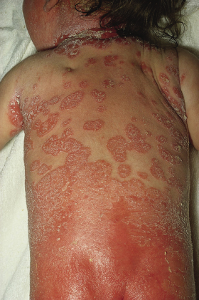
On rare occasions, a psoriasiform skin reaction (Fig. 20.8) may occur as a complication of severe candidal napkin rash. Shortly after therapy is initiated, scaly psoriasiform papules and plaques rapidly develop on the trunk, usually sparing the extremities. This eruption may last for days to weeks. Generally, C. albicans cannot be cultured from these plaques. However, active candidiasis of skinfolds in the neck and axilla, in addition to the inguinal area, may be seen. The pathogenesis of this allergic skin eruption is not well understood; it is presumed to be a response to an antigenic stimulus. Although it was once felt that this reaction suggested an underlying psoriatic or atopic diathesis, most affected infants do not go on to develop psoriasis or atopic dermatitis [2]. Confusion exists, especially in the UK literature, over the entity ‘napkin psoriasis’ [3]. Fergusson et al. [4] clearly showed that ‘napkin psoriasis’ is identical to this psoriasiform allergic skin reaction.
The rare congenital onset of candidal napkin dermatitis may represent a manifestation of congenital cutaneous candidiasis. Neonates with congenital cutaneous candidiasis may present with characteristic macerated areas of erythema in the anogenital region; however, infected infants may also present with papulopustules, widespread erosions or a burn-like dermatitis [5]. In term infants, congenital cutaneous candidiasis is typically a benign, self-limited eruption attributed to vertical transmission from the colonized mother. The condition responds to topical antifungal agents. However, preterm neonates, especially those manifesting signs of respiratory distress, are at risk for associated systemic candidiasis, which warrants systemic antifungal therapy.
Perianal Streptococcal Disease and Streptococcal Intertrigo
Stay updated, free articles. Join our Telegram channel

Full access? Get Clinical Tree


