18 Brachioplasty with an Inferior Scar

Abstract
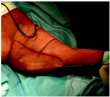
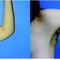
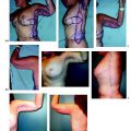
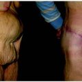
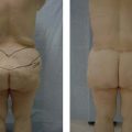
Complications associated with brachioplasty surgery, including the potential for neurovascular injury, mandate that the brachioplasty procedure be simple yet effective. Inexperienced surgeons are more prone to making skin resection errors with either over-or underresection as well as unnatural contours. An algorithm to mark the surgical plan during brachioplasty helps avoid many of these complications. The final scar is placed posterior to the me-dial bicipital groove, along the line between the posterior axillary line and the olecranon. This scar is not visible to the patient in front of a mirror. Simple skin undermining at the level of the superficial fascia is sufficient to correct the deformity of the arm. Unlike many other described techniques, we have not experienced neurovascular complications. The incidence of complications associated with our technique remains less than 7%, consisting mainly of persistent scar hypertrophy and scar contracture.
Introduction
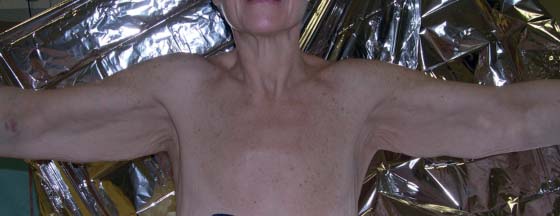
Ptosis of the inferior aspect of the arm, a “bat wing” appearance, is caused by inelasticity of the superficial fascia after loss of deep adipose tissue and by an excess of skin. These changes increase with age. The dermosenescence causes a skin excess and a loss of elasticity predominantly along the medial side of the proximal arm. Brachioplasty was initially described 80 years ago by Thorek.1 Since that description, there have been a large number of techniques described in the plastic surgery literature. Posse,2 in 1946, proposed an elliptical incision on the medial side of the arm to reduce the excess of the skin. In 1954, Correa-Iturraspe and Fernandez3 described correction of the brachial lipodystrophy. Clarkson and Jeffs4 corrected the thoracic wall deformity and the deformity of the arm by a curved suprascapular incision that passed around the posterior aspect of the arm. Pitanguy and Matta5 proposed an S-shaped incision extended onto the anterior axillary line to the inframammary crease. The scar was positioned at the level of the posterior border of the biceps muscle. Franco and Rebello6 published a similar approach to resculpt the arm using a separate L-shaped incision between and below the scapulae to contour the thorax. A quadrangular flap reduction with T-closure was alternatively proposed by Juri et al.7 For restoration of the axillary contour and to lengthen the scar, Guerrero-Santos8 described a small Z-plasty. Regnault9 successfully modified the Juri technique by using an M-shaped axillary incision to reduce the problems of scar retraction. A more complicated W-plasty technique was described by Borges.10
De-epithelialization of a posterior flap that is repositioned to create a bicipital sulcus was proposed by Goddio11 in 1989. Lockwood12 made a very important anatomic description of the superficial fascial system of the arm. On the basis of his observations, he proposed a brachioplasty procedure that provides secure anchoring of the arm flap to the axillary fascia. The scar is positioned anteriorly at the level of the medial bicipital sulcus.
The first classification of the upper arm deformity was described by Teimourian and Malekzadeh.13 This classification system describes four categories of arm deformations. Liposuction of the arms was proposed by Lillis,14 but the surgical indications were very limited. De Souza Pinto et al15 proposed an italic double S-shaped incision along the bicipital sulcus along with liposuction. Richards16 has described a minimal incision brachioplasty with adjunctive liposuction and “half-ellipse incision” at the level of the axilla. In 2004, Strauch et al17 conceived a treatment algorithm based on four treatment zones, helping the surgeon evaluate and treat upper extremity contour deformities. They presented a technique of sinusoidal excision along the posteromedial arm that provided good extremity contour with posterior scars, while simultaneously addressing axillary contour deformities with generous Z-plasty. Abramson18 has described treating moderate bat-wing deformities with short-scar brachioplasty or minibrachioplasty. Other authors have proposed surgical technique modifications of the previously described methods.19–23 As an alternative to brachioplasty, Gilliland and Lyos24 have proposed a circumferential paraaxillary superficial tumescent liposuction for aging women with excess arm fat and poor skin.
Despite the evolution in technique, current brachioplasty surgeries are somewhat unpredictable and are commonly associated with significant untoward results: misplaced, widened, or hypertrophic scars; contour deformities due to overcorrection centrally and underresection proximally and distally; transverse cutaneous folds; and delayed wound healing due to marginal skin necrosis and suture dehiscence. Goals of the surgery should be one preoperative marking that reduces overcorrection problems, making the surgery easier for young surgeons to learn, and determining an optimal position for the residual arm scar.
Indications
The arm in the massive weight loss patient can be considered as a sleeve that is too wide and too long. We consider it important to reduce the axillary diameter at the level of the posterior axillary line. The deformity of the upper arm can be divided into four general categories by the classification scheme of Teimourian and Malekzadeh:13
Group I: Minimal to moderate subcutaneous fat with minimal skin laxity
Group II: Important accumulation of subcutaneous fat with moderate skin laxity
Group III: Obesity and extensive skin laxity
Group IV: Minimal subcutaneous fat and extensive skin laxity
Group I patients are treated simply by liposuction to correct the deformity. For the patients in groups II and III, it is preferable to perform liposuction 3 to 5 months before the excisional surgery to reduce the excess of subcutaneous fat. This delay allows skin retraction and reduces the required amount of skin resection. Group IV patients are treated directly by skin resection. It is very important to perform this surgery only after the patient’s weight has stabilized.
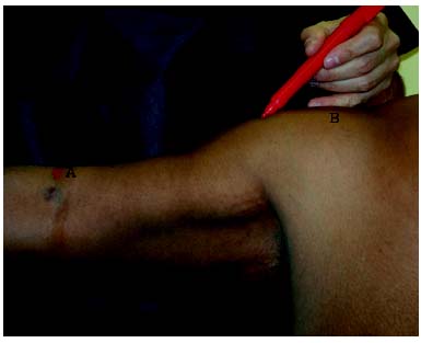
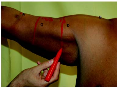
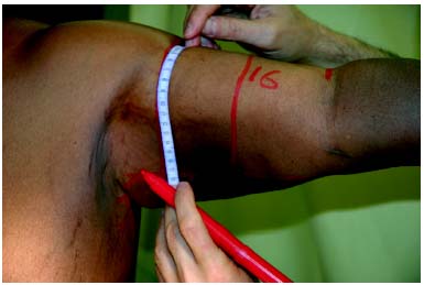
The upper inner arm, from the axillary area to the elbow, is very rich in superficial and deep lymphatic vessels. Large superficial veins, the cephalic and basilic veins, and an important sensory nerve, the medial cutaneous nerve, as well as large vascular trunks and nerves situated deeper immediately above the aponeurosis, are present on the anterior side of the inner arm. Severe lymphedema of the arm can occur in cases of axillary dissection. The most frequent problems are injuries of the lymphatics, which can lead to local seromas and poor healing, and nerve injury. The scar after skin resection is placed posterior to the medial bicipital groove, along the line between the posterior axillary line and the olecranon. This scar is not visible when the patient looks into a mirror.
Related posts:
 17 Brachioplasty with Interdigitation of the Posteromedial Scar
 19 Correction of Arm Ptosis with a Medial Bicipital Scar
 4 Anesthesia and Safety Considerations in the Perioperative Period
 12 Combination Abdominal Wall Hernia Repair and Mid-Body Contouring
 32 Combination Circumferential Abdominoplasty with Medial Thigh Lift
 36 Facial Rejuvenation: Open Technique
Stay updated, free articles. Join our Telegram channel

Full access? Get Clinical Tree


