Spontaneous improvement with complete resolution can occur. Surgical treatment produces the best results when performed before the age of 2 years [1].
Ingrown Nails
- Key diagnostic criteria: paronychia and pyogenic granuloma due to embedding of the nail edges into the lateral nailfold
- Key management features: remove the spicula and treat the inflammation; lateral nail matrix phenolization
- Differentials: nail pyogenic granulomas due to drugs or trauma
Ingrown nails are a common complaint and usually affect the great toe of teenagers and young adults. Predisposing factors include congenital malalignment of the great toenails and congenital hypertrophy of the lateral nailfolds. In the latter condition, the periungual soft tissues of the great toe are hypertrophic and partially cover the nail plate, favouring nail ingrowing [2]. The development of nail ingrowing is favoured by incorrect nail trimming, traumatic injuries and occlusive footwear.
The clinical manifestations of ingrown toenails can be divided into three stages.
- Stage 1. Embedding of the nail spicula within the lateral nailfold produces painful erythema and swelling of the nailfold. Treatment is conservative with extraction of the embedded spicula and introduction of a package of non-absorbent cotton under the lateral corner of the nail. This package should be replaced every few days.
- Stage 2. This stage is characterized by the formation of granulation tissue, which covers the nail plate. The affected nail is very painful and the nailfold presents a pyogenic granuloma with seropurulent exudation. In this stage, the topical application of high-potency steroids under occlusion for a few days can reduce the overgrowth of granulation tissue. Conservative treatment as for stage 1 can then be utilized.
- Stage 3. The granulation tissue becomes covered by newly formed epidermis of the lateral nailfold. This stage requires surgical treatment with selective destruction of the lateral horn of the nail matrix.
Newborns can develop multiple ingrown fingernails with paronychia as a result of the grasp reflex [3]. The pathogenesis of the condition is the repeated compression of the soft tissues of the lateral nailfold by the lateral edges of the nails during grasping. The condition regresses spontaneously when the grasp reflex disappears, at about 3 months of age.
Herringbone (Chevron) Nails
This is a very common finding in fingernails of young children. The nail plate surface presents longitudinal ridges that cross its surface diagonally from the lunula to the distal margin with a V-shaped pattern [4].
Acute Paronychia
- Key diagnostic criteria: acute painful periungual inflammation often with pus discharge
- Key management features: pus drainage, topical antibiotics
- Differentials: pustular psoriasis
Acute paronychia is usually caused by Staphylococcus aureus, although other bacteria and herpes simplex virus may be responsible. A minor trauma commonly precedes the development of the infection.
The affected digit shows acute inflammatory changes of the nailfolds, with erythema, swelling, pus formation and pain. Whenever possible, appropriate cultures should be taken to identify the responsible organism. Treatment includes prompt incision and drainage of the abscess, local medications with antiseptics and administration of systemic antibiotics or aciclovir, depending on the causative agent.
Warts
- Key diagnostic criteria: warty papules of the nailfolds or hyponychium
- Key management features: topical keratolytics
- Differentials: skin xerosis, subungual exostosis
Periungual and subungual warts are very common in children. Warts of this type often affect more than one digit and frequently recur. Nail biting facilitates the spread of periungual warts to several digits. Periungual warts may have the typical ‘warty’ exophytic appearance, or may present as a hyperkeratotic lesion of the nailfolds. Large subungual warts may cause lifting of the nail plate and result in pain.
In young children, surgical procedures should be avoided and treatment should be as conservative as possible [5]. Topical solutions containing salicylic and lactic acids are the treatment of choice. Topical immunotherapy with strong sensitizers (squaric acid dibutylester (SADBE) or diphenylcyclopropenone (DPCP)) is an effective and painless modality of treatment for multiple recalcitrant warts [6]. SADBE or DPCP 2% in acetone can be used for sensitization. After 21 days, weekly applications are carried out with dilutions selected according to the patient’s response. Complete cure usually requires 3–4 months. Similar to warts in other body sites, periungual warts may recur after cure.
Nail Biting and Onychotillomania
- Key diagnostic criteria: paronychia with peeling scales and blood crusts, shortened nails
- Key management features: topical unpleasant-tasting preparations
- Differentials: paronychia due to other causes
Nail biting is common in childhood and affects up to 60% of children. Conversely, onychotillomania is rather uncommon and usually associated with underlying psychological disorders.
Nail biting produces short and irregular nails that show depressions and scratches. Apical root resorption may occasionally occur.
The habit of picking, breaking or chewing the skin over the proximal nailfold produces paronychia and nail matrix injury with nail plate surface abnormalities. Secondary bacterial infections of the periungual tissues are common, as are periungual warts.
Most children discontinue nail biting when they grow up. Frequent application of unpleasant-tasting topical preparations on the nail and periungual skin can discourage patients from biting and chewing their fingernails.
Punctate Leuconychia
- Key diagnostic criteria: small white spots on the fingernails
- Key management features: treatment is not necessary
- Differentials: drug-induced leuconychia
Punctate leuconychia is a traumatic fingernail abnormality that is almost exclusively seen in children. It is usually caused by repetitive minor traumatic injuries to the nail matrix. This process produces a disturbance in nail matrix keratinization and the development of parakeratotic cells in the ventral nail plate. These modify the transparency of the nail plate and appear as white spots (Fig. 150.2). The affected nails show single or multiple small opaque white spots that move distally with nail growth and usually disappear before reaching the distal edge.
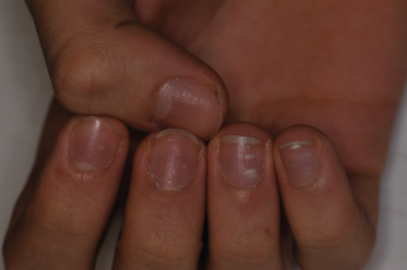
The condition may involve a few or all the fingernails and there may be a variable number of white opaque spots. Although punctate leuconychia is commonly believed to be caused by calcium deficiency, there is no known relationship between this condition and the calcium content of the nail. Punctate leuconychia spontaneously regresses by avoiding trauma.
Atopic Dermatitis
- Key diagnostic criteria: hand and fingernail dermatitis including the periungual skin
- Key management features: topical anti-inflammatory agents
- Differentials: contact eczema
The nails in children with atopic dermatitis may present nail plate surface abnormalities due to eczematous involvement of the nail matrix. These include irregular pitting and Beau’s lines. Onycholysis may occasionally occur as a consequence of eczematous involvement of the fingertips.
Parakeratosis Pustulosa
- Key diagnostic criteria: one digit showing onycholysis and pulp scaling
- Key management features: topical steroids
- Differentials: psoriasis, eczema
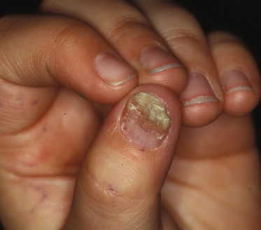
Parakeratosis pustulosa is a chronic condition that exclusively affects children and usually involves a single finger, most commonly the thumb or index finger [7]. In the early phases, the affected digit shows eczematous changes associated with mild distal subungual hyperkeratosis and onycholysis. Nail abnormalities are usually more marked on a corner of the nail. Pitting of the nail plate may be present (Fig. 150.3).
Whether parakeratosis pustulosa is a limited form of nail psoriasis or a clinical manifestation of other conditions, such as contact and atopic dermatitis, is a matter of controversy. In the authors’ experience, most children with parakeratosis pustulosa develop mild nail psoriasis when they become adults [8]. As parakeratosis pustulosa and nail psoriasis produce similar nail changes, the diagnosis of parakeratosis pustulosa is based on the localization of the disease to a single digit rather than on the morphology of the nail lesions. This diagnosis should always be considered in a child with psoriasiform nail changes limited to a single finger. Patch tests can be useful to rule out contact dermatitis.
The nail lesions usually resolve spontaneously. Topical treatment with steroids and/or retinoic acid may induce partial remission of the nail changes.
Psoriasis
- Key diagnostic criteria: irregular pitting, salmon patches of the nail bed, onycholysis with erythematous border
- Key management features: topical vitamin D derivatives or tazarotene on the nail bed after removal of the detached nail plate
- Differentials: onychomycosis, alopecia areata
The prevalence of nail involvement in children with psoriasis ranges from 7% to 39%, according to different studies. The clinical manifestations of nail psoriasis in children are quite similar to those of adults, except for the fact that nail bed involvement is usually absent or mild [9,10].
Related posts:


Stay updated, free articles. Join our Telegram channel

Full access? Get Clinical Tree


